Loading video…


 38 Concepts
 149.7K views

Video Summary: Veins of Head and Neck Explained
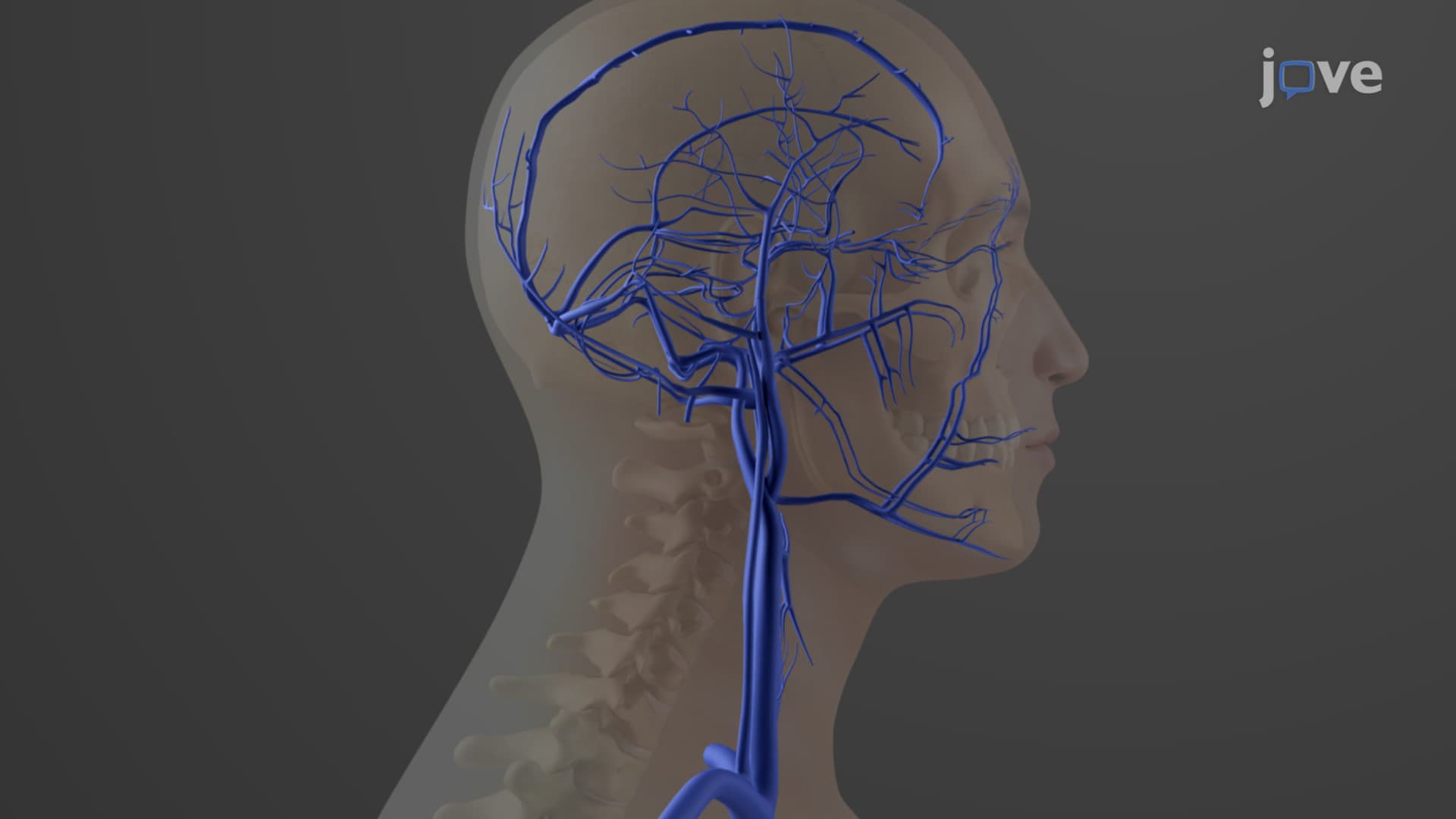
Did you know that your brain requires nearly 20% of your body's blood supply, yet relies on just three pairs of veins to drain this massive volume? Understanding head neck veins anatomy becomes crucial when considering conditions like stroke or brain trauma, which emergency room doctors across the US encounter daily. The veins of head and neck explained reveals how the internal jugular, external jugular, and vertebral veins work together as a sophisticated drainage system, with the internal jugular veins handling the heaviest workload by collecting blood from the brain's intricate dural venous sinuses. Watch the full video on JoVE Coach to master this concept with expert-led visuals and step-by-step explanations.
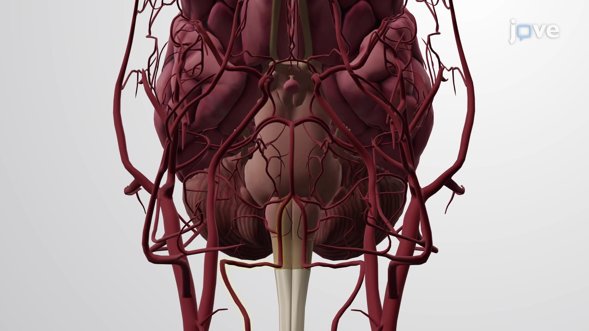
The venous drainage of the head and neck represents one of anatomy's most elegant examples of functional organization. Unlike other body regions where venous patterns mirror arterial supply, head neck veins anatomy follows a unique three-system design that prioritizes efficient blood return from the metabolically demanding brain and sensory organs.
The internal external jugular vein relationship begins with understanding that internal jugular veins serve as the major drainage route for intracranial blood. These vessels originate at the jugular foramina, where they collect blood from an intricate network of dural venous sinus head structures. The superior sagittal sinus, running along the brain's midline, collects blood from the cerebral cortex-the same regions tested extensively in AP Biology when students learn about brain function and blood-brain barrier concepts.
The cerebral vein head drainage system demonstrates remarkable efficiency through the sigmoid sinuses, which curve like the letter "S" before transforming into internal jugular veins. Medical students preparing for the MCAT often struggle with this transition point, but understanding that it occurs at the skull base helps visualize how intracranial pressure changes can affect this critical junction.
The neck venous drainage anatomy extends beyond just internal jugular function. External jugular veins handle superficial drainage from facial structures and scalp regions-areas frequently involved in trauma cases seen in US emergency departments. These vessels terminate in subclavian veins, creating an alternative pathway when internal jugular flow becomes compromised.
Vertebral veins represent the often-overlooked third component, draining the cervical spine and deep neck muscles. Physical therapy students studying neck anatomy for licensing exams like the NPTE recognize these veins' importance in understanding cervical spine pathology and treatment approaches.
For students preparing for healthcare careers, what are the veins of the head and neck becomes more than academic knowledge. NCLEX and HESI A2 exams frequently test venous anatomy in contexts of IV access, recognizing signs of increased intracranial pressure, or understanding stroke pathophysiology. The internal jugular vein's accessibility makes it a common site for central venous catheter placement in intensive care units across major US hospitals like Mayo Clinic or Johns Hopkins.
Understanding venous drainage patterns also proves essential for USMLE Step 1 preparation, where questions often integrate anatomy with pathophysiology, asking students to predict consequences of venous obstruction or explain why certain symptoms occur with specific vascular injuries.
Related Micro-courses