Loading subject page…


149,645 views
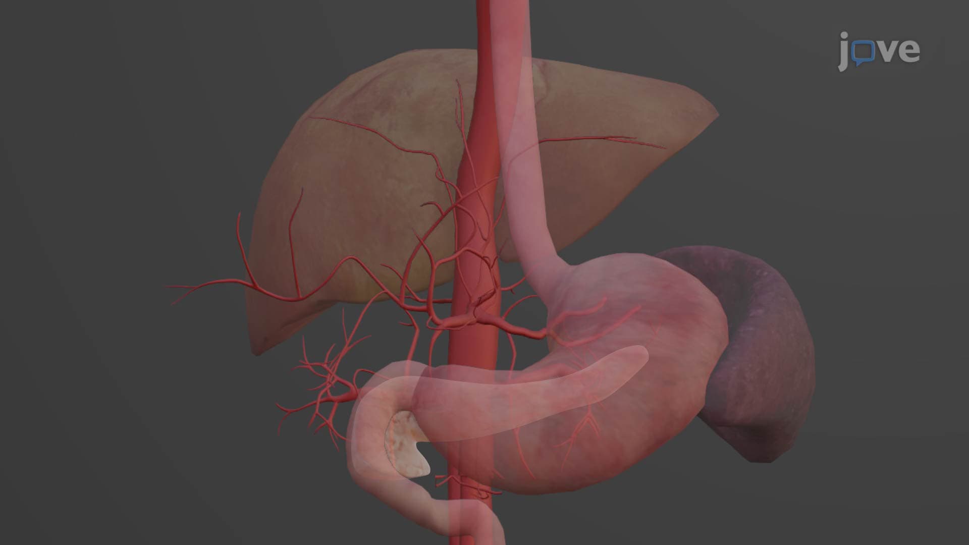

Video Summary: What Is Abdominal Aorta
Did you know that your abdominal aorta anatomy is like a major highway system delivering blood to every organ below your heart? This critical vessel, starting at the diaphragm's aortic hiatus and extending to the L4 vertebra, branches into an intricate network that supplies your digestive organs, kidneys, and reproductive systems. Medical students at Johns Hopkins University study these complex branching patterns as essential knowledge for surgery and radiology. Watch the full video on JoVE Coach to master this concept with expert-led visuals and step-by-step explanations.
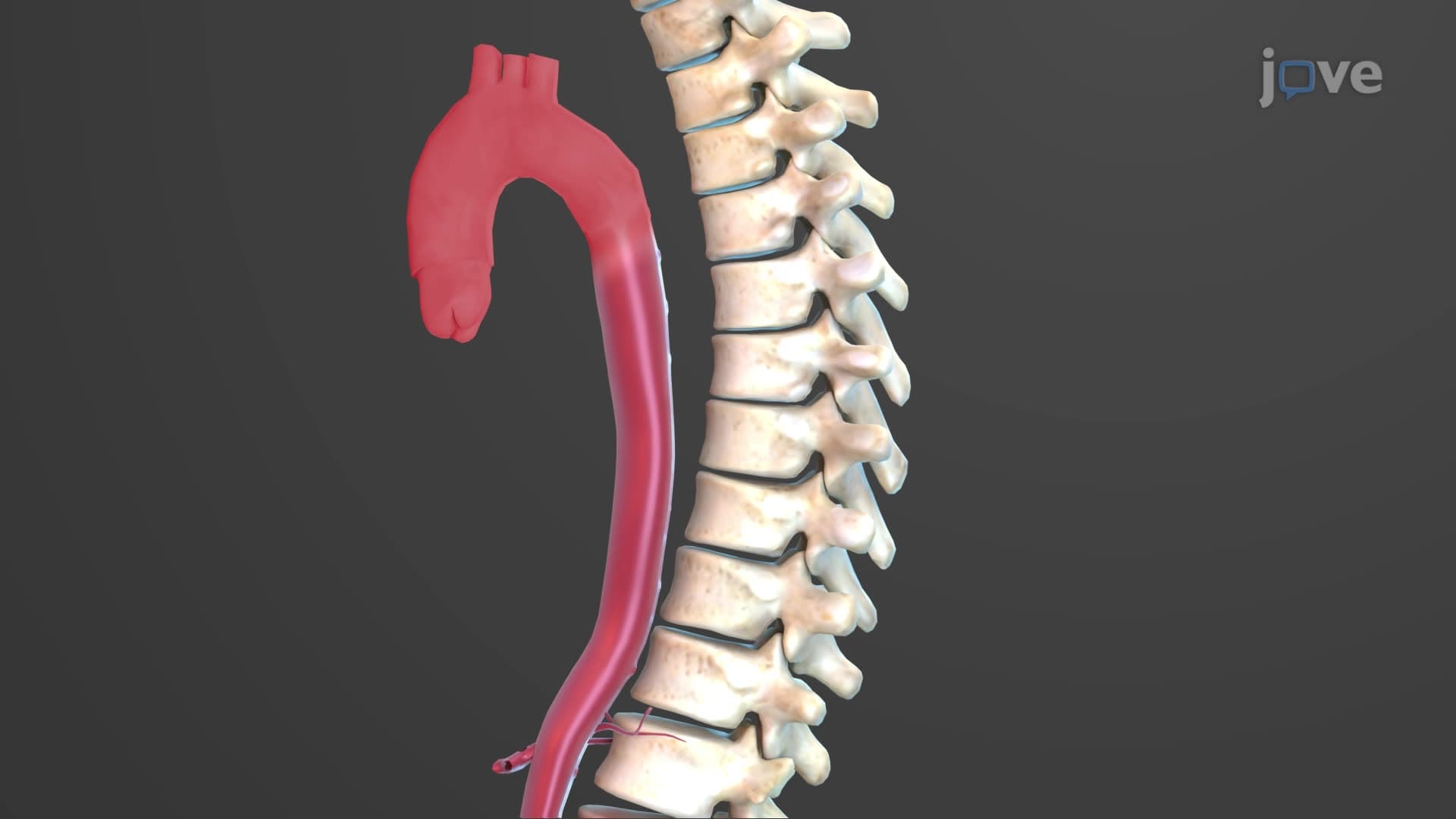
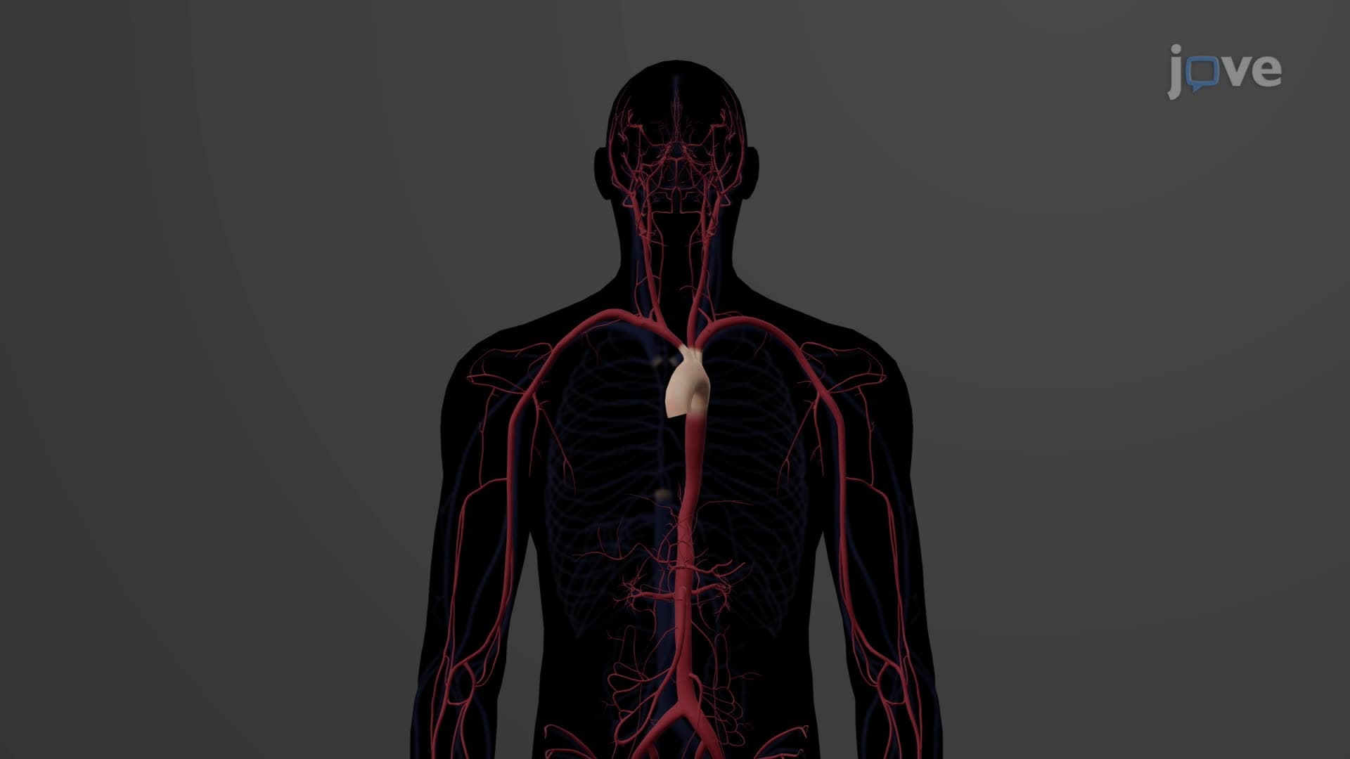
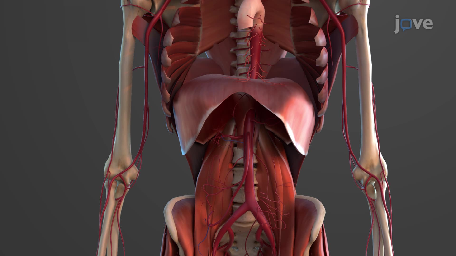
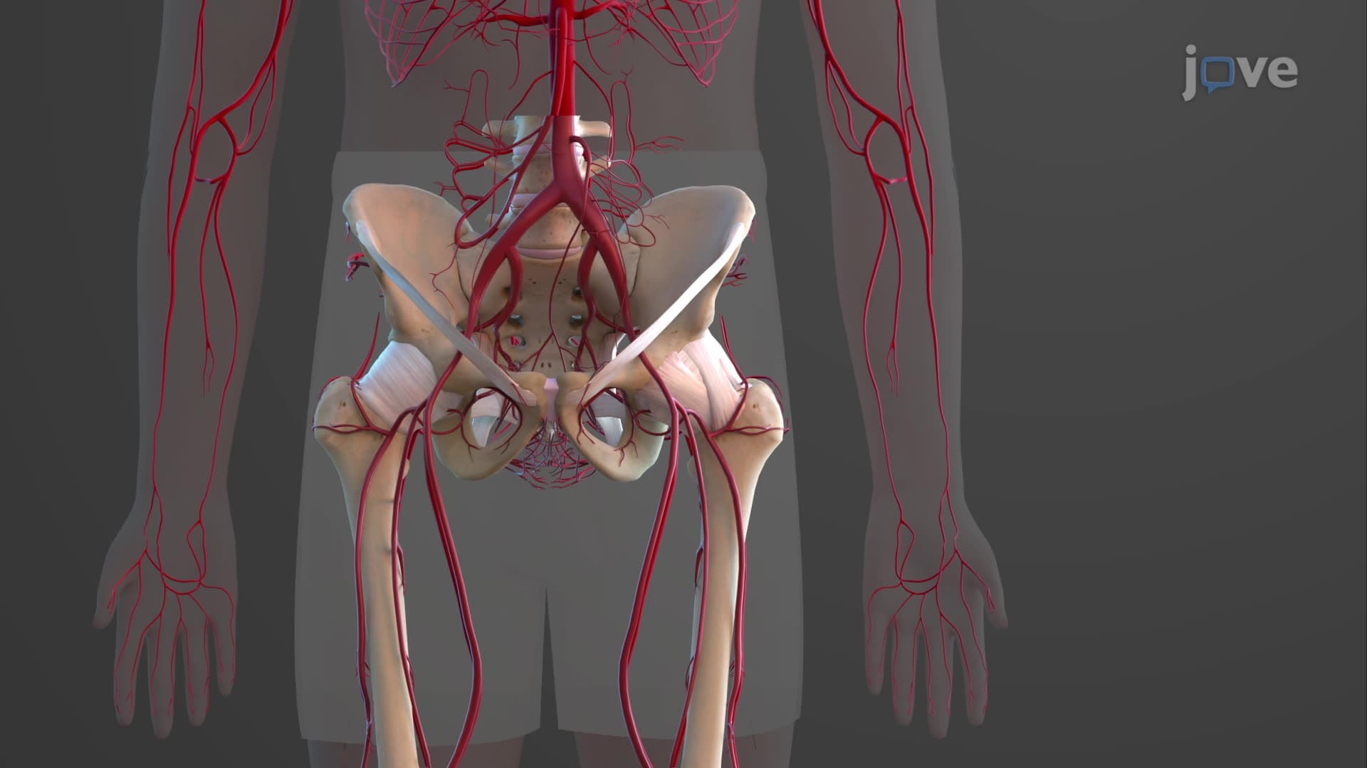
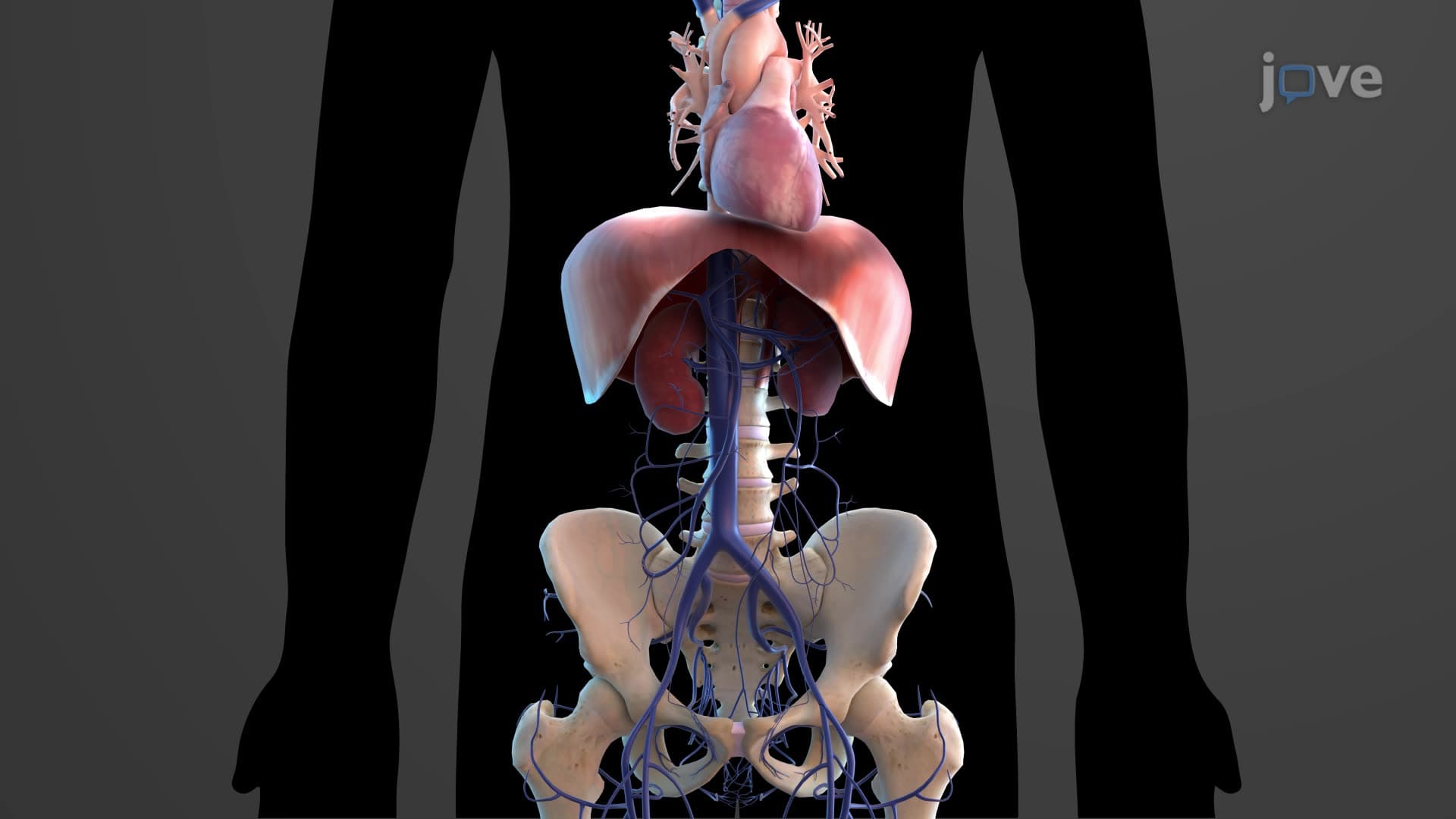
The abdominal aorta represents the continuation of the thoracic aorta as it passes through the diaphragm at the T12 level via the aortic hiatus. This major vessel descends along the posterior abdominal wall, slightly left of the midline, terminating at the L4 vertebral level where it bifurcates into the common iliac arteries. Understanding this precise anatomical pathway is crucial for medical students preparing for the MCAT or anatomy courses at institutions like UCLA Medical School, where cadaveric dissection reveals these relationships in detail.
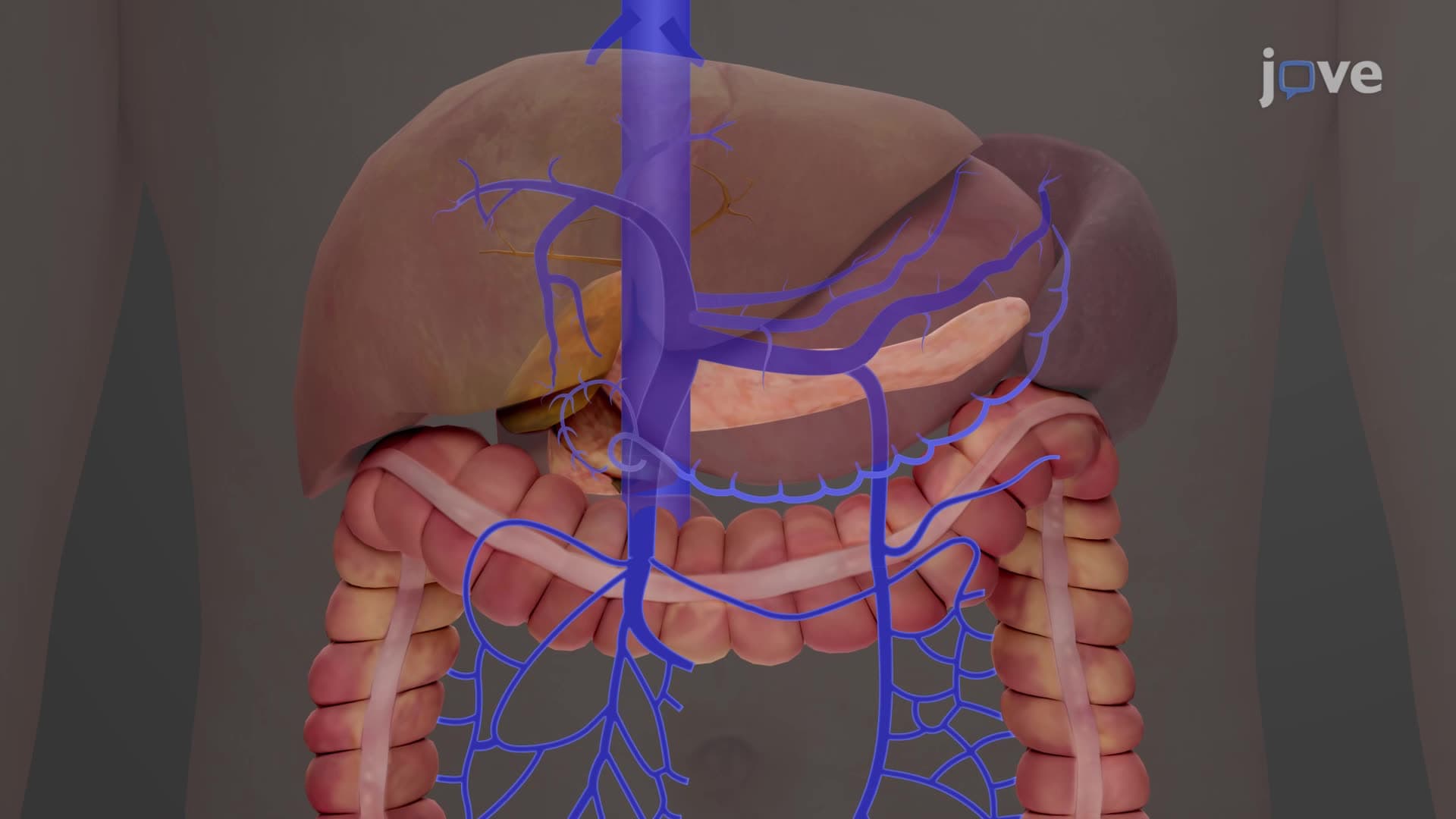
The celiac mesenteric abdominal aorta system demonstrates remarkable organization through its visceral branches. The unpaired visceral arteries emerge at specific levels: the celiac trunk at T12, superior mesenteric artery at L1, and inferior mesenteric artery at L3. The celiac trunk's three-branch pattern-left gastric, splenic, and common hepatic arteries-creates what surgeons call the "tripod of Haller," supplying the foregut derivatives including stomach, liver, spleen, and proximal duodenum.
The superior mesenteric artery serves as the primary blood supply for most small intestine structures and the proximal large intestine, while the inferior mesenteric artery supplies the distal colon and rectum. This anatomical distinction becomes clinically significant in conditions like mesenteric ischemia, commonly studied in pathophysiology courses at medical schools across the United States.
The renal artery aorta branch relationship exemplifies the paired visceral pattern, with renal arteries emerging at L1-L2 to supply the kidneys-organs requiring approximately 20% of cardiac output. The suprarenal and gonadal arteries complete the paired visceral group, demonstrating the abdominal aorta's role in endocrine and reproductive organ perfusion.
Parietal branches serve the body wall and vertebral structures. The inferior phrenic arteries supply the diaphragm's inferior surface, while four pairs of lumbar arteries provide blood to posterior abdominal wall muscles and vertebrae. The median sacral artery, though small, represents an important unpaired parietal branch supplying sacrococcygeal structures.
The abdominal aorta bifurcation iliac relationship at L4 serves as a crucial landmark for clinical examination and imaging interpretation. Abdominal aortic aneurysms, affecting approximately 200,000 Americans annually, most commonly occur in the infrarenal segment. This knowledge proves essential for AP Biology students studying cardiovascular pathology and pre-med students preparing for standardized examinations. Understanding these branching patterns also facilitates comprehension of referred pain patterns and surgical approaches taught in advanced anatomy courses at institutions like Harvard Medical School.
Related Micro-courses