Loading video…


 38 Concepts
 149.7K views

Video Summary: What Is Arteries and Arterioles
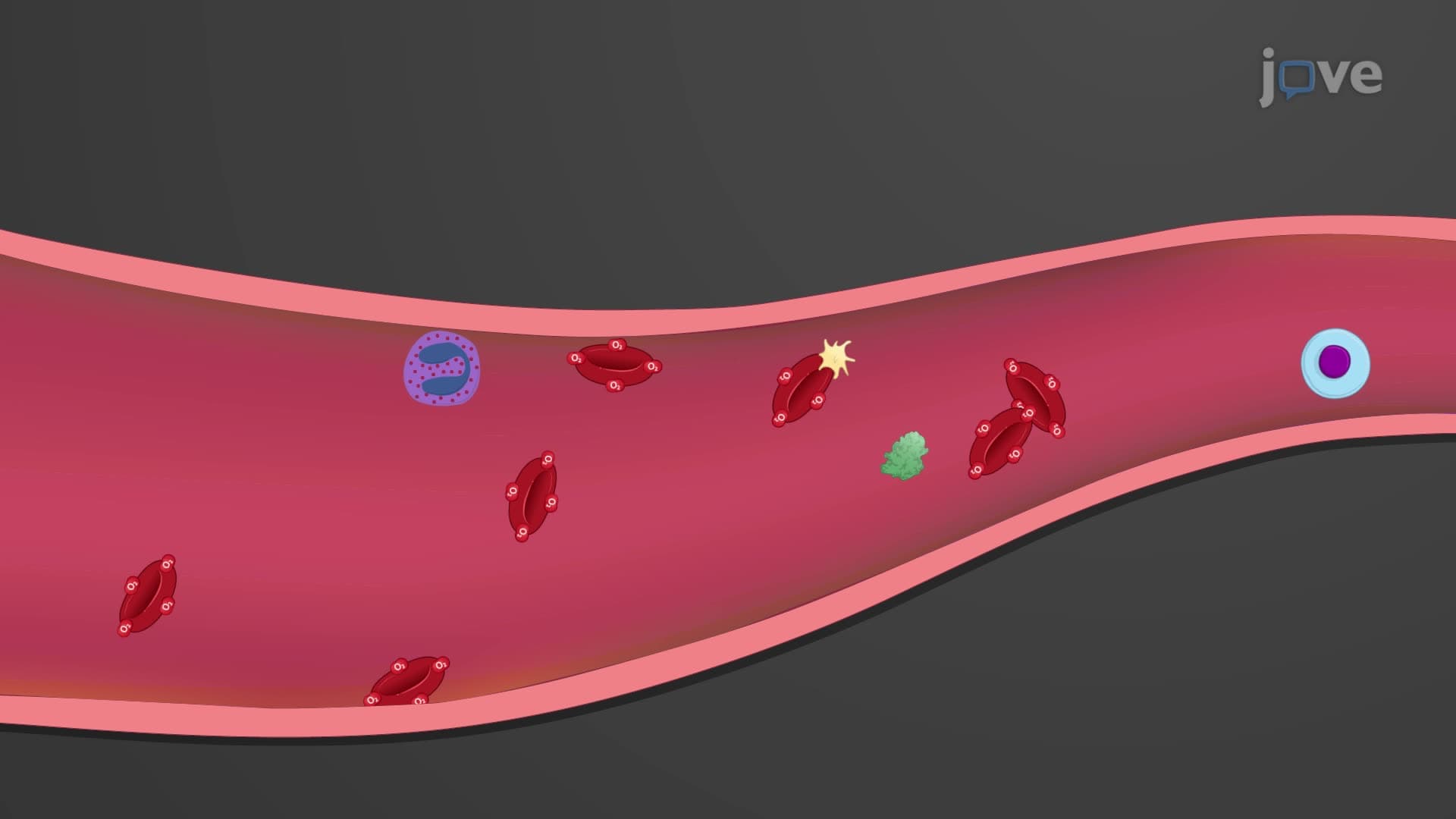
Did you know your aorta can stretch up to 10% with each heartbeat, acting like a biological balloon? Understanding arteries arterioles anatomy reveals how your cardiovascular system maintains steady blood pressure despite your heart's pulsing rhythm. From the elastic aorta near your heart to tiny arterioles controlling blood flow to your biceps during exercise, these vessels form a sophisticated distribution network. What is Arteries And Arterioles becomes clear when you examine their distinct structural adaptations for specific functions. Watch the full video on JoVE Coach to master this concept with expert-led visuals and step-by-step explanations.
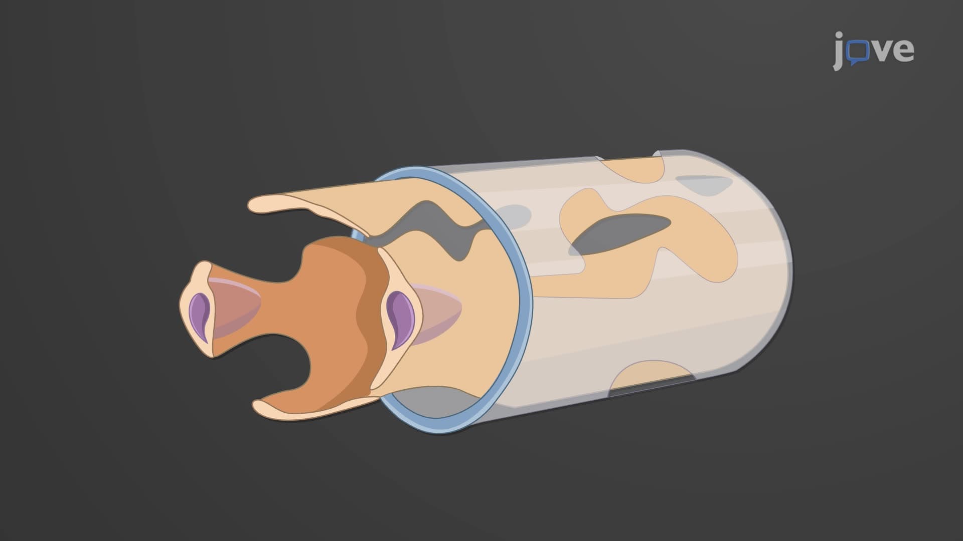
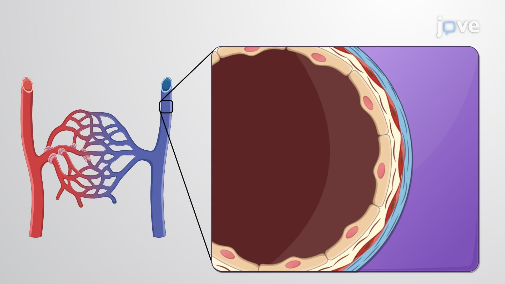
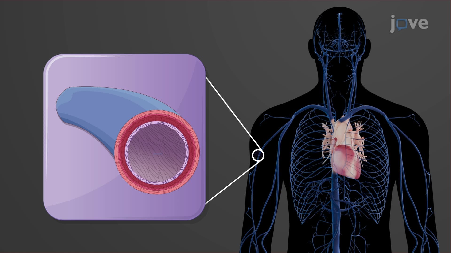
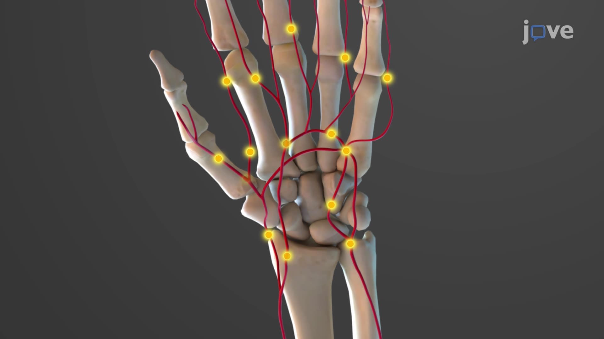
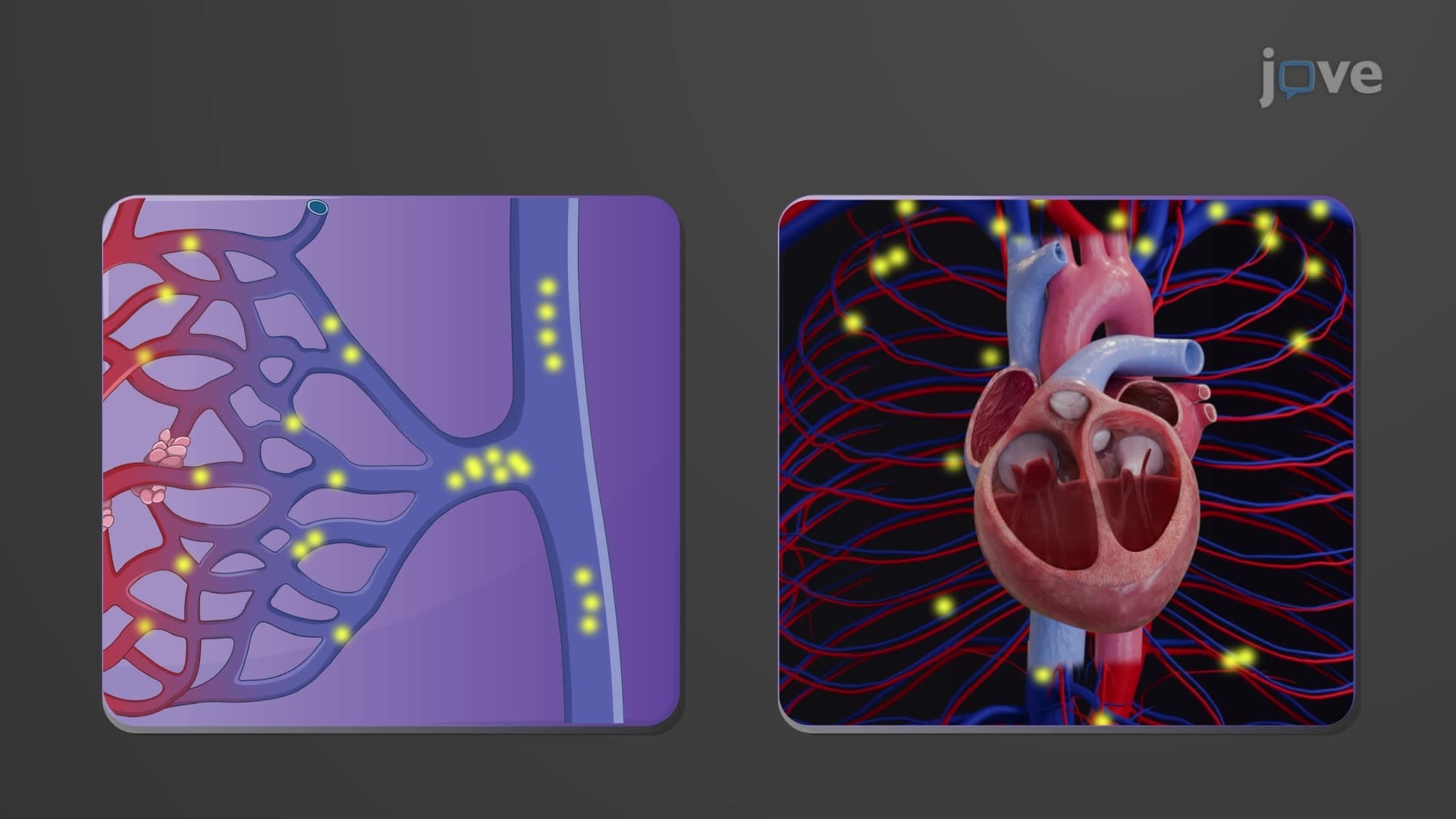
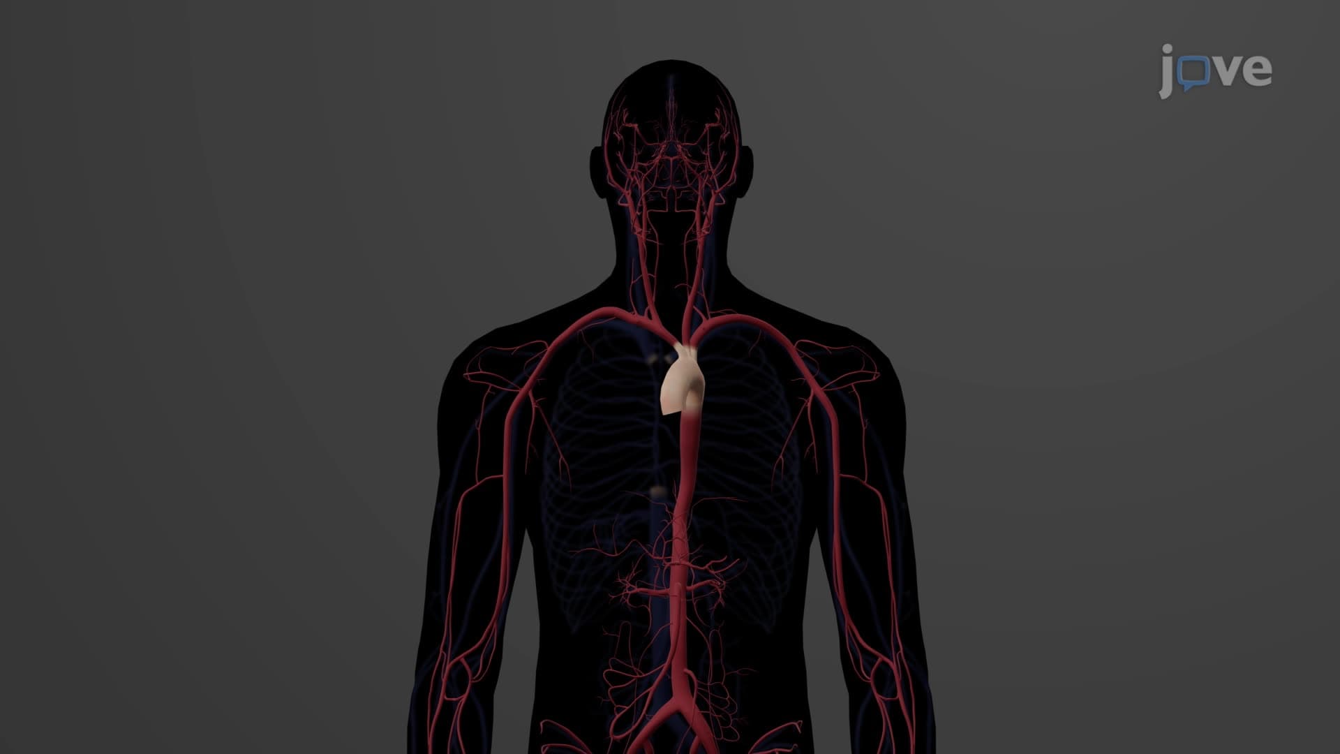
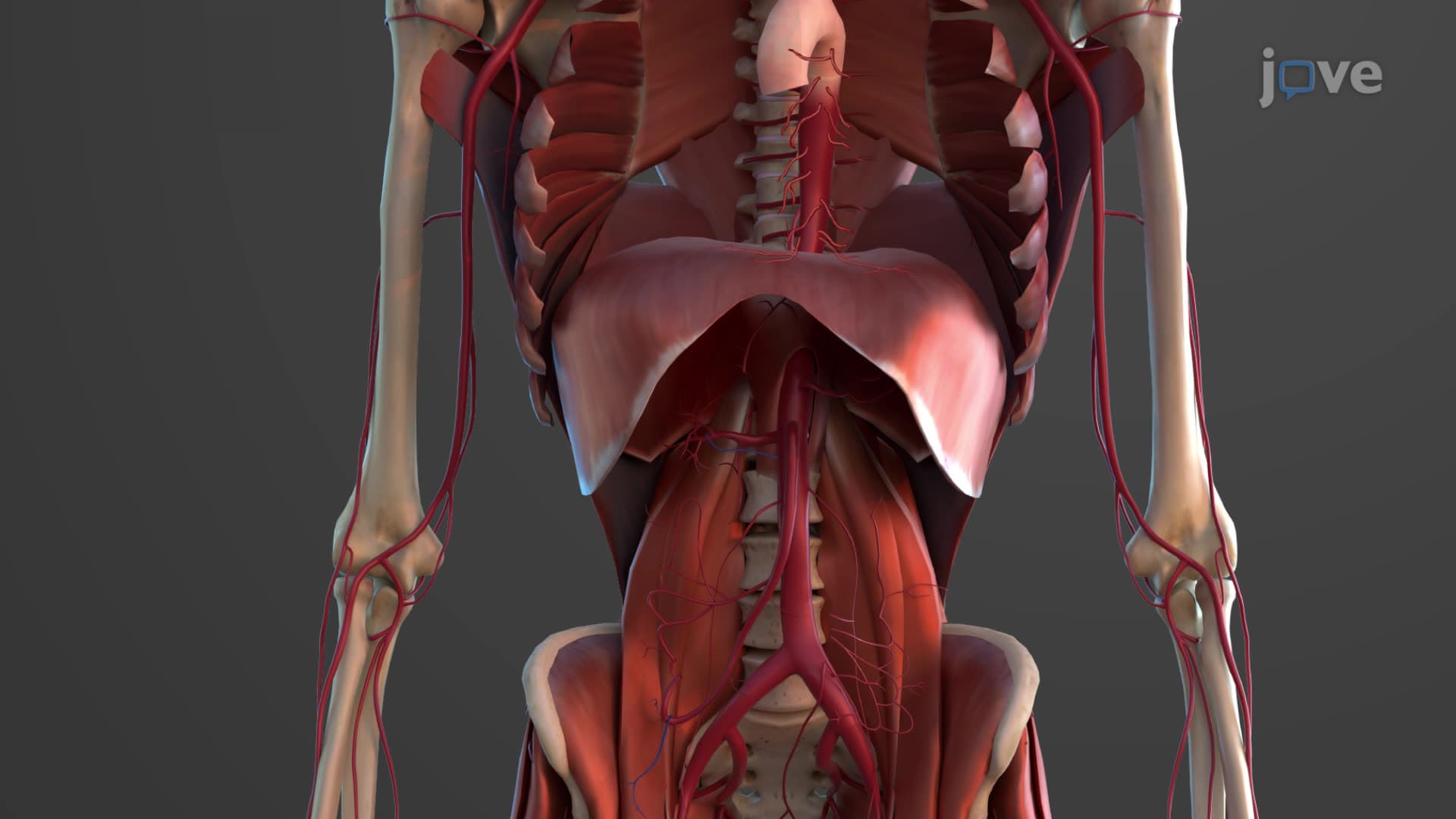
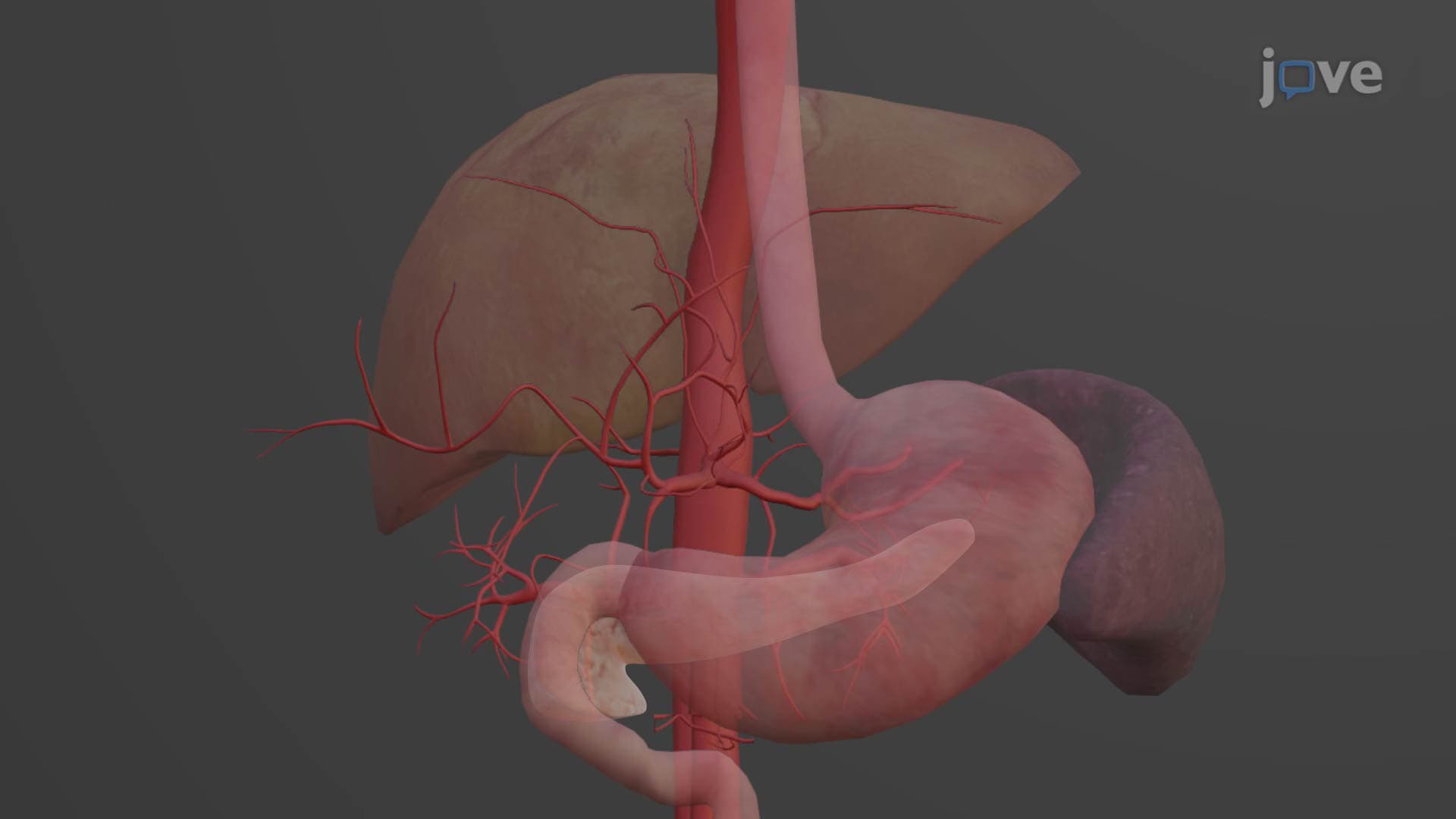
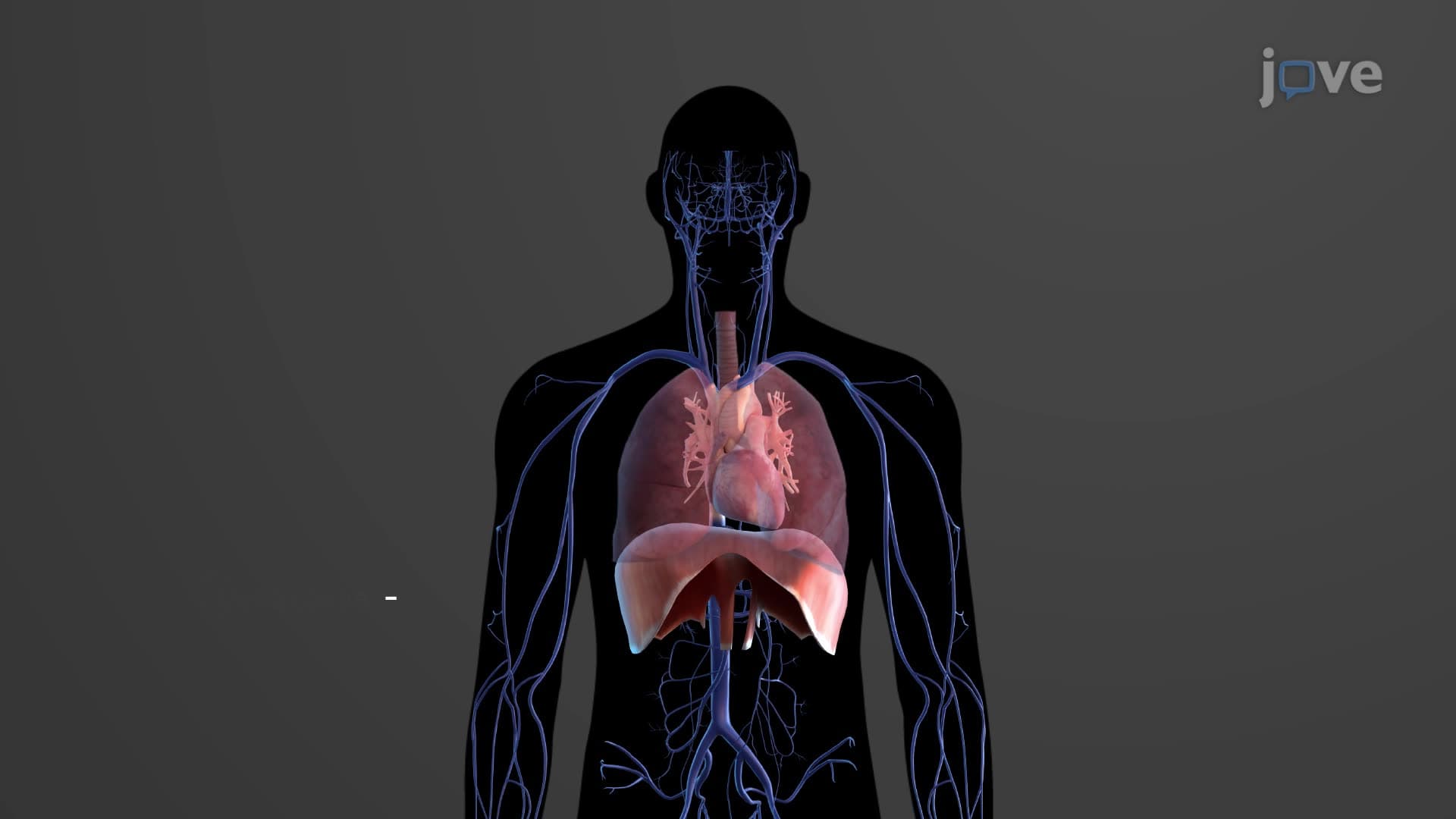
The arteries arterioles anatomy forms a sophisticated distribution network that ensures oxygen-rich blood reaches every tissue in your body. Think of this system like America's interstate highway network-major highways (elastic arteries) carry high volumes of traffic from central hubs, while smaller roads (muscular arteries) distribute traffic regionally, and local streets (arterioles) control access to individual neighborhoods (capillary beds).
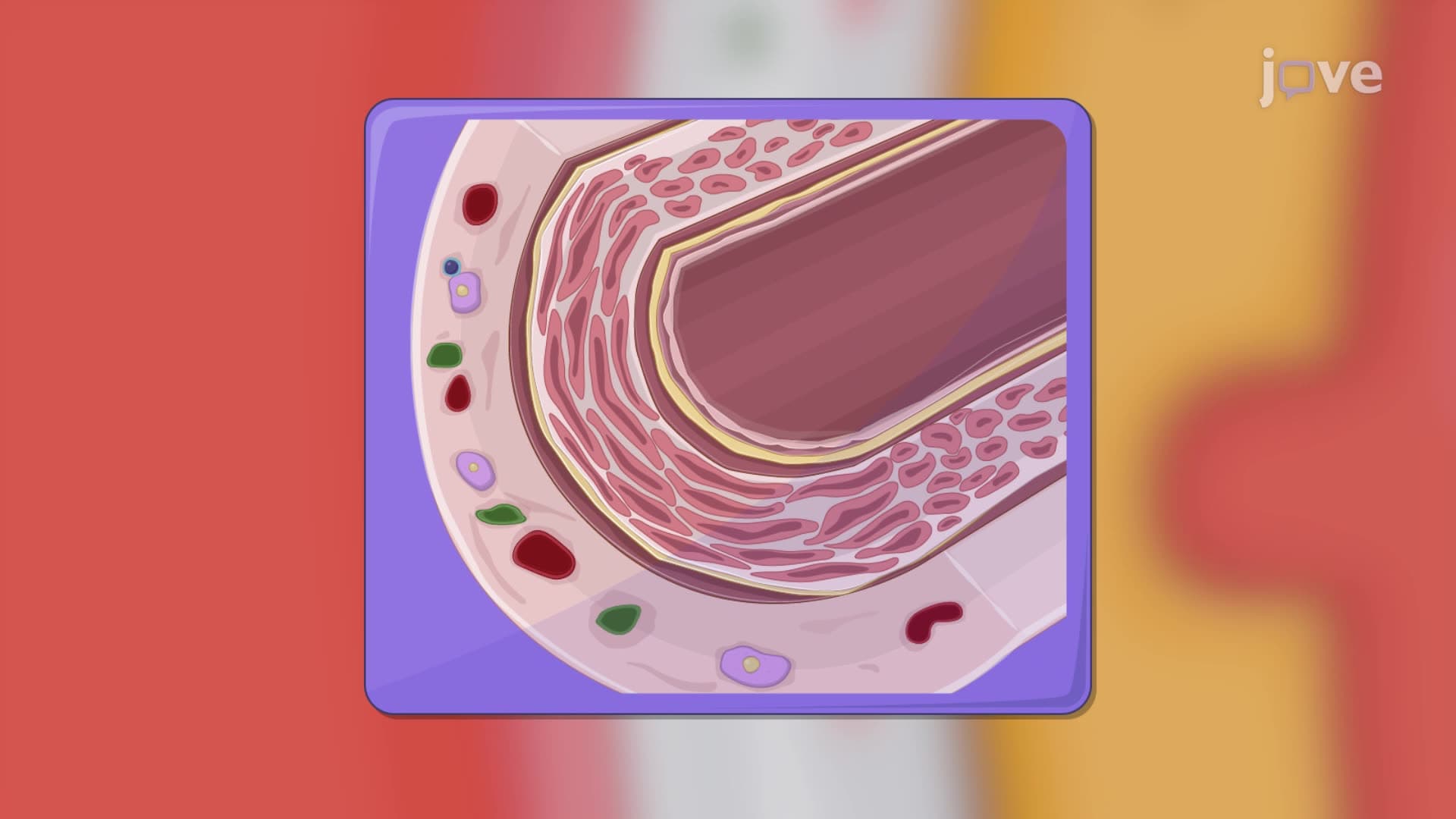
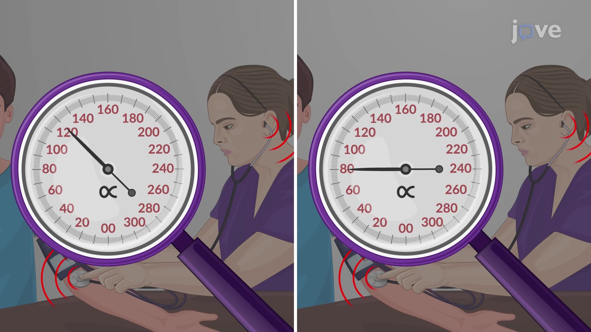
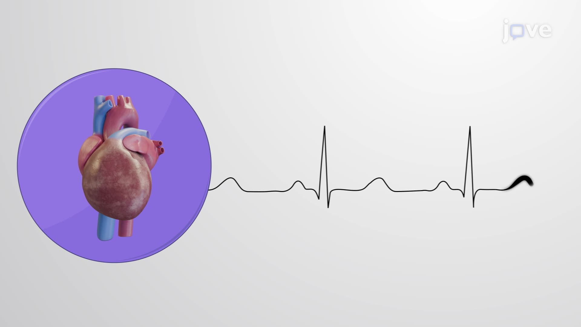
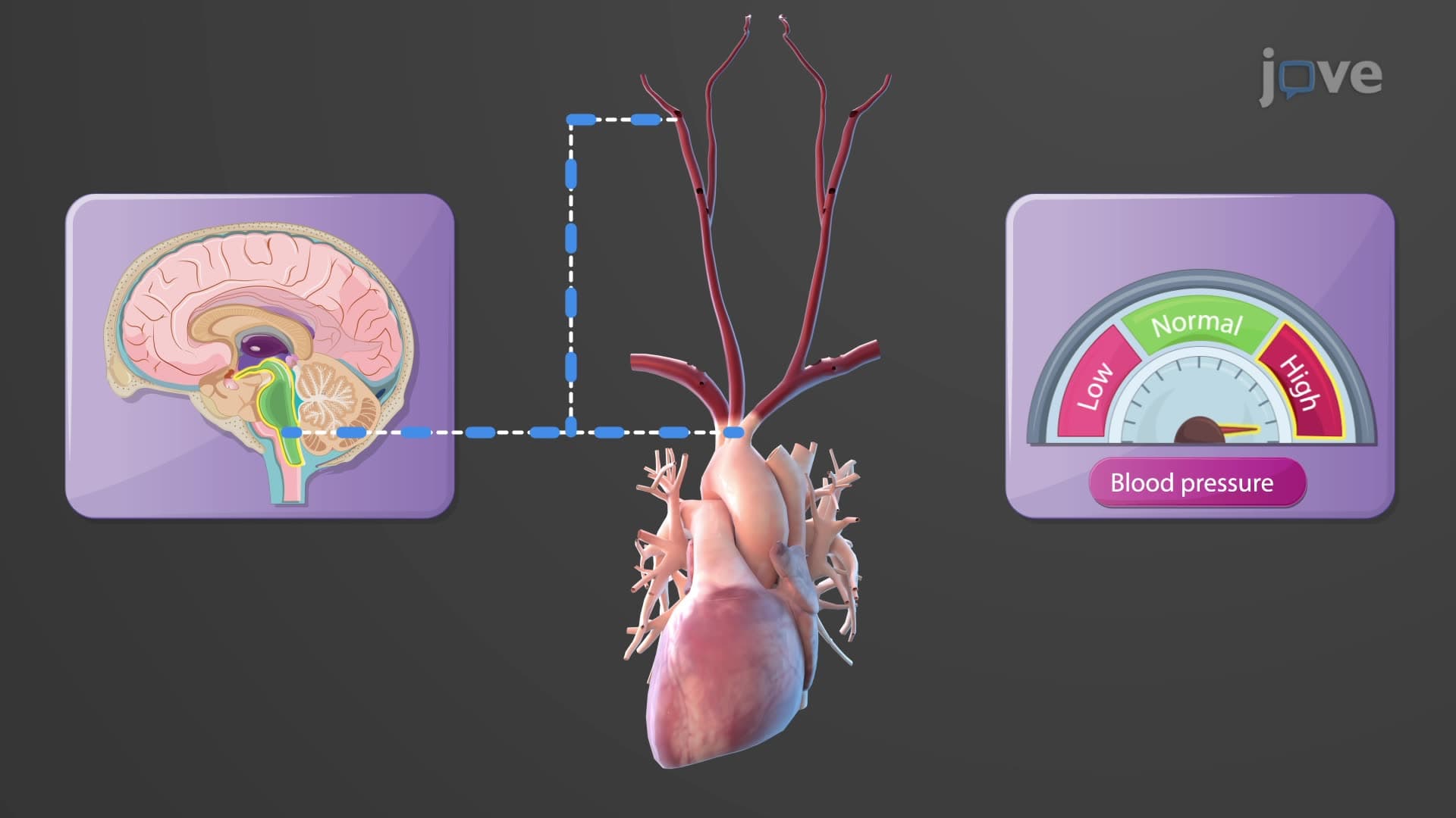
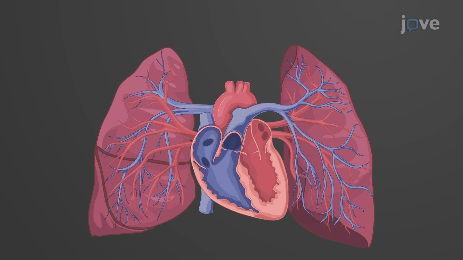
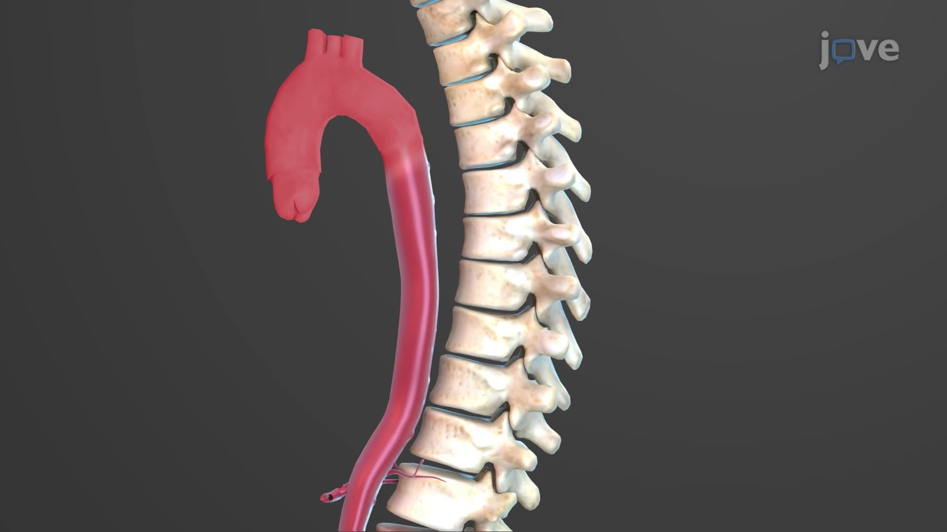
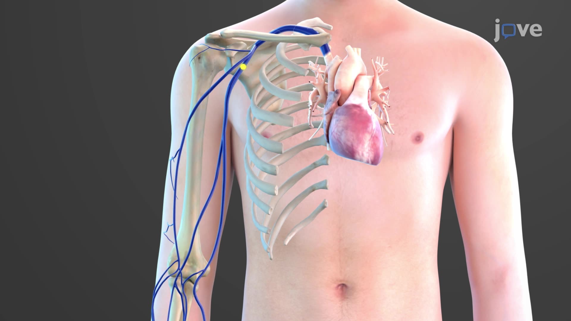
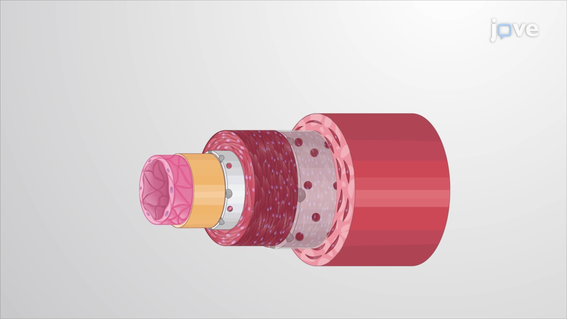
Elastic arteries, including the aorta and pulmonary artery, represent the body's largest vessels with diameters reaching 2.5 centimeters. Their artery wall thick tunica media contains abundant elastic fibers arranged in concentric layers, creating what physiologists call "windkessel vessels." During systole, when your left ventricle contracts with pressures reaching 120 mmHg, these arteries stretch to accommodate the surge of blood. During diastole, the elastic recoil maintains arterial blood flow continuity-crucial for coronary perfusion when your heart muscle relaxes. This mechanism appears frequently on AP Biology exams and MCAT cardiovascular passages, where students must explain how elastic arteries maintain diastolic pressure.
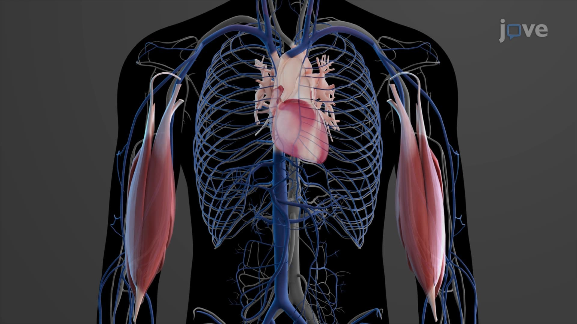
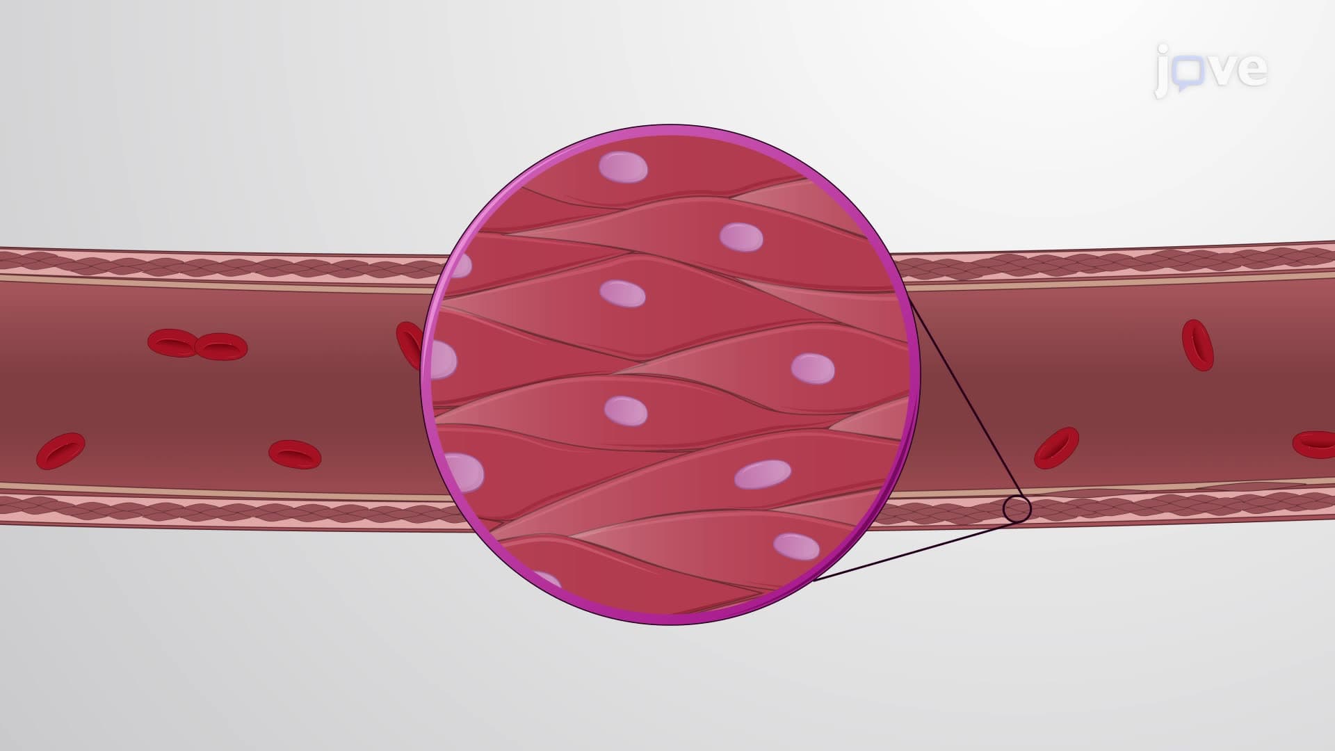
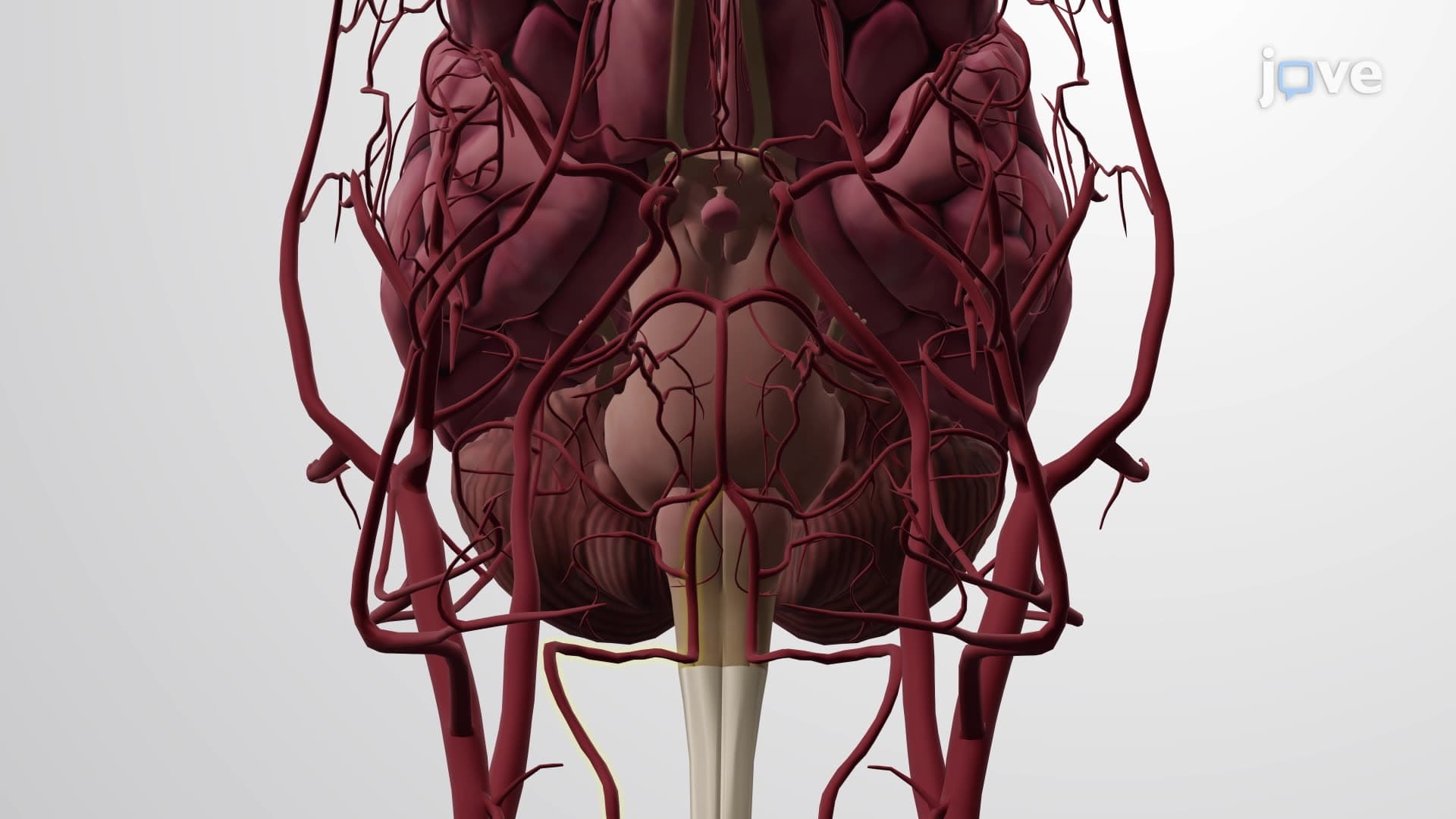
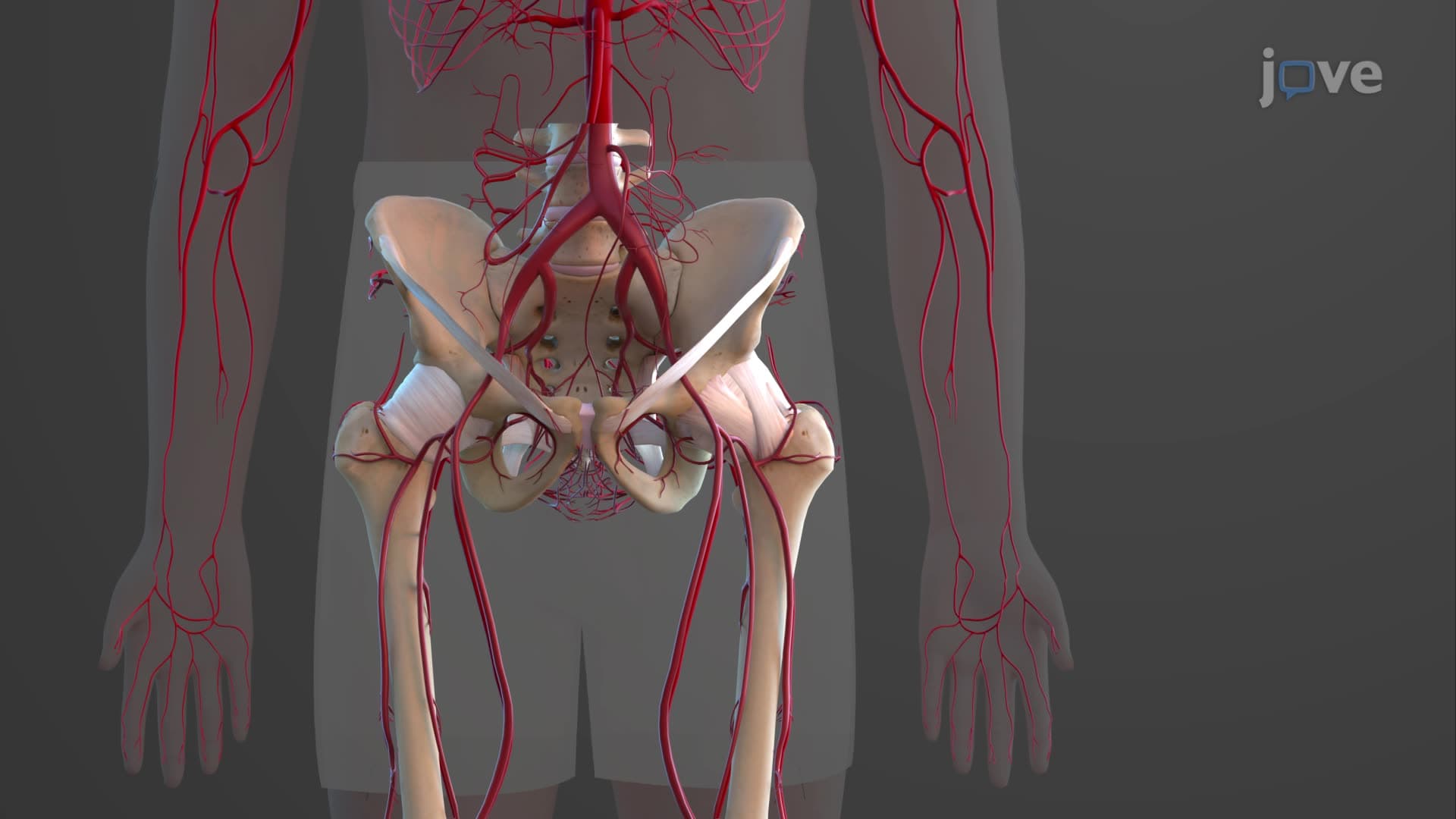
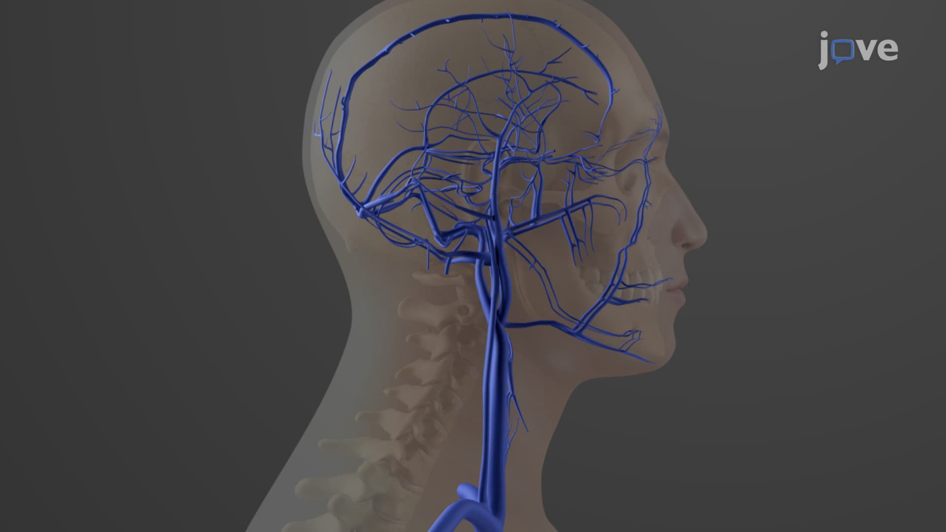
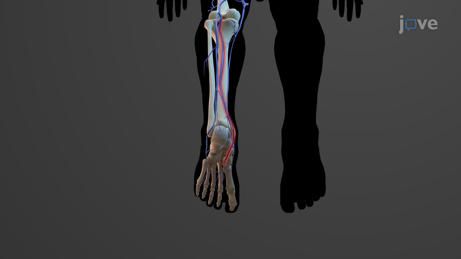
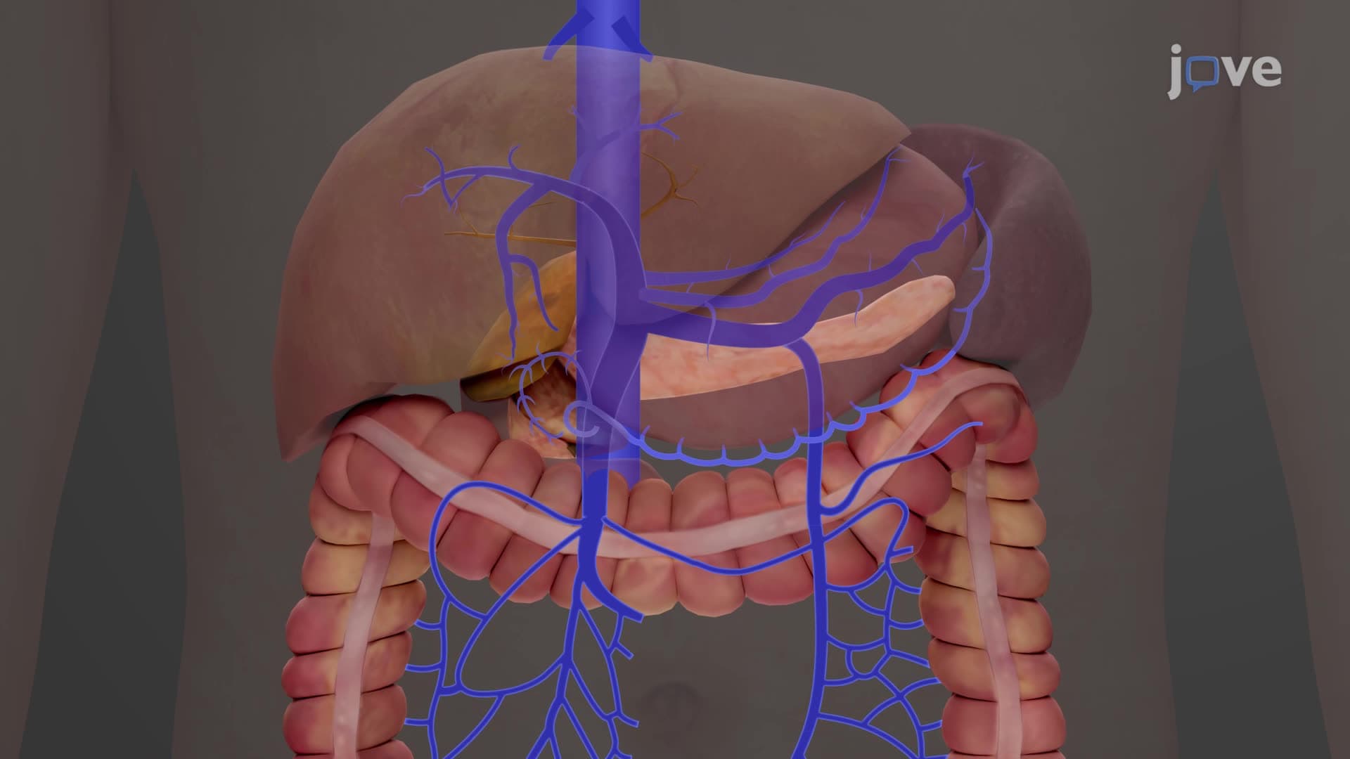
Muscular arteries like the radial, femoral, and carotid arteries feature artery wall thick tunica media dominated by smooth muscle cells comprising 75% of the vessel wall mass. Unlike elastic arteries, these vessels cannot significantly expand or recoil due to limited elastic tissue. Instead, their thick smooth muscle layers enable active vasoconstriction and vasodilation in response to sympathetic nervous system signals and local metabolic demands. During exercise, muscular arteries supplying skeletal muscles dilate while those serving digestive organs constrict-a concept tested extensively in college physiology courses and USMLE Step 1 examinations.
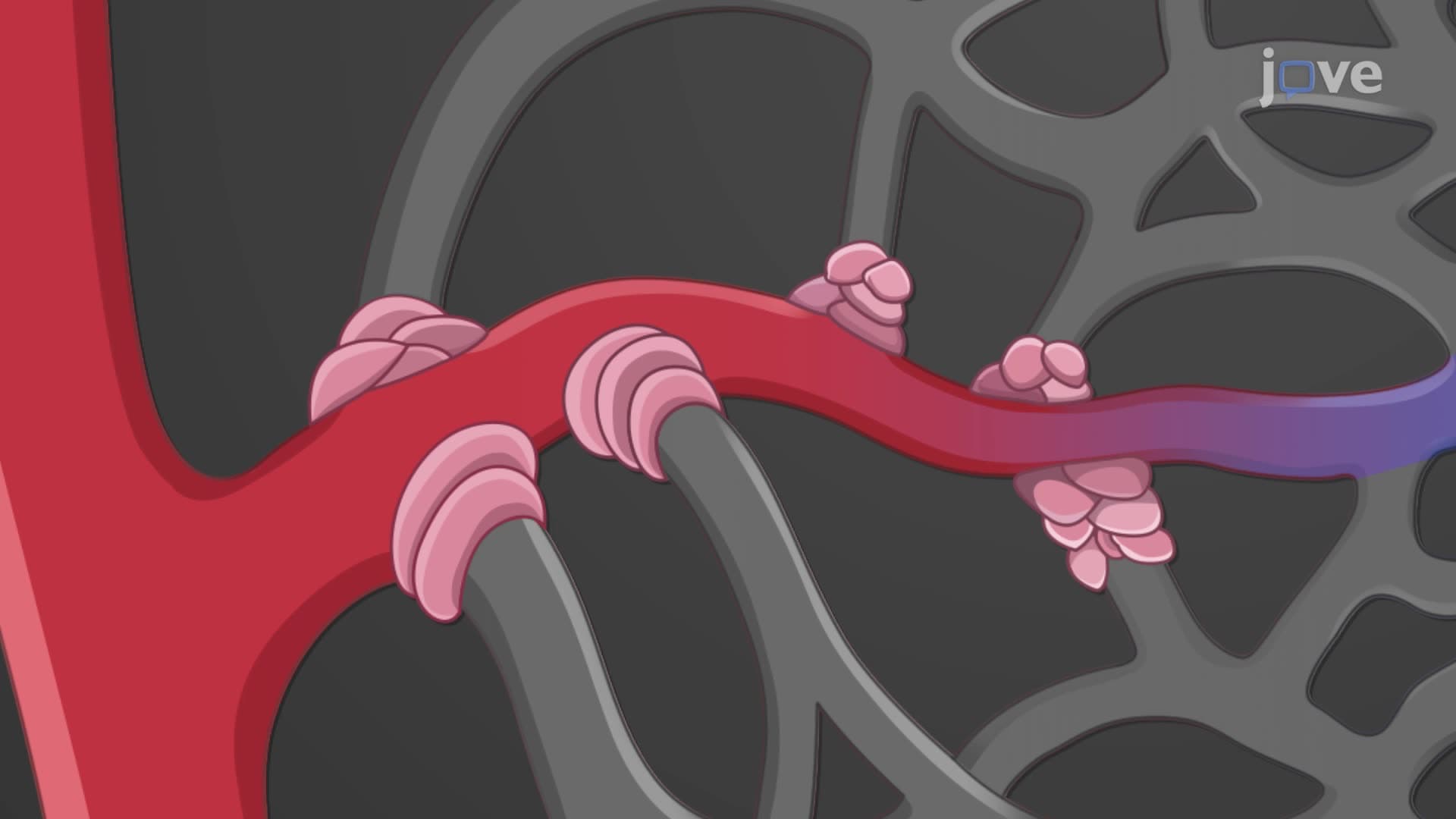
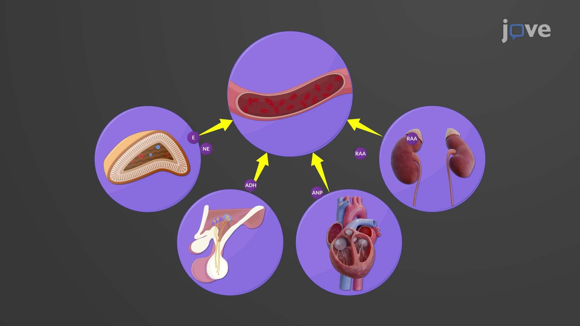
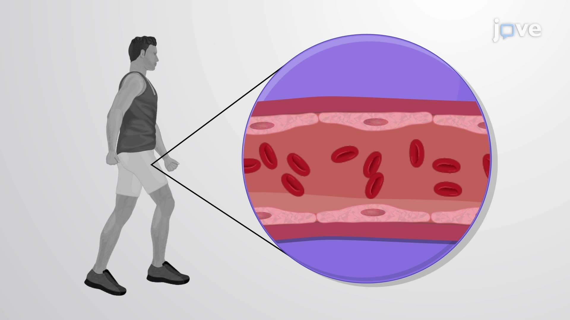
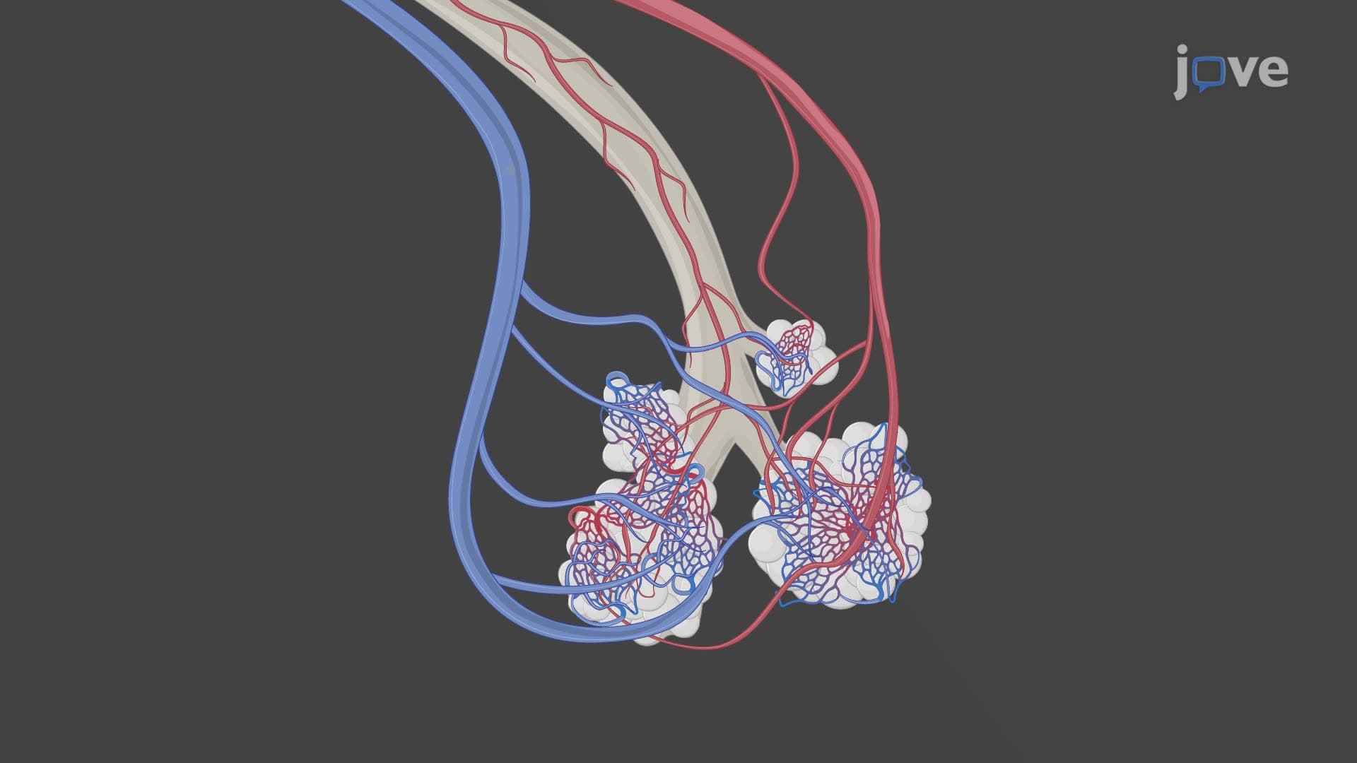
Arteriole resistance vessel function makes these tiny arteries (10-300 micrometers) the primary controllers of systemic vascular resistance. Their 1-2 smooth muscle cell layers enable rapid arteriole vasoconstriction regulation in response to local factors like oxygen tension, pH, and temperature. In clinical medicine, arteriole dysfunction contributes to hypertension development-a topic emphasized in NCLEX cardiovascular questions and medical school pathophysiology courses. Understanding what are arteries and arterioles in anatomy helps explain why antihypertensive medications often target arteriolar smooth muscle or the regulatory mechanisms controlling these vessels.
Related Micro-courses