Loading video…


 38 Concepts
 149.7K views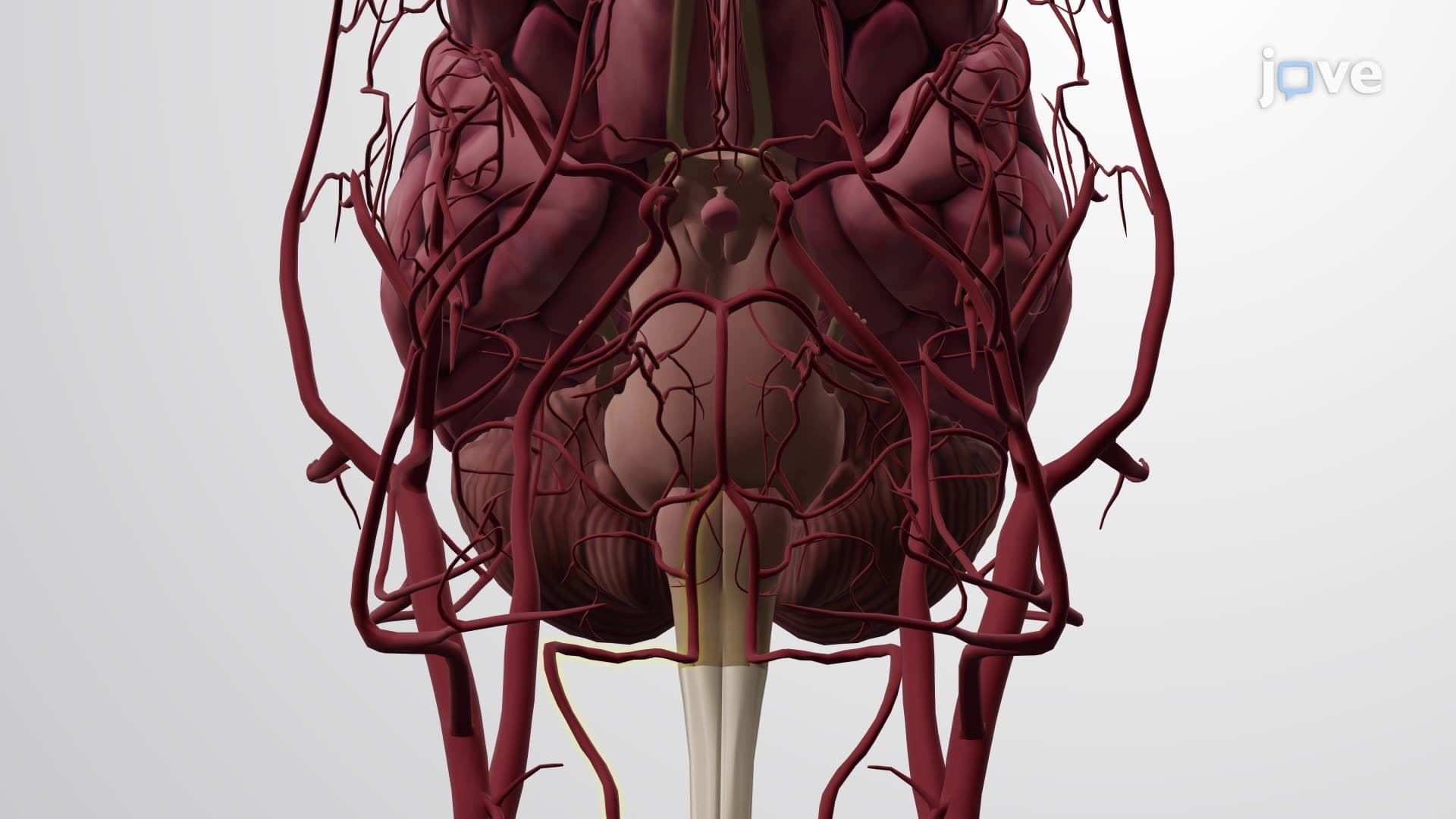

Video Summary: What Is Arteries Head and Neck
Did you know that a stroke occurs every 40 seconds in the United States, often due to blocked arteries supplying the brain? Understanding head neck arteries anatomy is crucial for recognizing how blood reaches every structure from your brain to your jaw. The complex network includes major vessels like the carotid and vertebral arteries that ensure oxygen delivery to critical areas. For instance, Mayo Clinic surgeons rely on detailed arterial maps during carotid endarterectomy procedures to prevent strokes. Watch the full video on JoVE Coach to master this concept with expert-led visuals and step-by-step explanations.
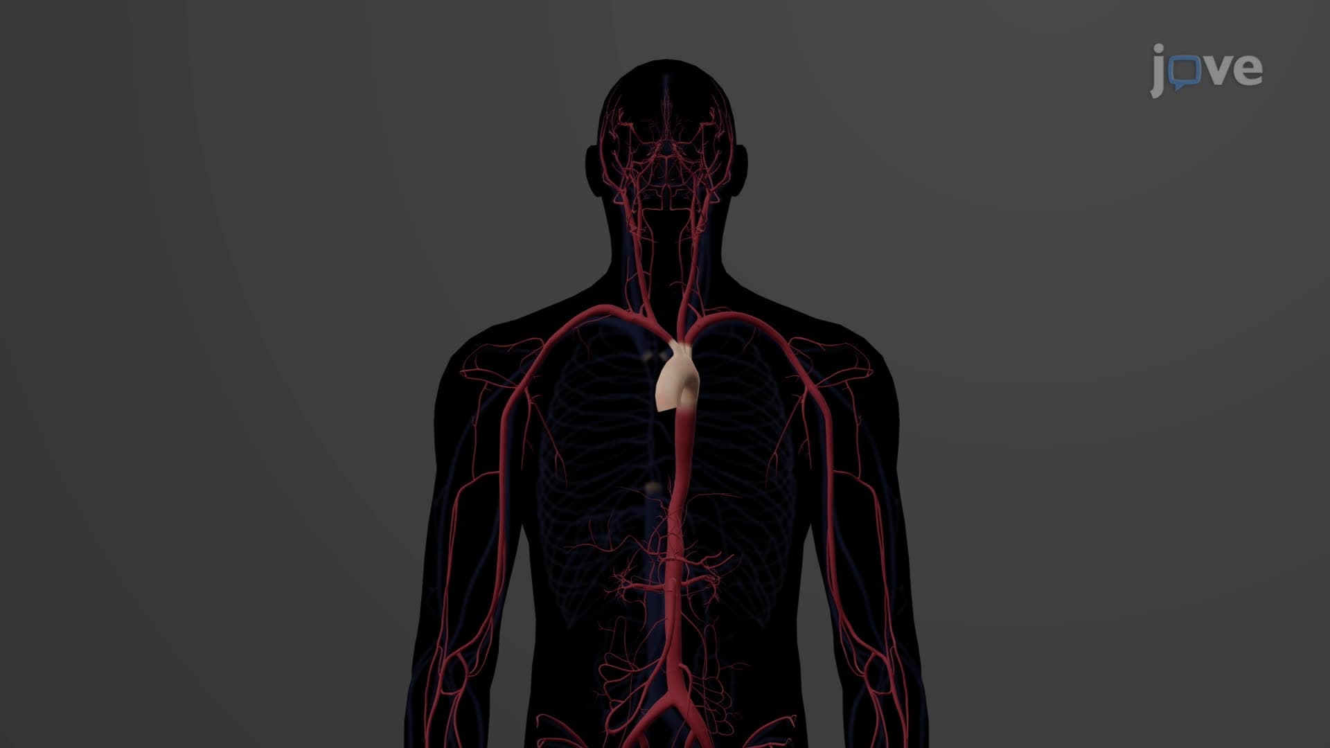
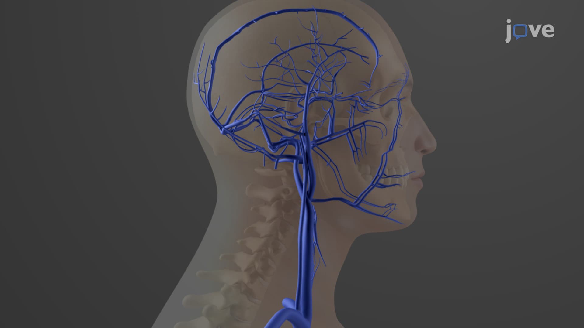
The arterial supply to the head and neck represents one of the most complex vascular networks in human anatomy, critical for maintaining brain function and sensory organ vitality. This intricate system ensures that oxygen-rich blood reaches every tissue from the scalp to the deep cervical structures, making it essential knowledge for students pursuing healthcare careers or advanced biology studies.
The head neck arteries anatomy centers on two primary sources. The common carotid arteries serve as the main highways, with the right arising from the brachiocephalic trunk and the left directly from the aortic arch. These vessels bifurcate at the level of the thyroid cartilage into external and internal branches, a landmark frequently tested on the MCAT and in anatomy courses at institutions like Johns Hopkins University.
External carotid arteries function as the supply network for facial structures, branching into eight named vessels including the superior thyroid, facial, and superficial temporal arteries. Medical students at Harvard Medical School often study these branches when learning about facial reconstruction surgery, as each vessel supplies specific regions that surgeons must preserve during procedures.
Internal carotid arteries carry the enormous responsibility of supplying over 80% of cerebral blood flow. After entering the skull through the carotid canal, these vessels give rise to the ophthalmic arteries (supplying the eyes) and terminate as the anterior and middle cerebral arteries. This distribution pattern becomes crucial during USMLE Step 1 preparation, as students must recognize stroke syndromes based on affected arterial territories.
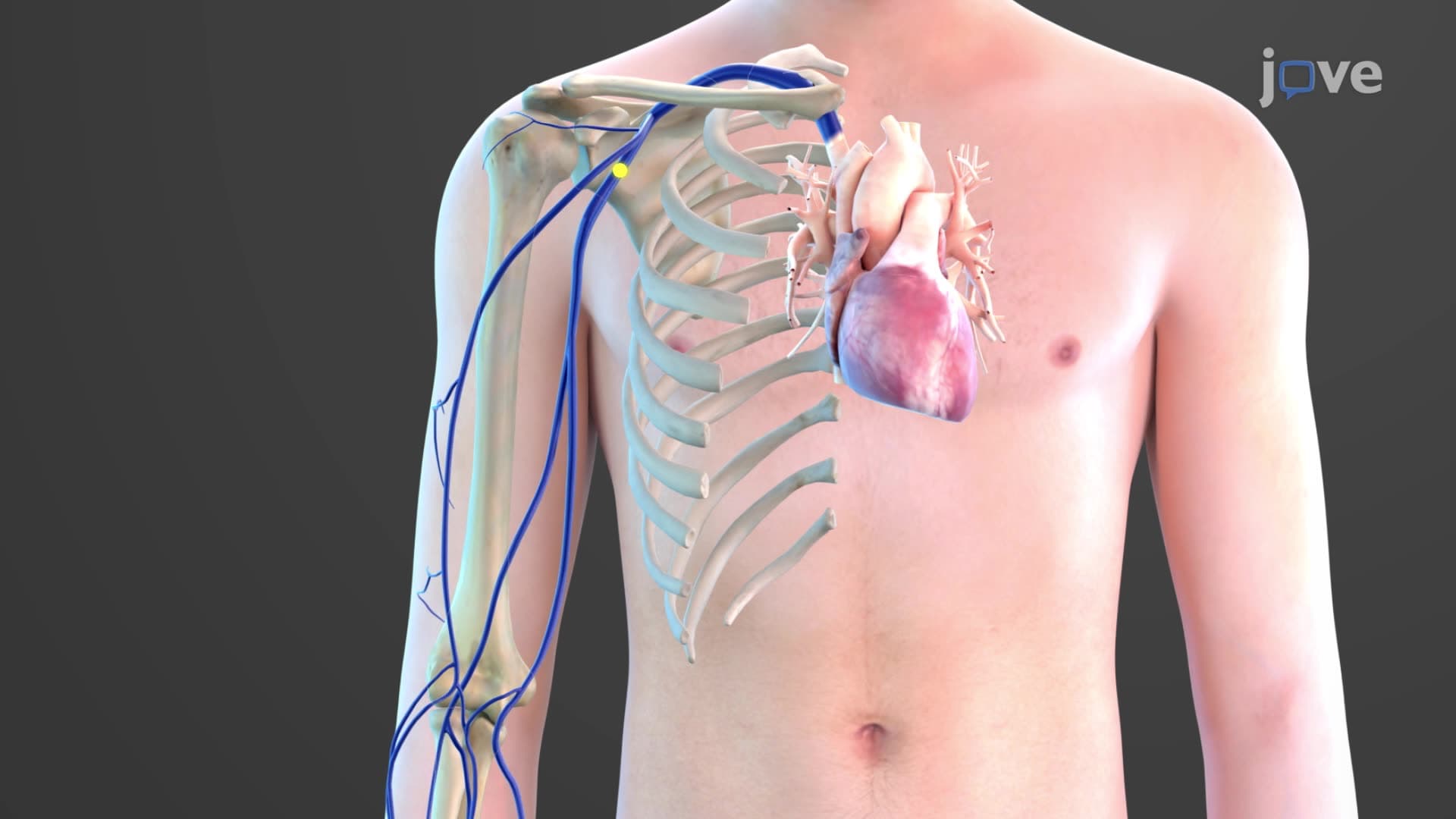
The vertebral arteries, arising from subclavian vessels, ascend through cervical vertebrae transverse foramina before merging intracranially to form the basilar artery. This posterior circulation supplies the brainstem, cerebellum, and occipital lobes, regions frequently damaged in vertebrobasilar strokes that emergency physicians at Cleveland Clinic encounter regularly.
Understanding these arterial patterns proves invaluable in clinical practice. Radiologists use this knowledge when interpreting CT angiograms, while surgeons rely on arterial anatomy during carotid endarterectomies-procedures performed over 100,000 times annually in US hospitals to prevent strokes. The thyrocervical and costocervical trunks, though smaller, supply critical neck muscles and spinal structures, making them important during cervical spine surgeries performed at institutions like Mayo Clinic.
Related Micro-courses