Loading subject page…


149,645 views

Video Summary: What Is Fetal Circulation
Ever wondered how a developing baby gets oxygen without breathing? Fetal circulation anatomy biology reveals a remarkable system where babies rely on their mother's lungs and liver through an intricate network of specialized blood vessels. Unlike adult circulation, fetal blood flow creates unique bypasses-including the foramen ovale and ductus arteriosus-that redirect blood away from non-functioning lungs. For example, premature babies in US NICUs often require careful monitoring of these fetal structures as they transition to independent breathing. Watch the full video on JoVE Coach to master this concept with expert-led visuals and step-by-step explanations.
What is fetal circulation represents one of biology's most elegant adaptations. Unlike adults who rely on lungs for oxygenation, developing fetuses obtain oxygen and nutrients directly from maternal blood through the placenta. This creates a completely different circulatory pattern that medical students encounter extensively in anatomy courses and healthcare professionals see daily in obstetric units across US hospitals.
The umbilical cord contains three vessels that form the foundation of fetal blood flow explained. Two umbilical arteries carry deoxygenated blood from the fetus to the placenta, where waste products are exchanged for oxygen and nutrients from maternal circulation. The single umbilical vein returns this oxygen-rich blood to the fetus. This arrangement appears counterintuitive since arteries typically carry oxygenated blood, making it a frequent AP Biology exam question and MCAT topic.
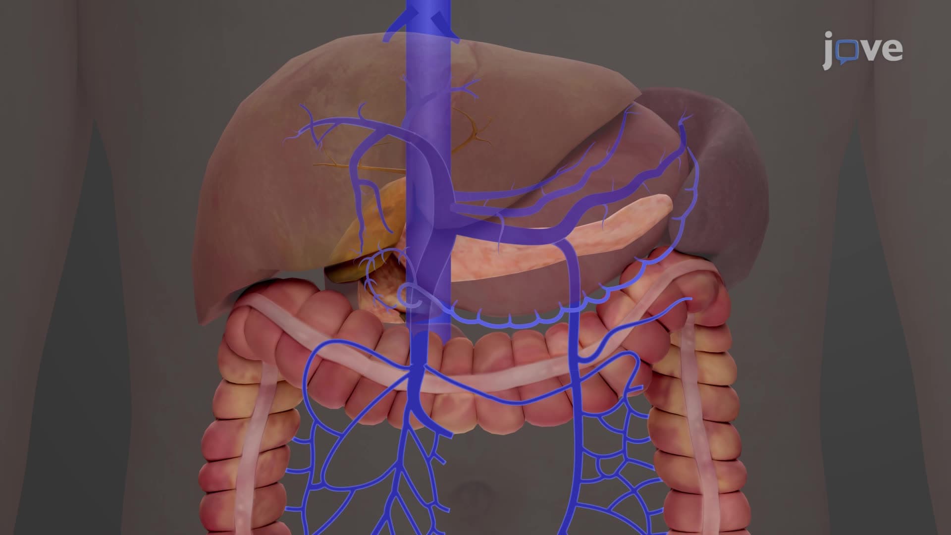
In US medical schools, students learn that the umbilical vein splits upon entering the fetal liver. Part of the blood flows through the hepatic portal circulation to nourish the developing liver, while the remainder bypasses the liver entirely through the ductus venosus-a unique fetal structure that connects directly to the inferior vena cava.
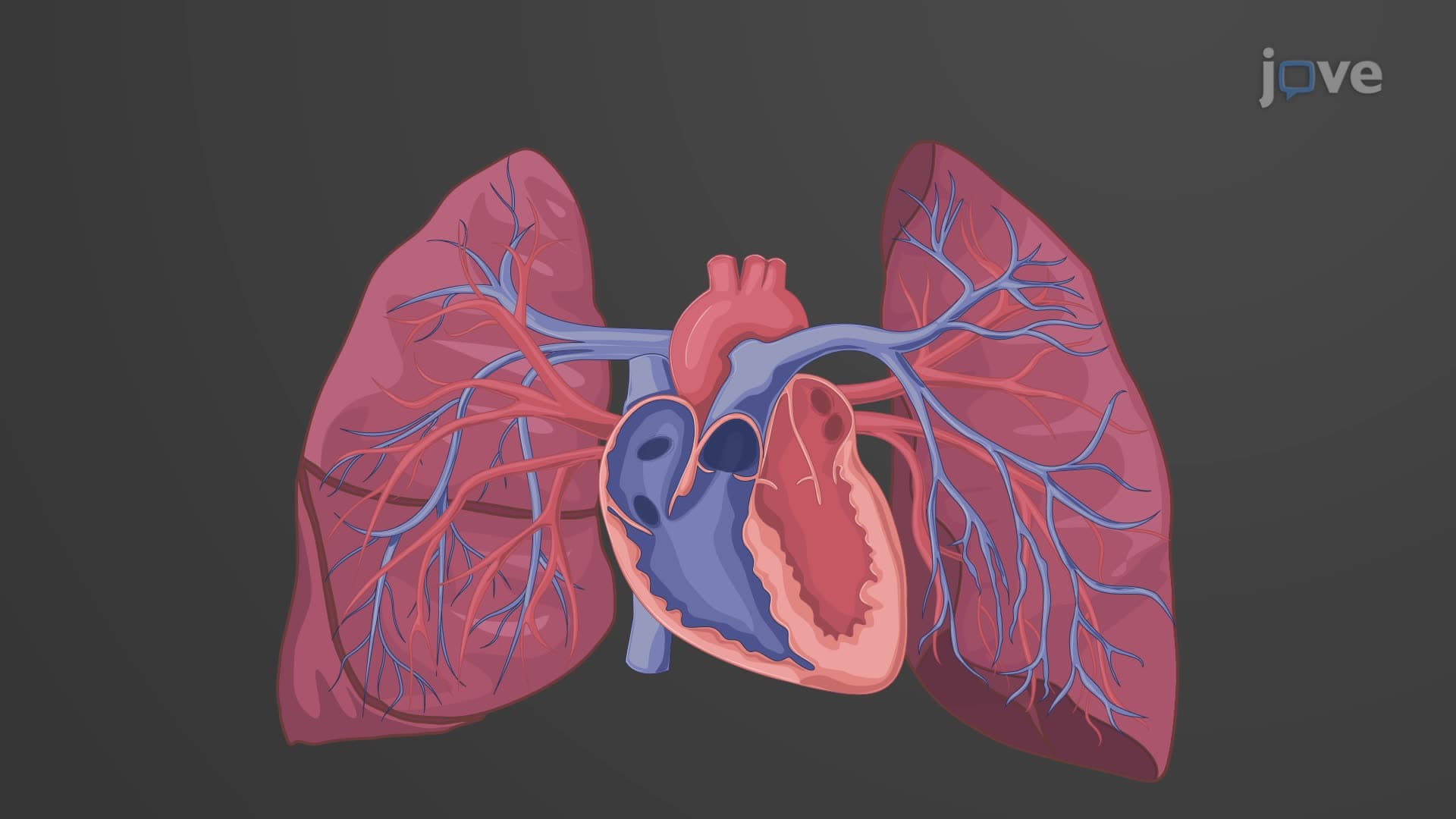
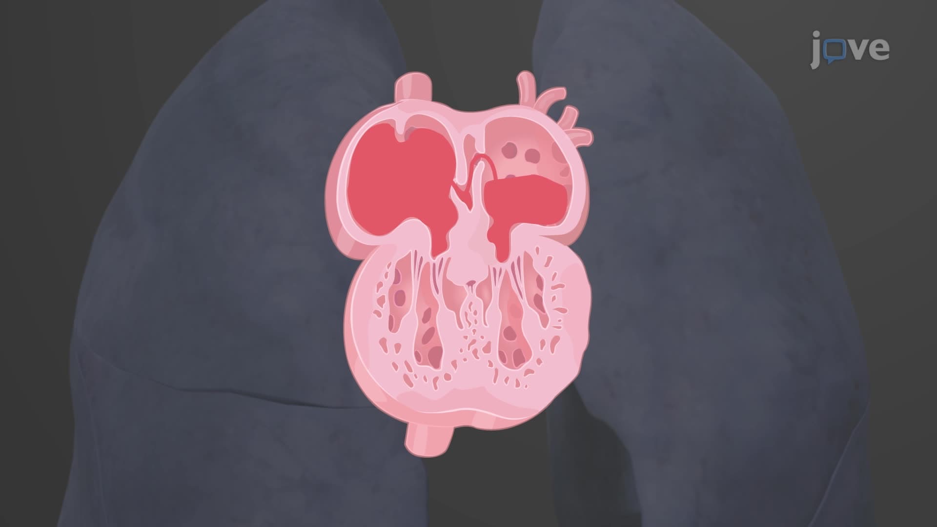
What makes fetal circulation in developmental anatomy so fascinating are the three major bypass structures. The foramen ovale fetal opening allows blood to flow directly from the right atrium to the left atrium, essentially bypassing the lungs. Similarly, the ductus arteriosus connects the pulmonary artery to the aorta, redirecting blood away from non-functional fetal lungs.
These bypasses create a system where the most oxygenated blood preferentially flows to the brain and heart-crucial for proper development. Students preparing for the USMLE or NCLEX often struggle with this concept because it reverses many assumptions about normal circulation patterns.
The transition from fetal to adult circulation occurs rapidly at birth when the baby takes their first breath. US neonatal intensive care units regularly monitor this transition, particularly in premature infants born before 37 weeks gestation. When fetal bypass structures fail to close properly-such as patent ductus arteriosus affecting approximately 1 in 2,000 US births-surgical intervention may be necessary.
Understanding these concepts proves essential for healthcare careers and appears prominently on nursing entrance exams like HESI A2 and TEAS, where students must demonstrate knowledge of both normal fetal development and potential complications.
Related Micro-courses