Loading subject page…


149,645 views

Video Summary: What Is the Aorta
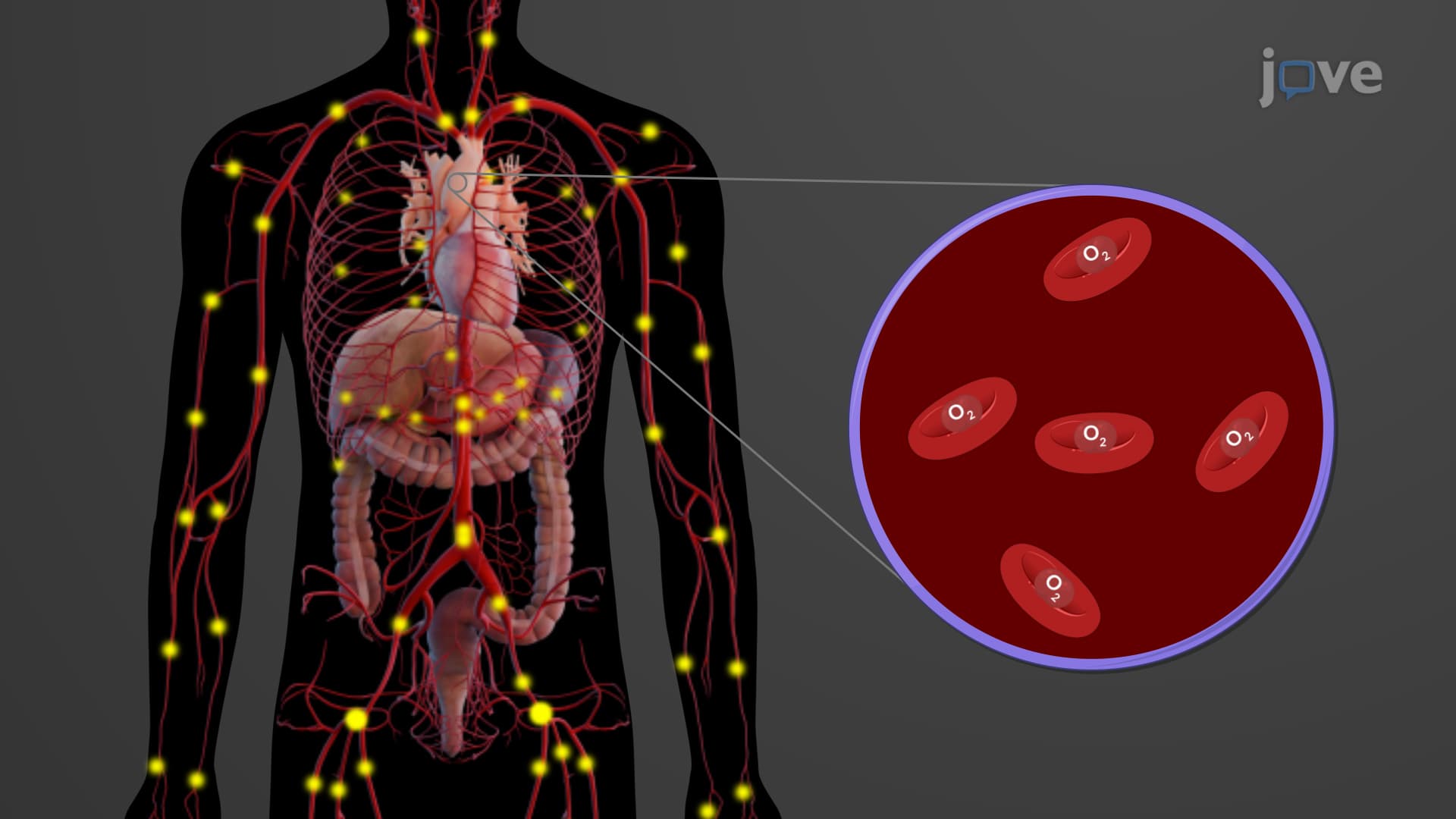
Did you know your aorta anatomy biology carries the equivalent of 5 liters of blood every minute-enough to fill a large soda bottle? The aorta structure function explained reveals how this massive 2.5 cm diameter vessel serves as your body's main highway, pumping oxygenated blood from your heart to every organ system. Consider how emergency room doctors at Cleveland Clinic immediately assess aortic function when treating chest pain patients, as this largest artery determines whether vital organs receive life-sustaining oxygen. Watch the full video on JoVE Coach to master this concept with expert-led visuals and step-by-step explanations.
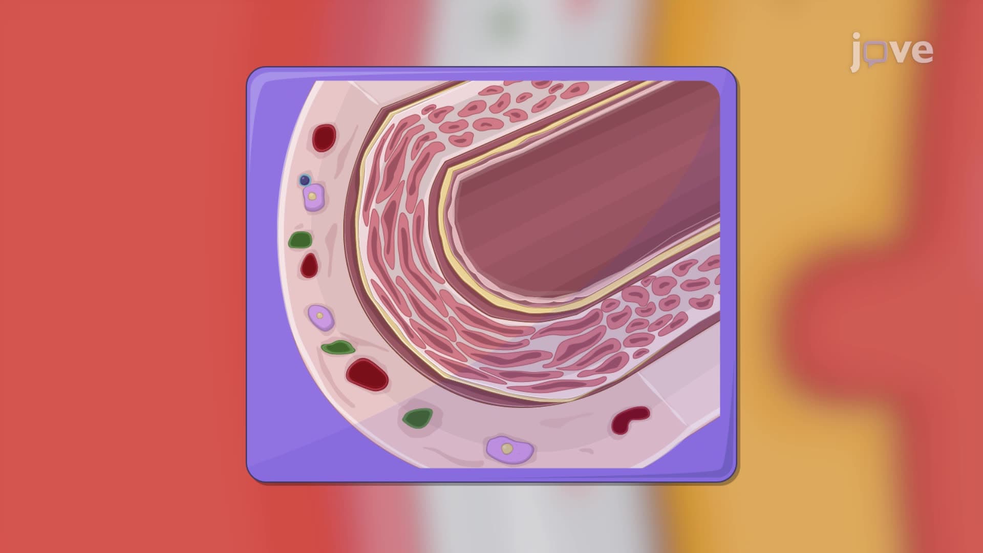
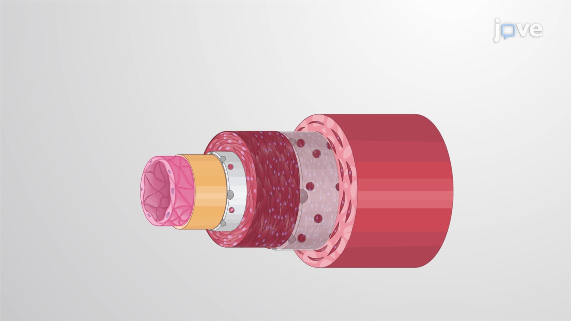
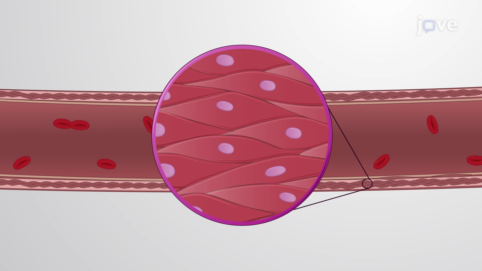
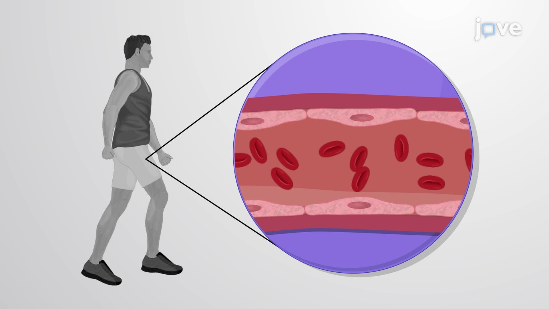
The aorta represents the cardiovascular system's primary distribution center, functioning as a high-pressure conduit that delivers oxygenated blood throughout the body. With its impressive 2.5 cm diameter and 2 mm wall thickness, this aorta largest artery must withstand systolic pressures reaching 120 mmHg while maintaining structural integrity over billions of cardiac cycles. The vessel's elastic properties, derived from smooth muscle and elastin fibers, enable it to expand during systole and recoil during diastole, creating the crucial Windkessel effect that maintains continuous blood flow.
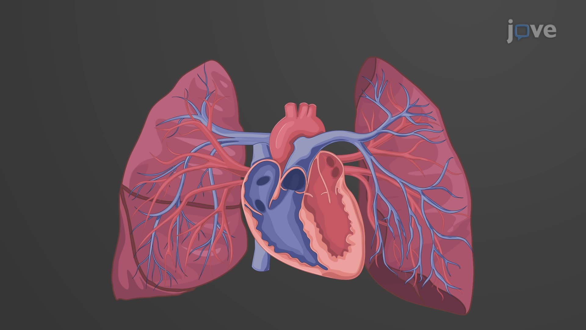
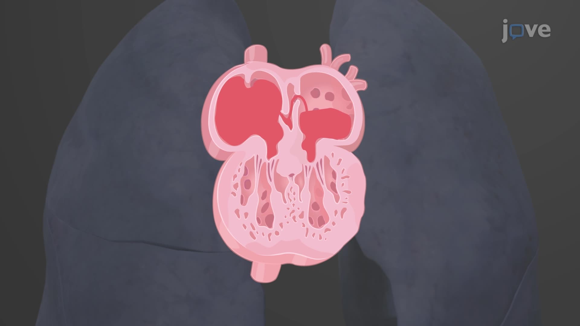
The aortic root valve complex initiates the aorta's journey, featuring three semilunar cusps that prevent retrograde flow during ventricular relaxation. This mechanism proves essential for maintaining coronary perfusion pressure, as the right and left coronary arteries originate from the ascending aorta immediately above the valve. Medical students studying for the MCAT frequently encounter questions about this anatomical relationship, particularly how aortic stenosis can compromise coronary blood flow and lead to angina symptoms.
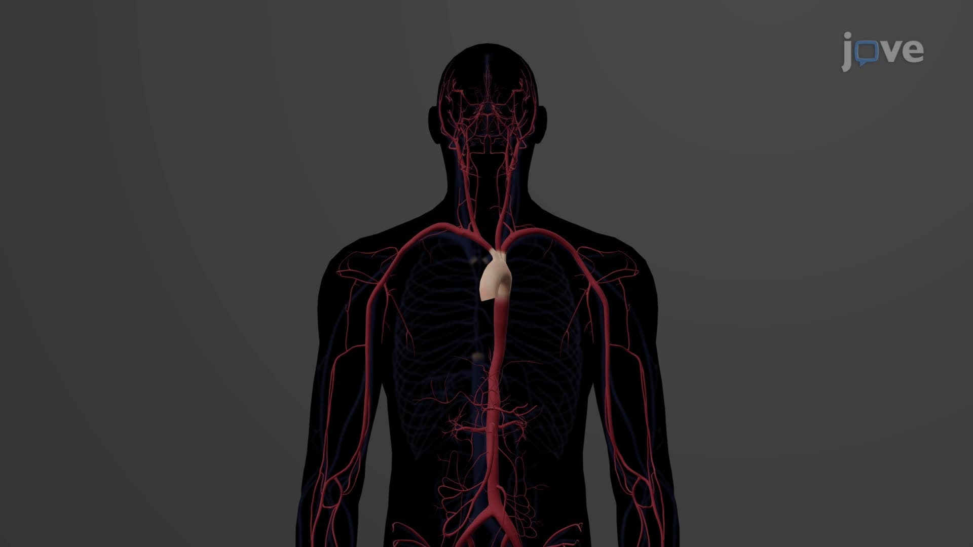
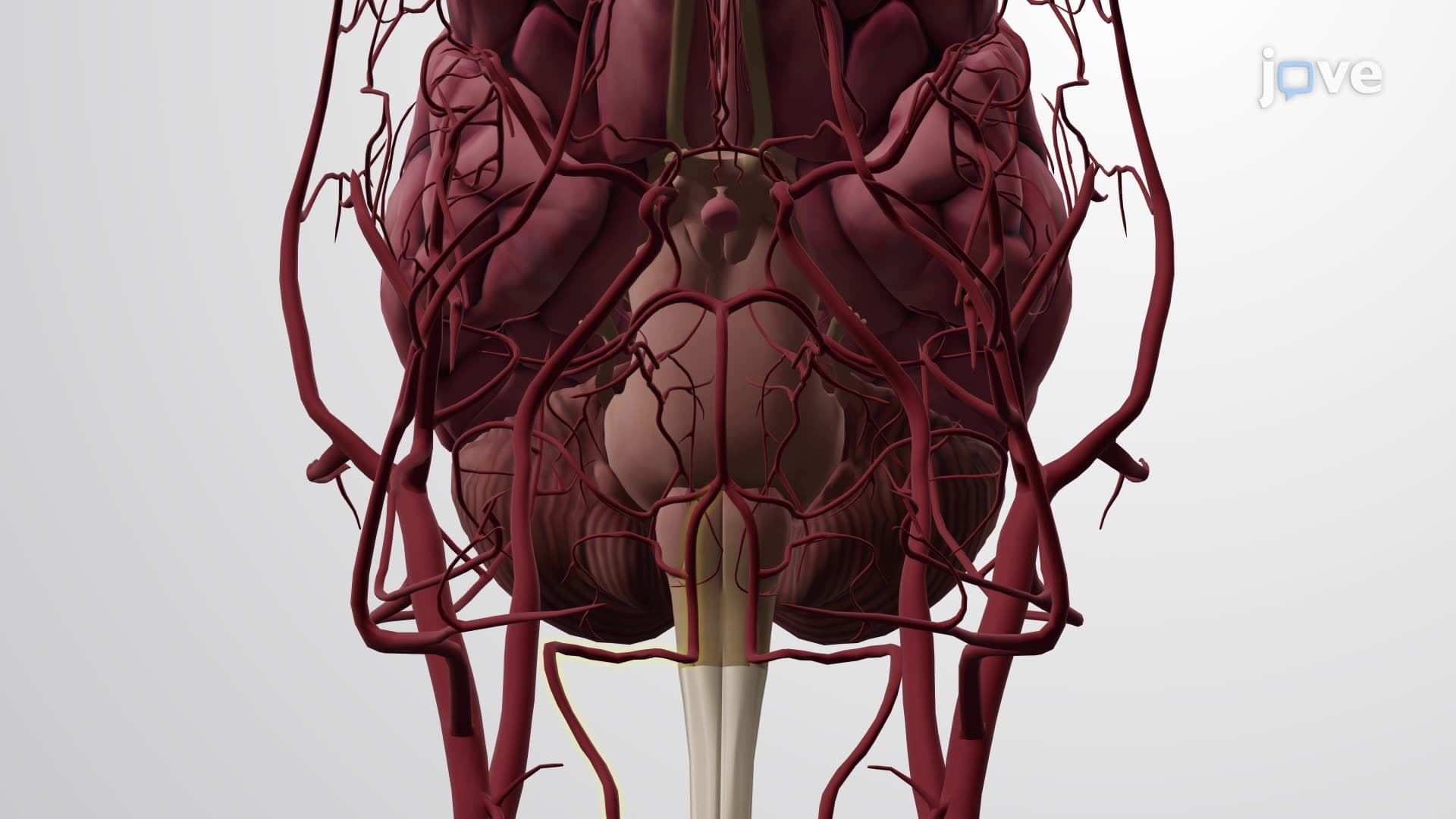
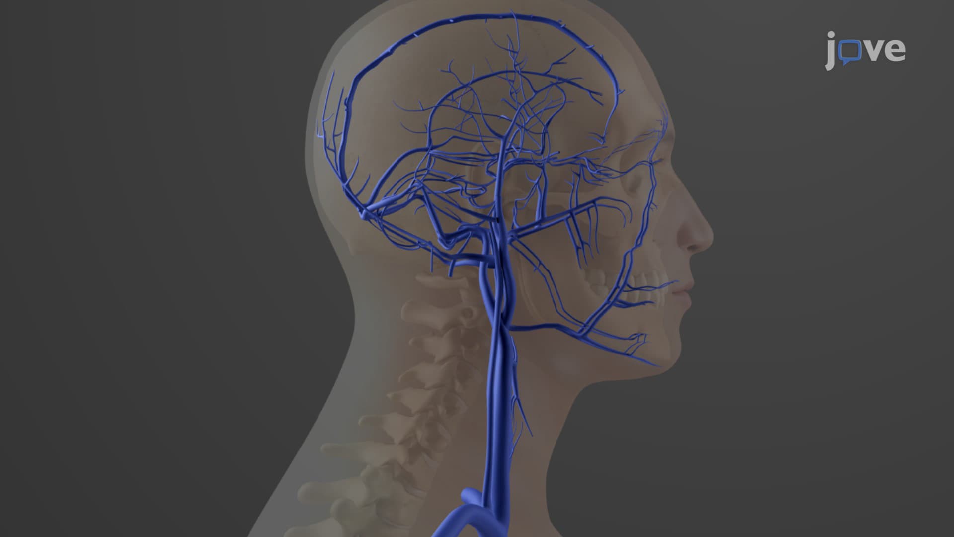
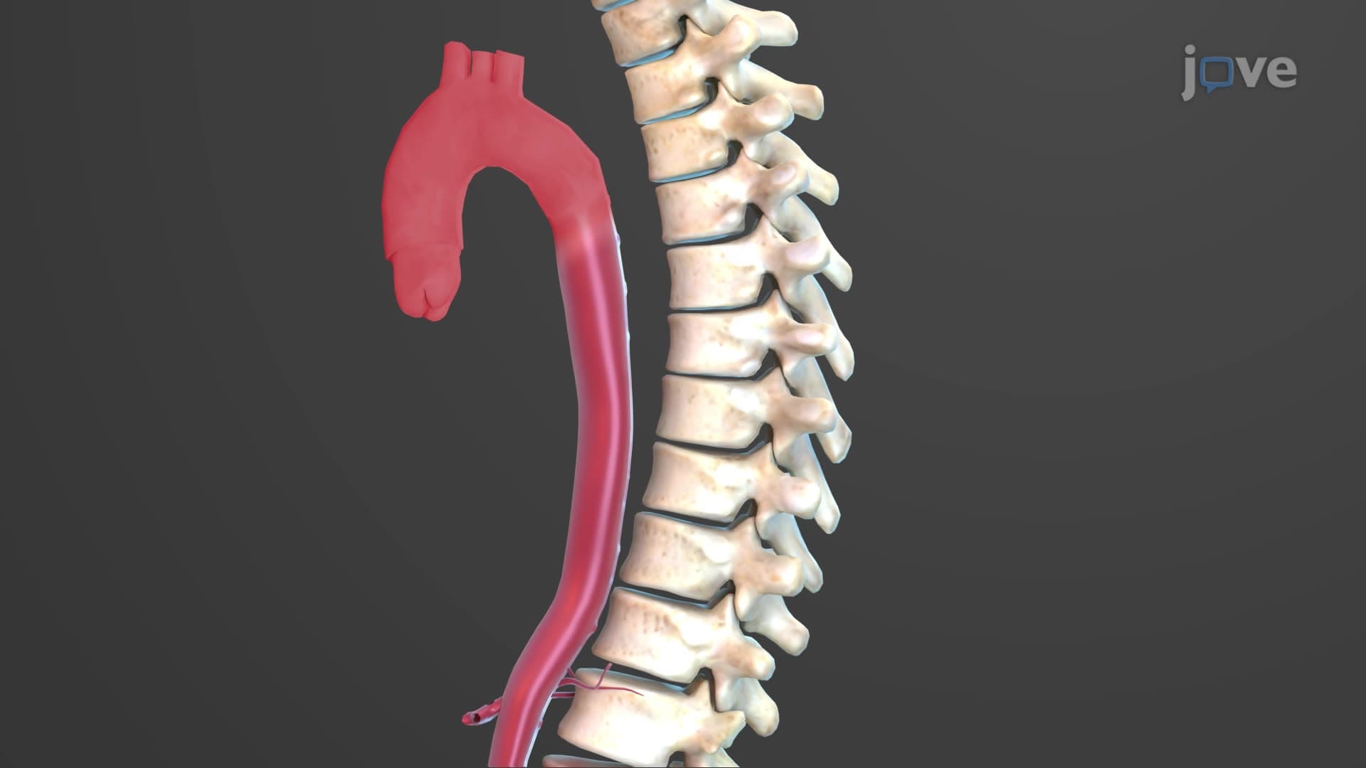
The ascending descending aorta transition occurs at the aortic arch, which gives rise to three major vessels: the brachiocephalic trunk, left common carotid artery, and left subclavian artery. This branching pattern, crucial for AP Biology students, demonstrates how evolutionary development optimized blood distribution to the head, neck, and upper extremities. The arch's leftward curvature around the heart creates the anatomical landmark that thoracic surgeons at institutions like Johns Hopkins use during cardiac procedures.
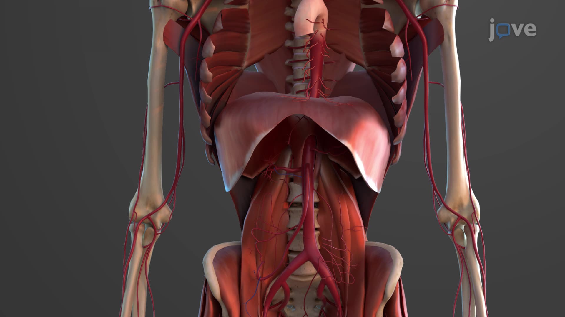
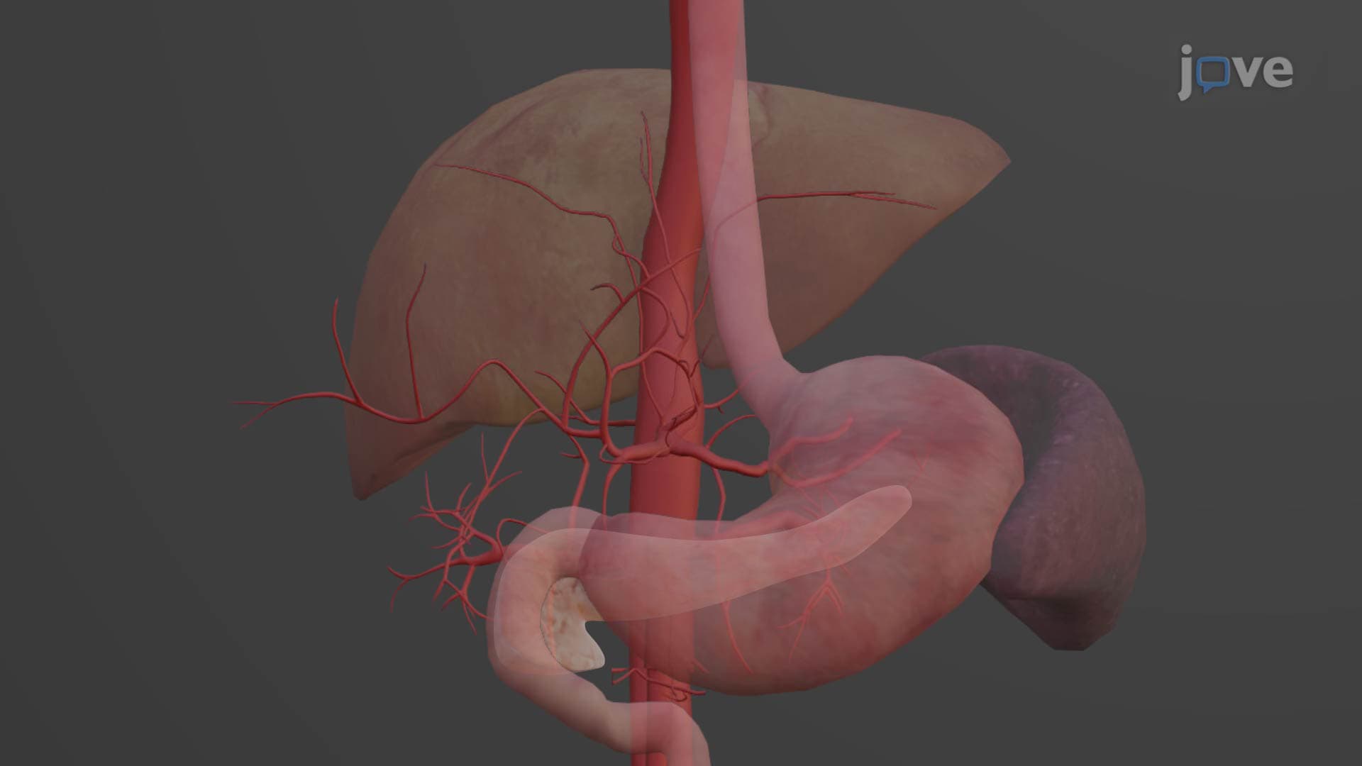
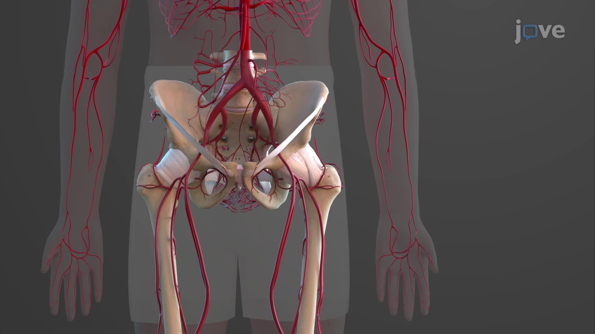
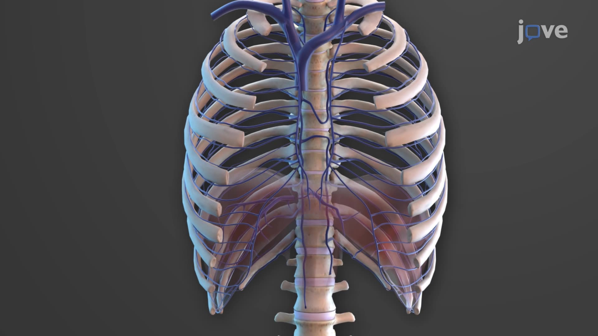
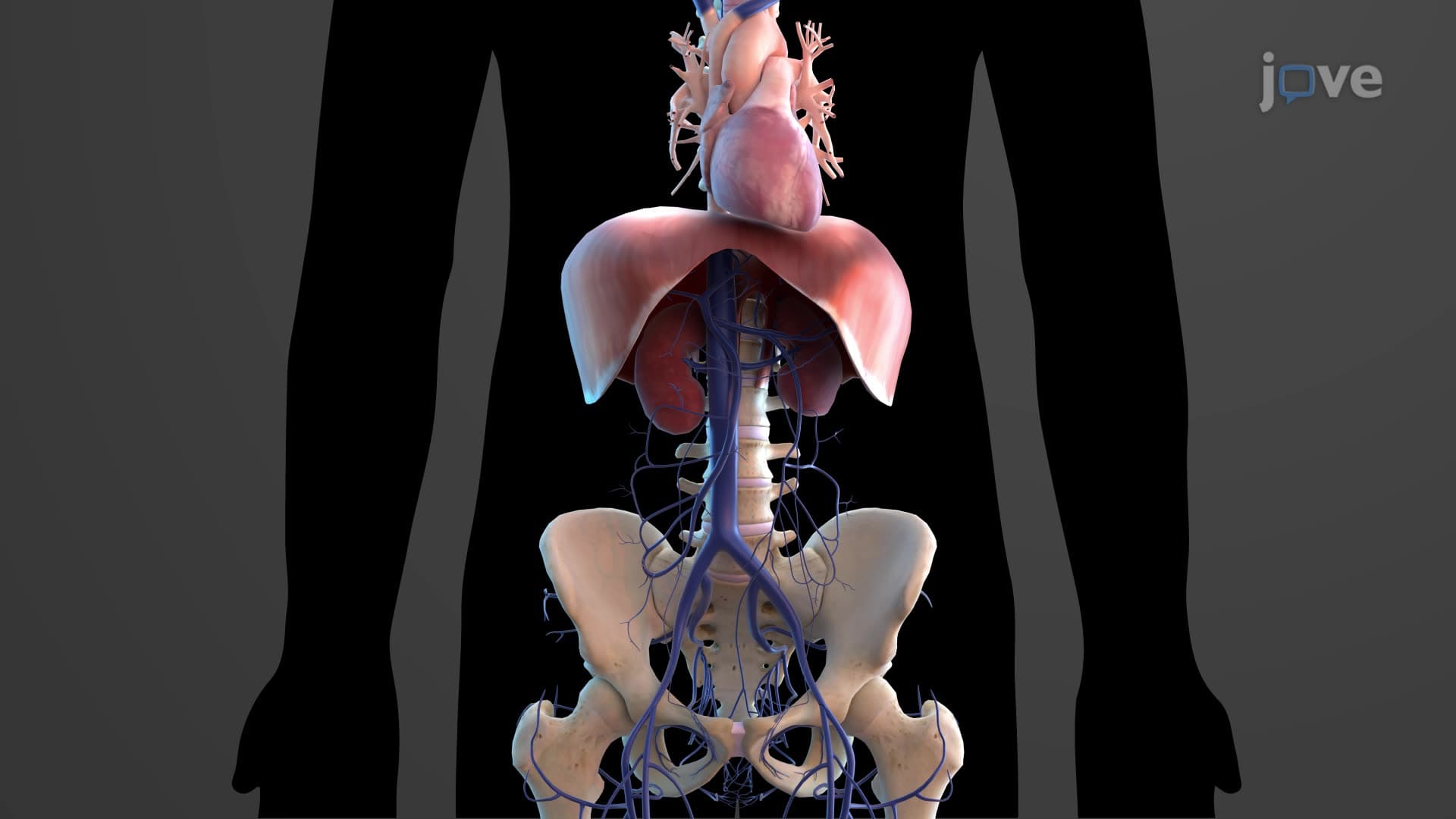
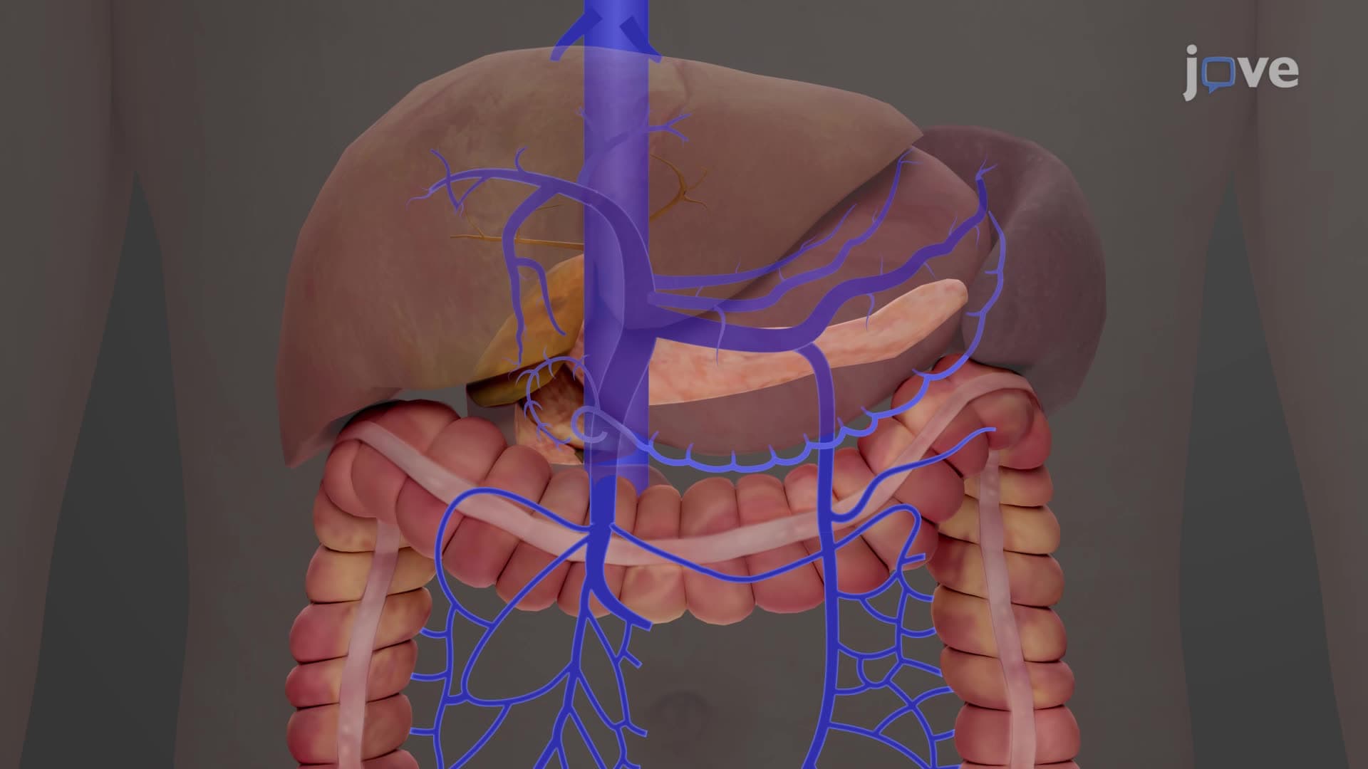
The descending aorta's division into thoracic (T5-T12) and abdominal segments reflects distinct functional territories. The thoracic aorta supplies intercostal arteries for chest wall perfusion and bronchial arteries for lung tissue, while the abdominal aorta branches extensively to serve gastrointestinal organs through the celiac trunk, superior mesenteric artery, and renal arteries. At the L4 vertebral level, the aorta's terminal bifurcation into common iliac arteries marks the transition to lower extremity circulation-a relationship that appears regularly on USMLE Step 1 examinations.
Related Micro-courses