Loading subject page…


149,645 views

Video Summary: What Is Thoracic Aorta
Did you know that your thoracic aorta anatomy includes a 20-centimeter powerhouse that pumps blood to vital organs like your lungs and heart lining every single heartbeat? This descending thoracic aorta explained reveals how this critical vessel branches into multiple arteries, similar to how major highways split into smaller roads to serve different neighborhoods in cities like Chicago or Los Angeles. Watch the full video on JoVE Coach to master this concept with expert-led visuals and step-by-step explanations.
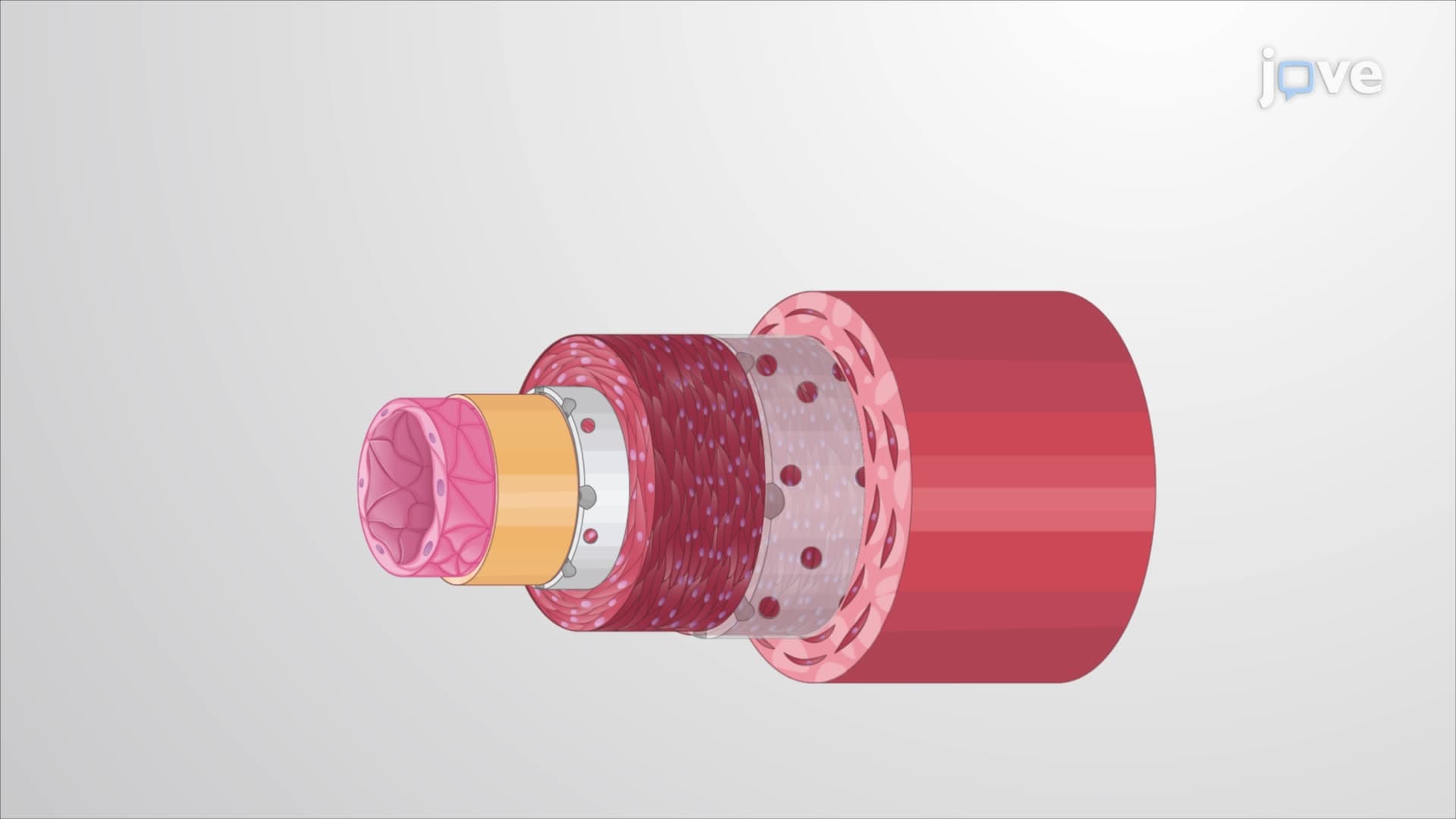
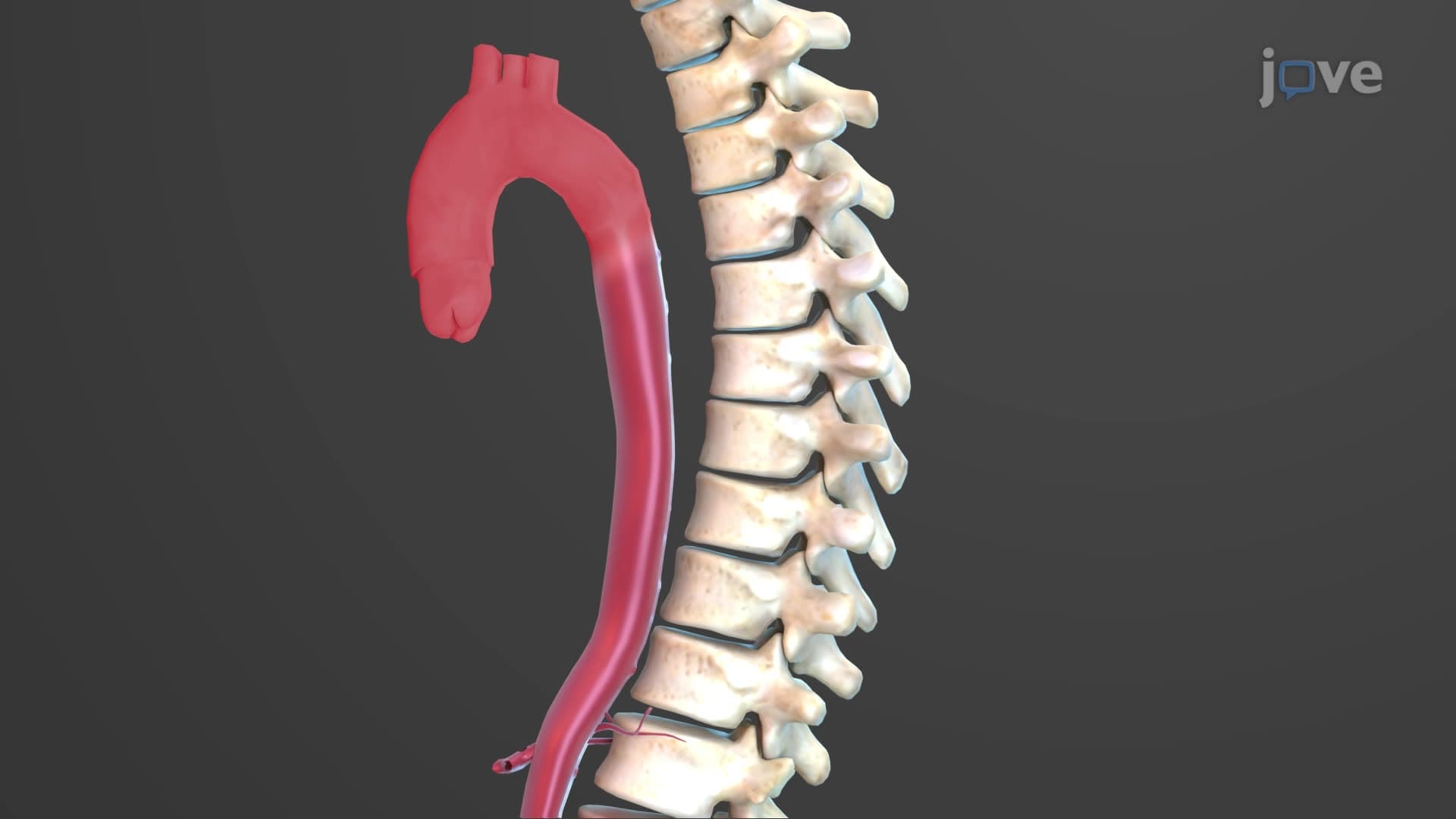
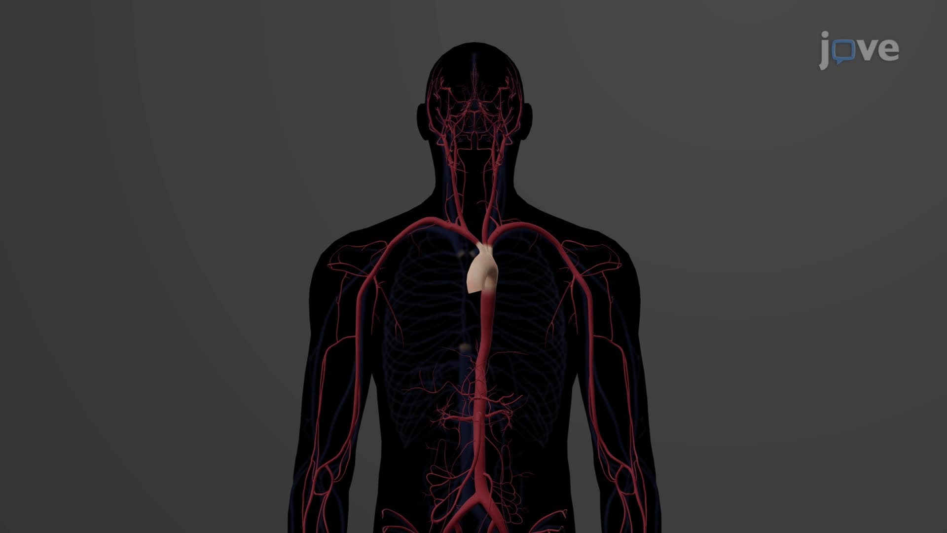
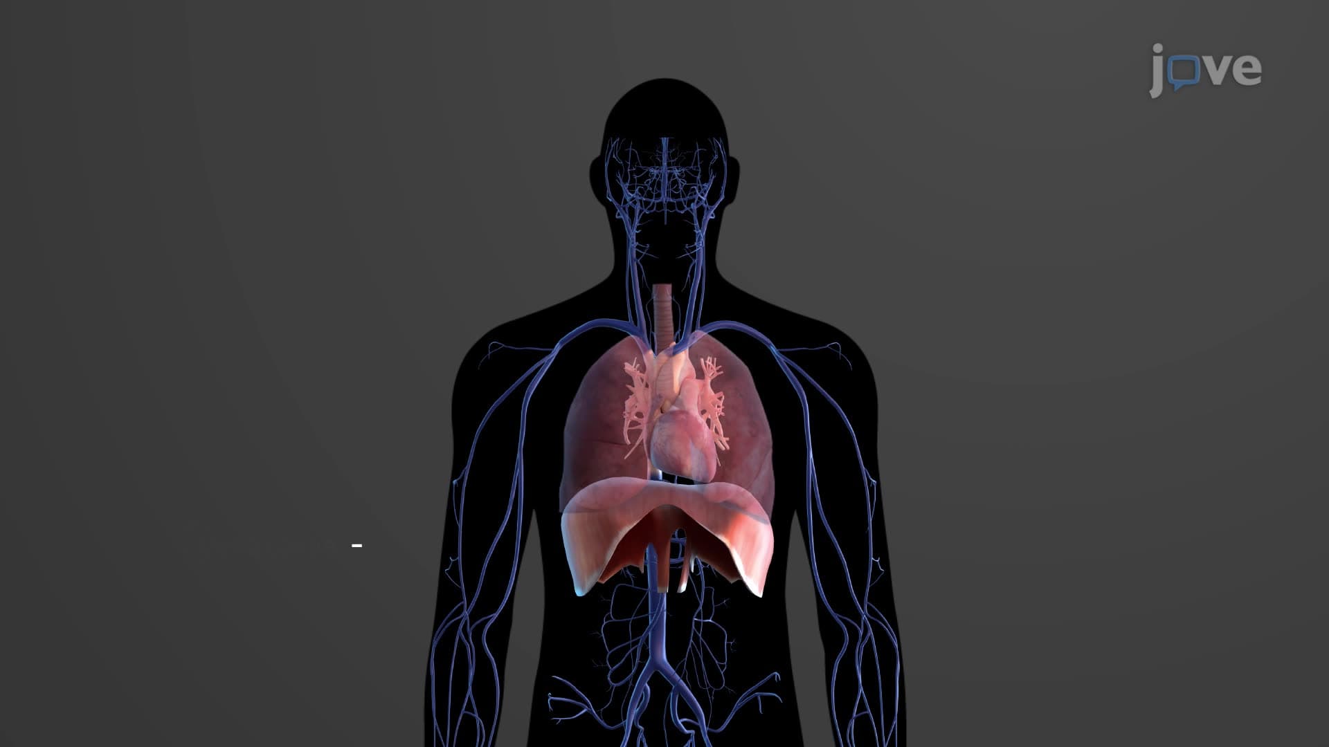
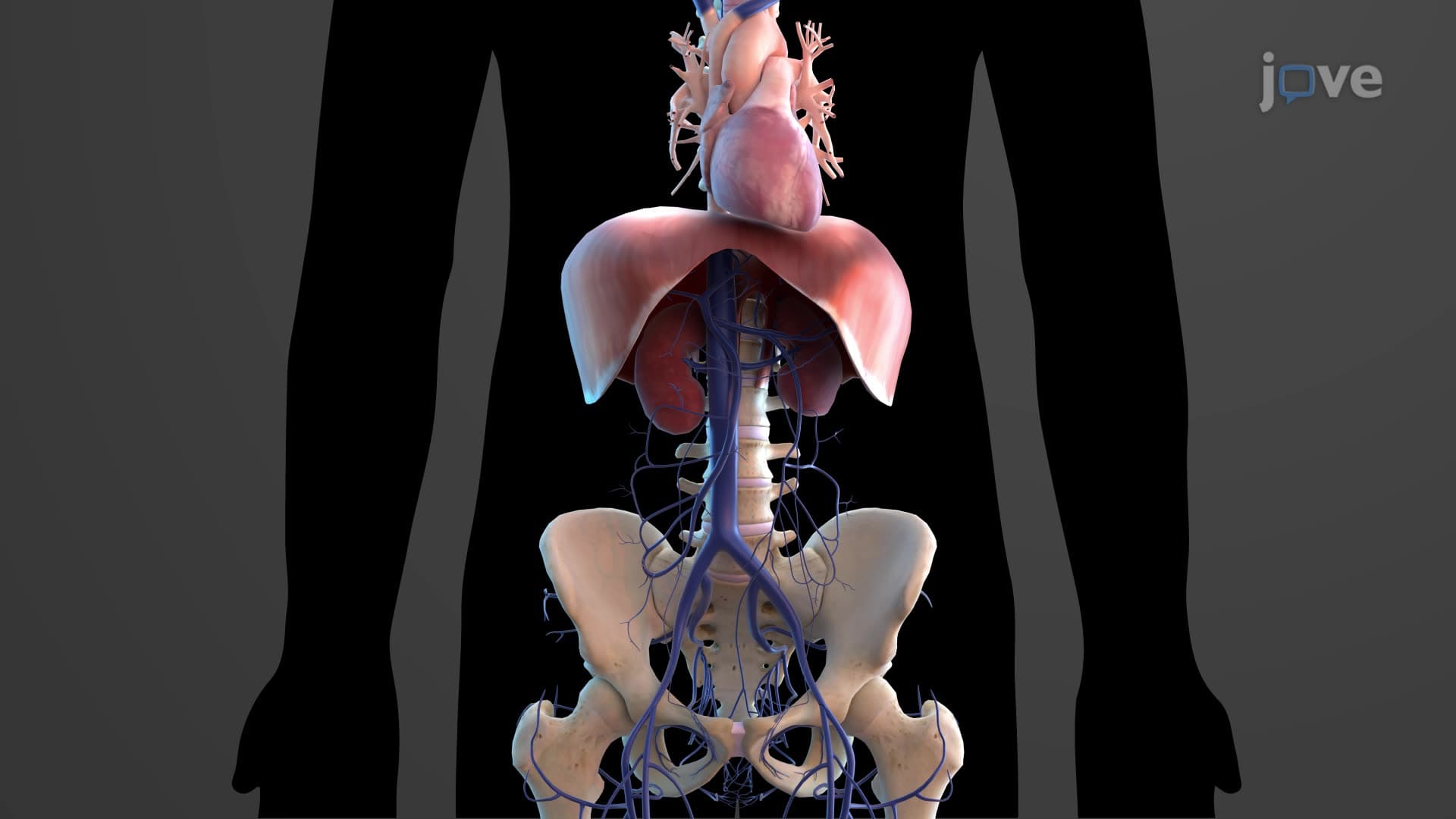
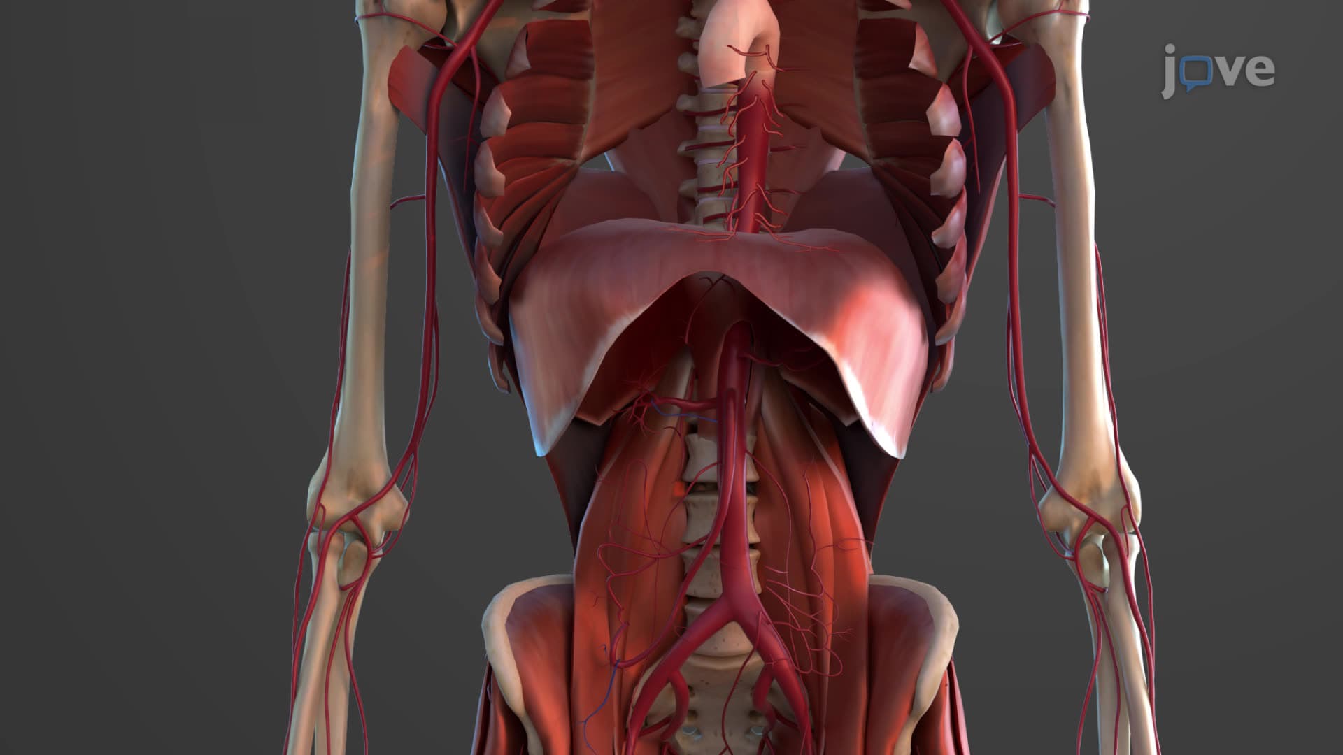
The thoracic aorta represents a critical 20-centimeter segment of the body's main arterial highway, strategically positioned along the left side of the vertebral column. This descending thoracic aorta explained begins its journey at the T4-T5 vertebral level and terminates when it passes through the aortic hiatus-a specialized opening in the diaphragm. Think of it like Interstate 95 running along the East Coast: just as this major highway connects multiple states while branching into smaller roads, the thoracic aorta connects the heart to various organs through its intricate branching system.
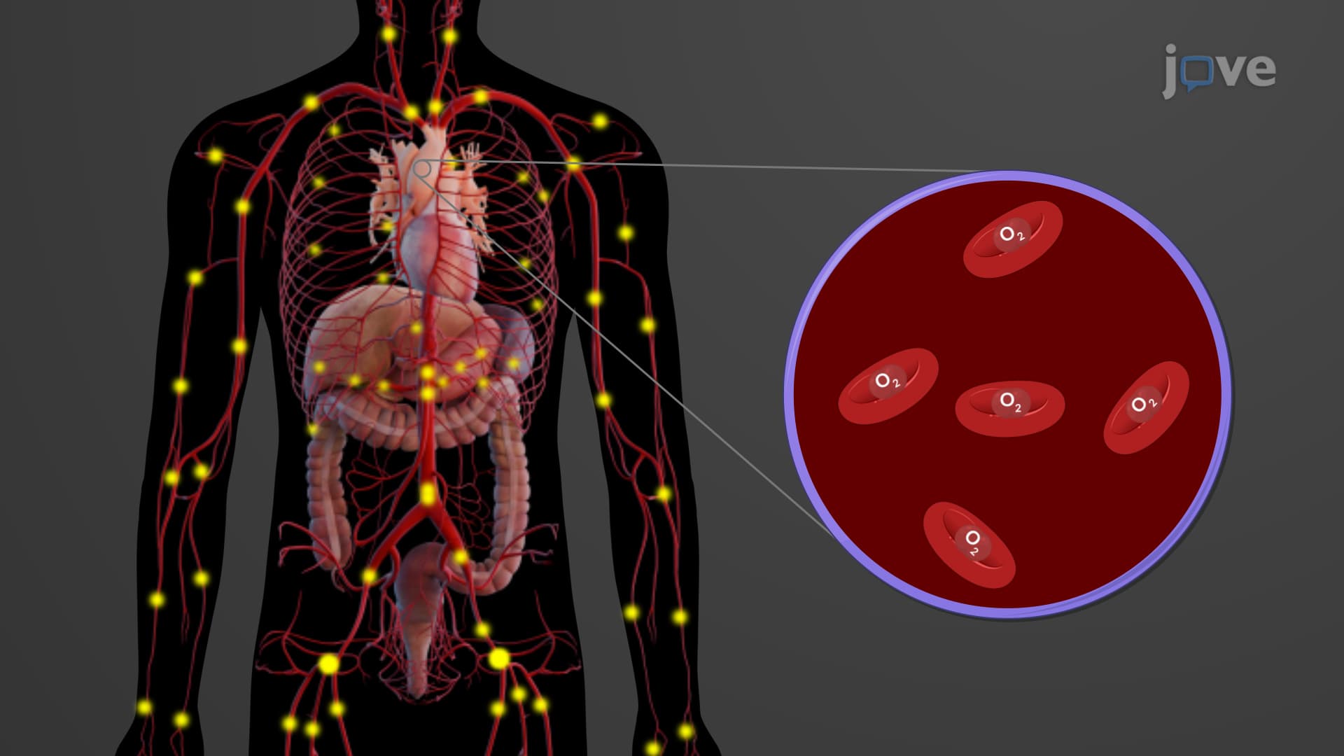
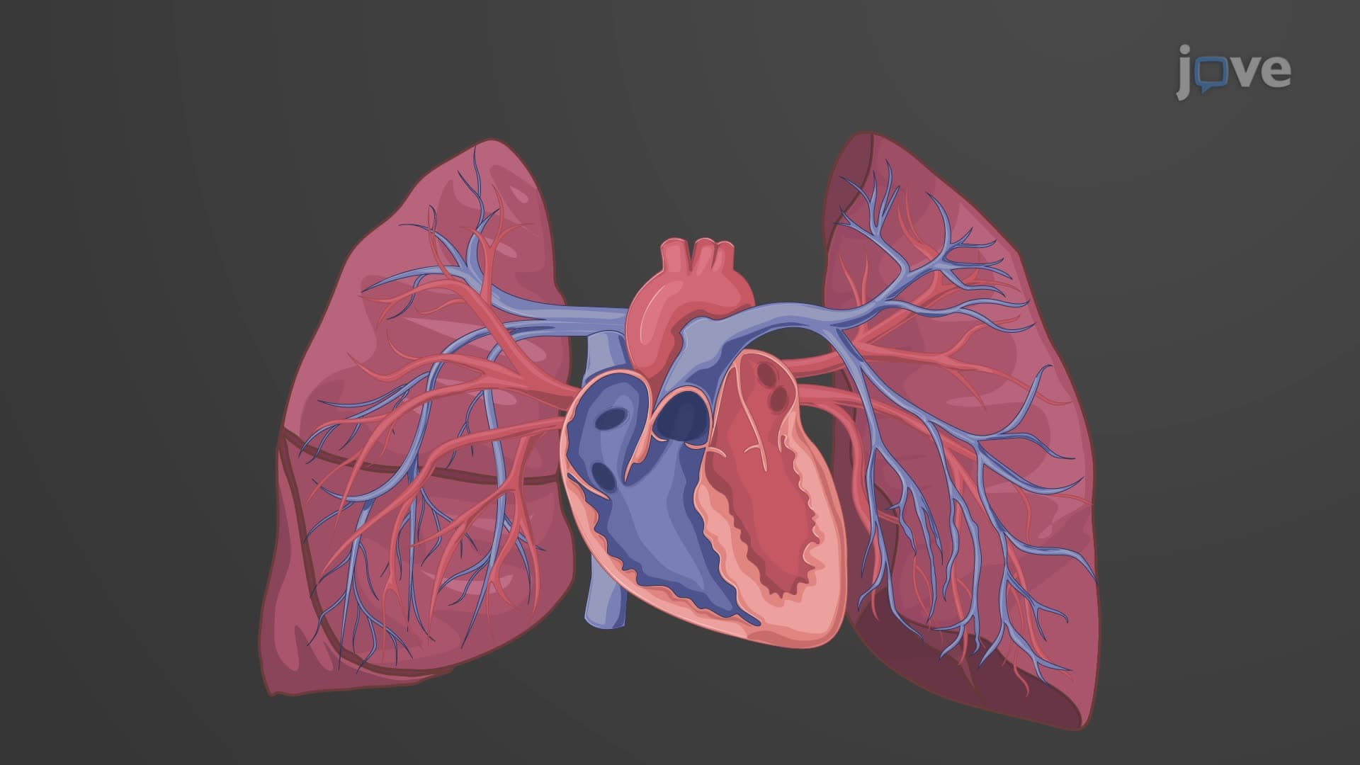
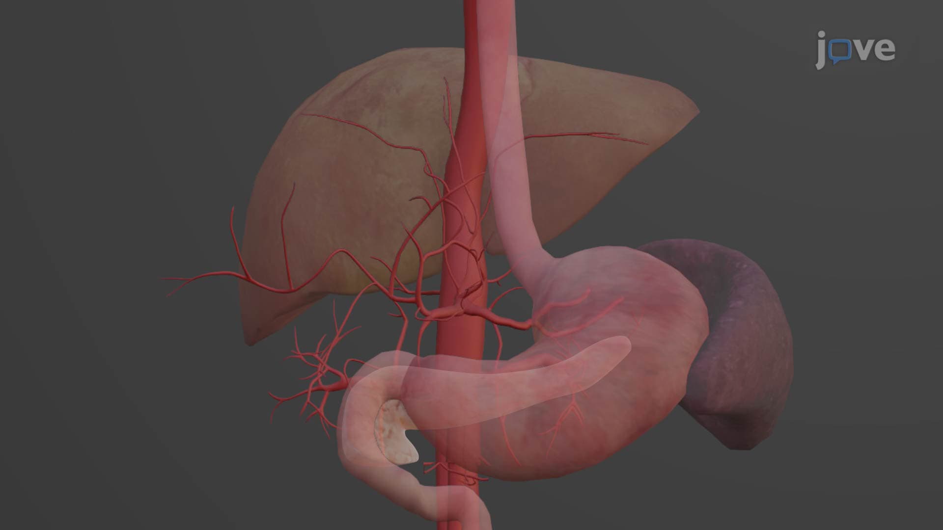
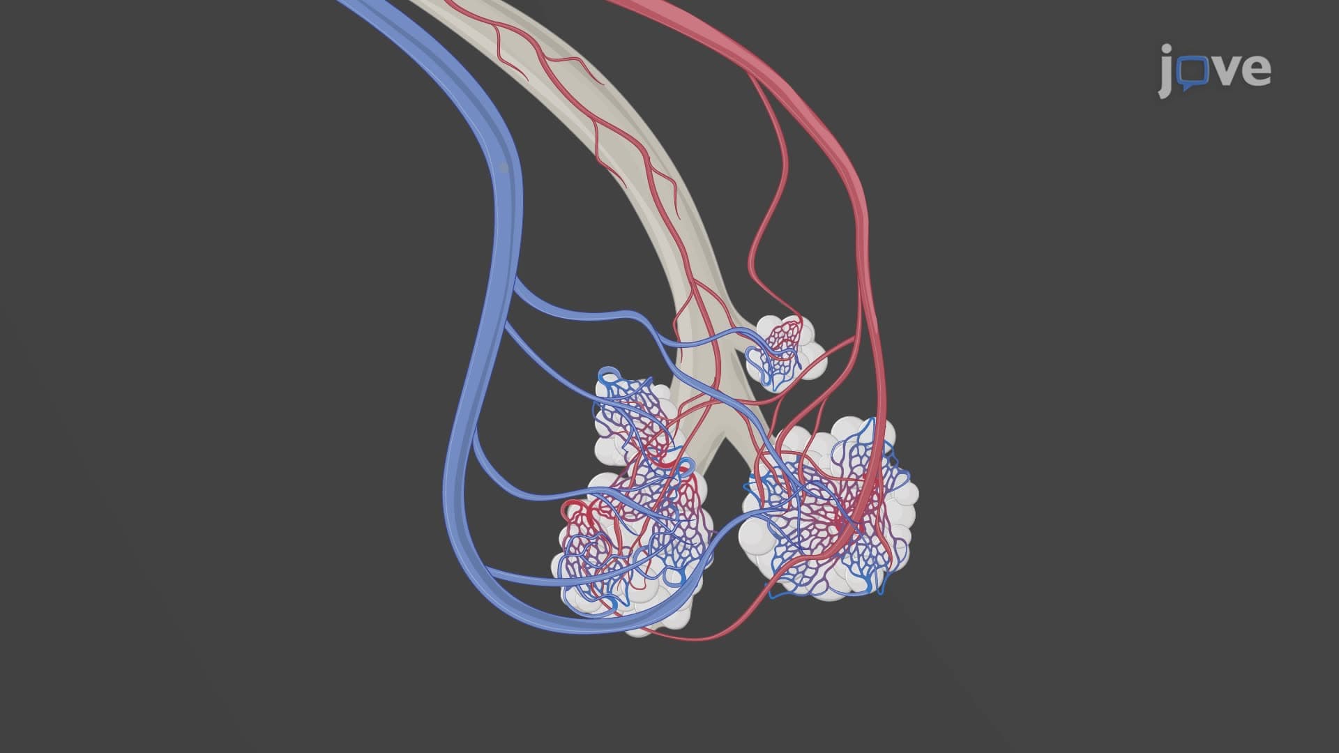
What is the thoracic aorta and its branches reveals a sophisticated dual-branching system. The visceral branches function like a specialized delivery network, targeting internal organs with precision. The pericardial artery nourishes the heart's protective sac, while the bronchial arteries serve as the lungs' dedicated blood supply-distinct from the pulmonary circulation. The esophageal thoracic branch ensures your swallowing tube receives adequate oxygenation, and the mediastinal artery supports the chest cavity's central compartment, including crucial lymph nodes that filter immune system threats.
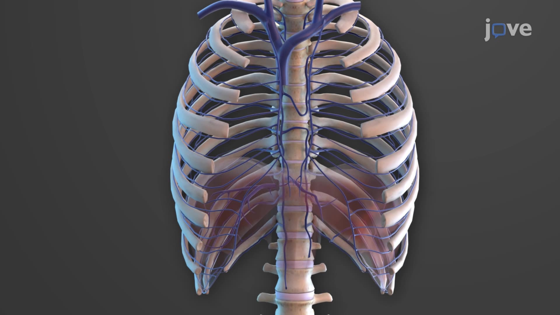
The parietal branches demonstrate nature's engineering excellence through their systematic organization. Nine pairs of thoracic aorta bronchial intercostal arteries create a ladder-like pattern, supplying the ribs, intercostal muscles, and chest wall skin. This arrangement proves essential during medical procedures-thoracic surgeons at institutions like Mayo Clinic and Johns Hopkins rely on this anatomical knowledge when performing chest surgeries. The subcostal and superior phrenic arteries complete this network by serving the lower chest wall and diaphragm respectively.
Understanding thoracic aorta branch anatomy becomes crucial for students preparing for advanced coursework and professional exams. On the MCAT, questions frequently test knowledge of aortic branching patterns and their physiological significance. AP Biology students encounter this topic when studying circulatory system organization, while pre-nursing students preparing for the HESI A2 or TEAS exams must master cardiovascular anatomy. In clinical settings, radiologists use this knowledge to interpret chest imaging, while cardiac surgeons depend on precise anatomical understanding during complex procedures involving aortic pathology.
Related Micro-courses