Loading subject page…

- Biology
- Osmoregulation and Excretion
- Kidney Structure
Micro-course: Osmoregulation and Excretion
338,101 views
Learn key concepts and earn your certificate.



Kidney Structure

00:00
01:45
Video Summary: What Is Kidney Structure
Every day, your kidneys filter approximately 50 gallons of blood-equivalent to filling a standard bathtub! Understanding kidney structure is essential for grasping how these remarkable organs maintain your body's chemical balance. From the Mayo Clinic's nephrology department to medical schools nationwide, kidney structure forms the foundation of renal physiology education. Watch the full video on JoVE Coach to master this concept with expert-led visuals and step-by-step explanations.
- Understand what is kidney structure and identify the major anatomical components of the renal system
- Recognize the pathway of blood flow from the renal artery through nephrons to the renal vein
- Analyze the functional relationship between the renal cortex, medulla, and collecting duct system
- Compare the roles of different nephron segments in filtration and reabsorption processes
- Identify the structural components of the renal corpuscle, including glomerulus and Bowman's capsule
- Evaluate how tubular structures (PCT, loop of Henle, DCT) contribute to urine formation
- Apply knowledge of kidney structure to understand osmoregulation and waste elimination
- Explore the connection between structural organization and functional efficiency in renal physiology
Understanding Kidney Structure: The Foundation of Renal Function
The human kidney represents one of biology's most elegant examples of structure-function relationships. These bean-shaped organs, each roughly the size of a computer mouse, demonstrate how anatomical organization directly supports physiological processes. In AP Biology and college-level anatomy courses, kidney structure serves as a prime example of how complex organ systems maintain homeostasis.
Gross Anatomy: External Structure and Blood Supply
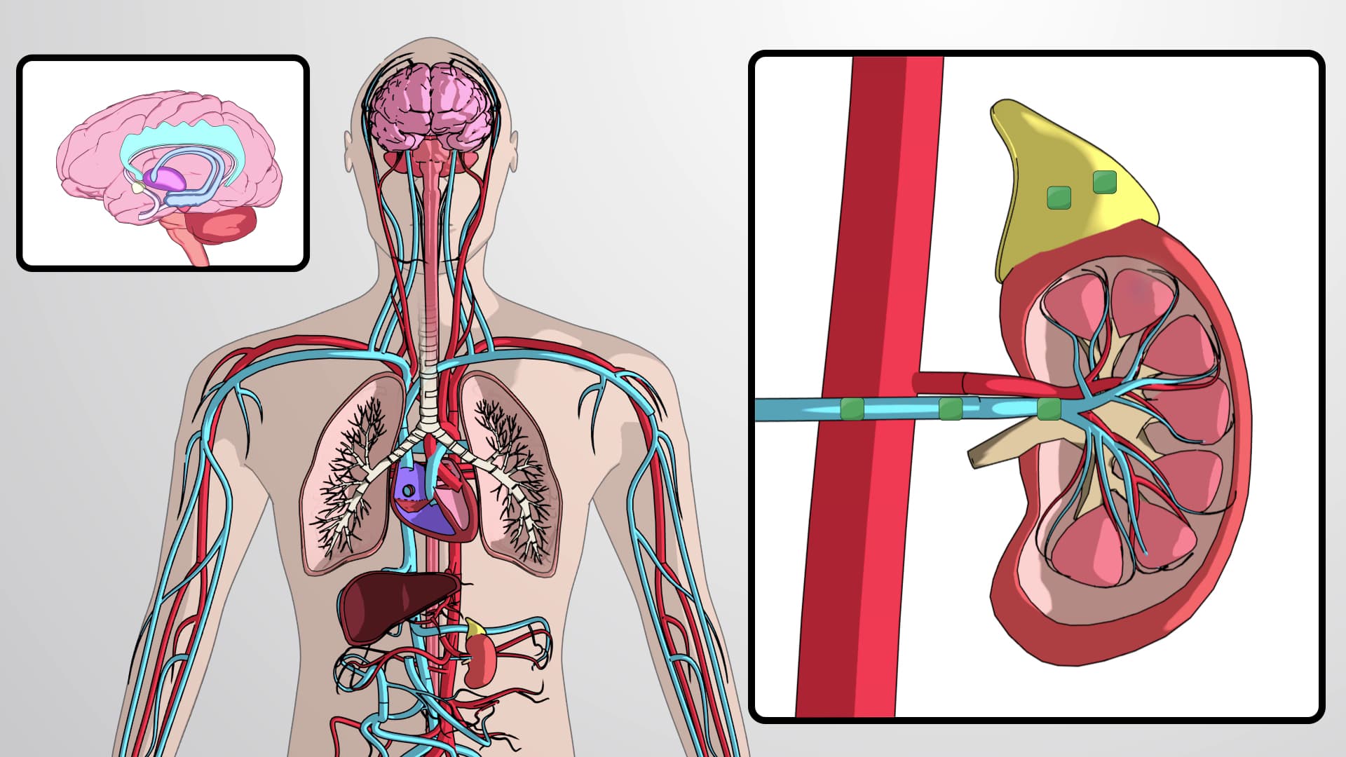
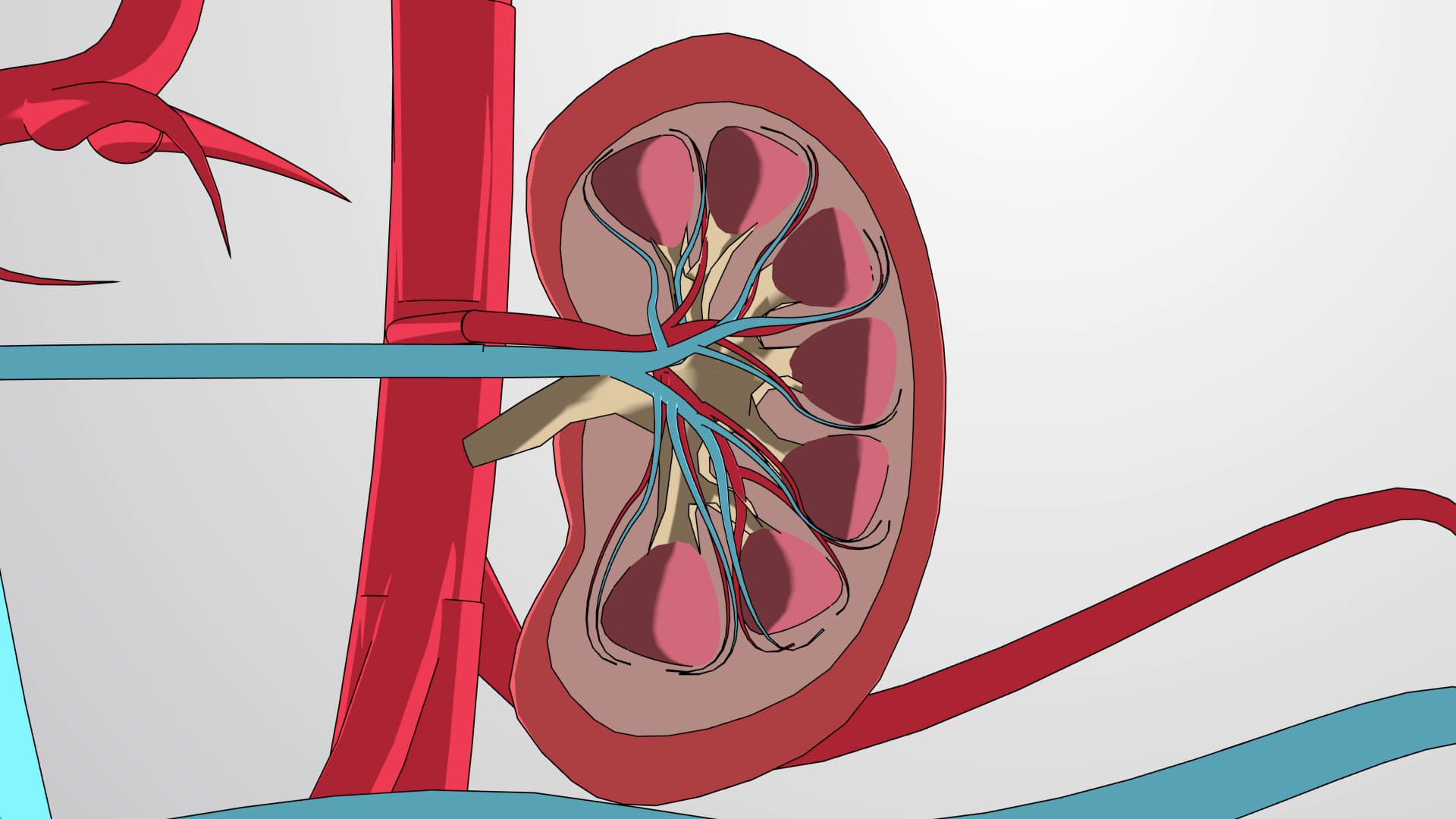
Each kidney's external structure reveals important functional zones. The hilum, located on the concave medial surface, serves as the entry and exit point for major vessels and the ureter. The renal artery enters here, branching into segmental, interlobar, and arcuate arteries before forming the afferent arterioles that supply individual nephrons. This vascular arrangement ensures that blood reaches every functional unit efficiently-a critical concept tested on the MCAT and medical school exams.
The kidney's internal architecture divides into two distinct regions: the outer cortex and inner medulla. The cortex houses the renal corpuscles and convoluted tubules, while the medulla contains the loops of Henle and collecting ducts arranged in pyramid-shaped structures. This organization isn't random-it reflects the kidney's evolutionary adaptation to concentrate urine effectively, a process crucial for terrestrial life.
Microscopic Structure: The Nephron as the Functional Unit
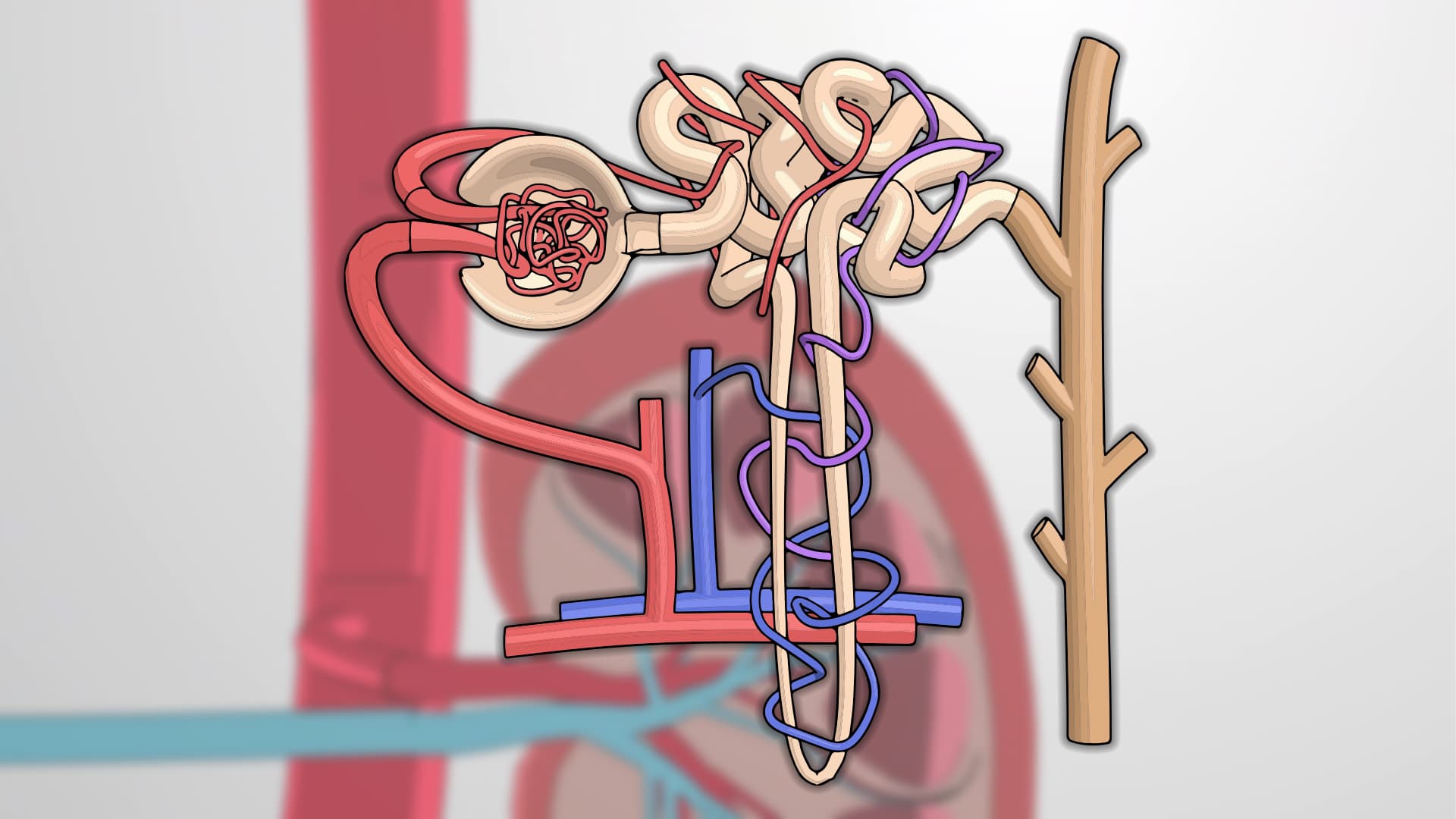
Each kidney contains approximately one million nephrons, the microscopic functional units responsible for filtration, reabsorption, and secretion. The nephron's structure directly correlates with its function, beginning with the renal corpuscle where initial filtration occurs. The glomerulus, a cluster of specialized capillaries, sits within Bowman's capsule like a fist in a balloon, creating the filtration barrier that allows water and small molecules to pass while retaining proteins and blood cells.
The tubular system-comprising the proximal convoluted tubule, loop of Henle, and distal convoluted tubule-demonstrates remarkable structural specialization. The PCT's brush border increases surface area for reabsorption, while the loop of Henle's countercurrent structure enables concentration of urine. Understanding these relationships proves essential for success in courses like Human Physiology and helps students tackle challenging questions on standardized exams.
Clinical Relevance and Exam Applications
Kidney structure knowledge directly applies to understanding diseases and medical interventions. For instance, diabetic nephropathy affects glomerular structure, while kidney stones can obstruct the collecting system. These connections frequently appear on nursing exams like the NCLEX and HESI A2, where students must link anatomical knowledge to patient care scenarios.
Frequently Asked Questions
Kidney structure refers to the anatomical organization of the kidneys, including their gross anatomy and microscopic components like nephrons. Understanding this structure is crucial because it directly explains how kidneys filter blood, regulate fluid balance, and produce urine. This knowledge forms the foundation for studying renal physiology, kidney diseases, and medical treatments in healthcare fields.
The MCAT frequently tests kidney structure through passages about osmoregulation, nephron function, and disease processes. AP Biology includes kidney structure in Unit 5 (Heredity) when discussing homeostasis and feedback mechanisms. Expect questions linking structural features to functional outcomes, such as how glomerular structure enables selective filtration or how tubule organization supports concentration gradients.
Prioritize understanding the nephron's components and their specific functions, the relationship between cortex and medulla, and blood flow pathways. Many professors emphasize structure-function relationships, so practice explaining how anatomical features support physiological processes. Laboratory practical exams often require identifying structures on models and microscope slides, so visual familiarity is essential.
Healthcare professionals use kidney structure knowledge daily when interpreting diagnostic tests, understanding medication effects, and educating patients. For example, nurses monitoring patients with chronic kidney disease must understand how structural damage affects filtration rates. Medical imaging specialists rely on structural knowledge to identify abnormalities in CT scans and ultrasounds of the renal system.
Kidney structure builds logically from basic cell and tissue concepts taught in introductory biology courses. Students with high school biology background typically succeed by focusing on understanding function before memorizing detailed anatomy. The key is recognizing patterns-how structure supports function-rather than memorizing isolated facts.
Create concept maps linking structure to function, use labeled diagrams to trace blood and filtrate flow pathways, and practice explaining processes aloud. Focus on understanding rather than memorization-exam questions often require applying structural knowledge to novel scenarios. Form study groups to teach concepts to peers, as explaining helps solidify understanding.
Progress to renal physiology processes like glomerular filtration rate, tubular transport mechanisms, and hormonal regulation of kidney function. Understanding acid-base balance and electrolyte regulation builds naturally from structural knowledge. Advanced topics include pathophysiology of kidney diseases and pharmacology of diuretics and other renal medications.
The HESI A2 and TEAS exams include kidney structure within anatomy and physiology sections, often focusing on basic organization and function. Questions may ask about nephron components, urine formation pathways, or the relationship between structure and disease processes. These exams emphasize practical applications relevant to patient care rather than detailed molecular mechanisms.
Related Micro-courses