Loading subject page…


152,815 views

Video Summary: What Is Neuron Structure
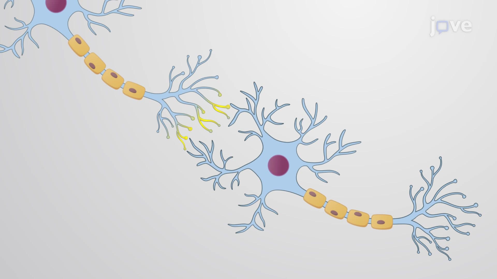
Ever wondered how a single brain cell can transmit information faster than a Ferrari on the Indianapolis 500 speedway? Neuron structure forms the foundation of all nervous system communication, from reflexes to complex thoughts. This specialized cellular architecture includes dendrites that receive signals, a cell body housing the nucleus, and an axon that transmits electrical impulses across synapses. Medical students at Harvard Medical School study these same components when learning how anesthesia works during surgery. Watch the full video on JoVE Coach to master this concept with expert-led visuals and step-by-step explanations.
Neuron structure represents one of biology's most elegant examples of form following function. These specialized cells have evolved unique architectural features that enable rapid, precise communication across vast networks. Understanding neuron structure is essential for AP Biology students and forms a cornerstone of pre-med coursework at universities like Stanford and Johns Hopkins.
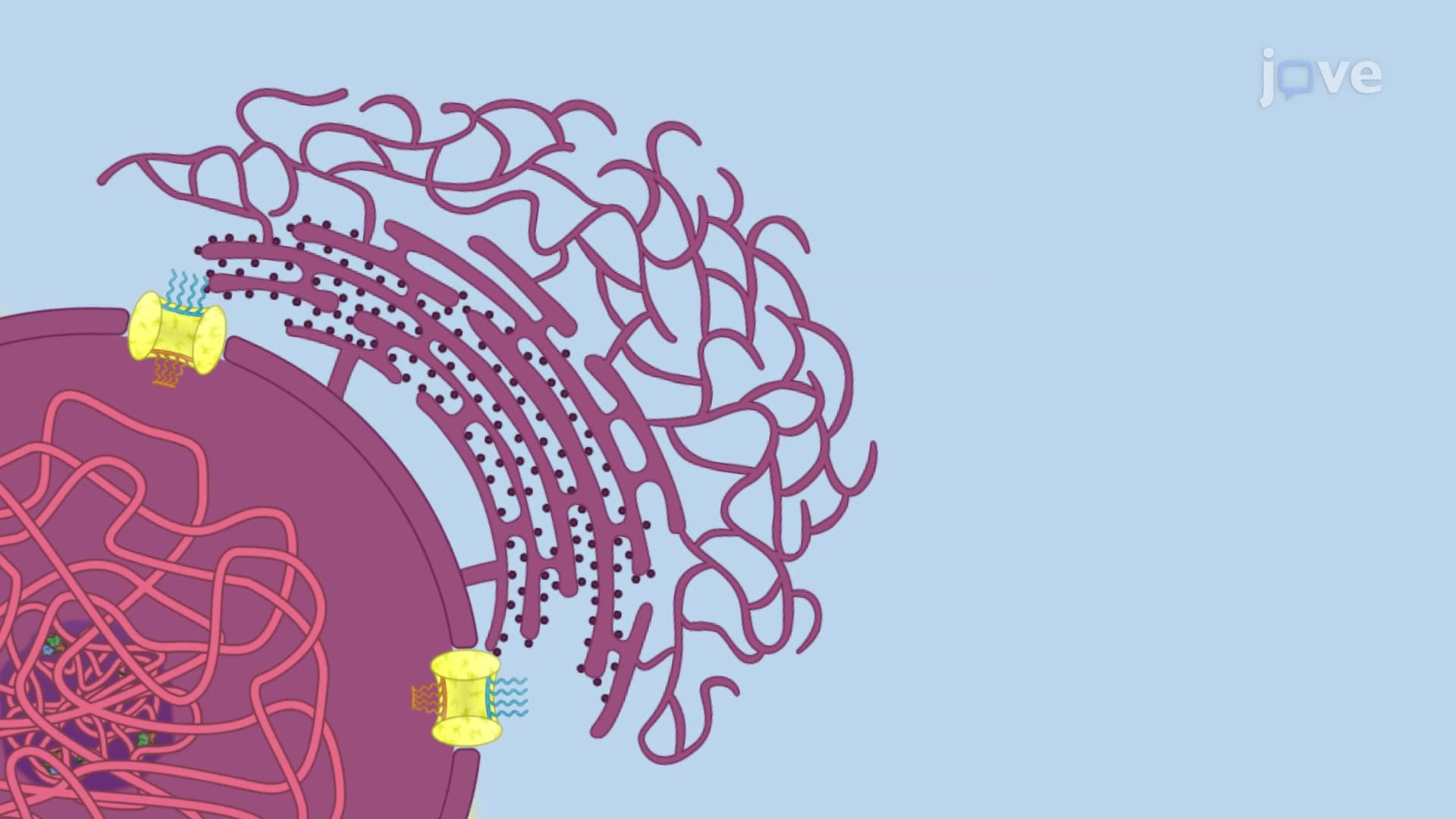
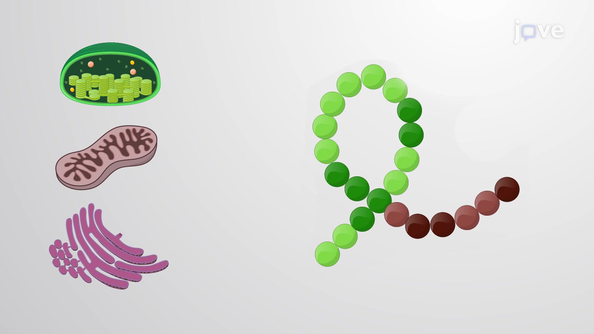
The soma, or cell body, functions as the neuron's metabolic headquarters. Unlike typical cells, the neuronal soma must support extremely long projections-some motor neuron axons extend from the spinal cord to the toes, spanning over three feet. The nucleus contains DNA that codes for specialized proteins like ion channels and neurotransmitter enzymes. Rough endoplasmic reticulum, called Nissl bodies in neurons, produces these critical proteins. This knowledge proves vital for MCAT test-takers when analyzing neurological disorders like ALS, where motor neuron cell bodies deteriorate.
Dendrites create the neuron's receiving apparatus through their highly branched, tree-like structure. A single cortical neuron may have thousands of dendritic branches, each forming synaptic connections with other neurons. This branching pattern determines how many inputs a neuron can integrate simultaneously. Medical students at UCLA study how dendritic damage in Alzheimer's disease disrupts memory formation. The dendritic spine density directly correlates with cognitive function-a concept frequently tested on neuroscience exams.
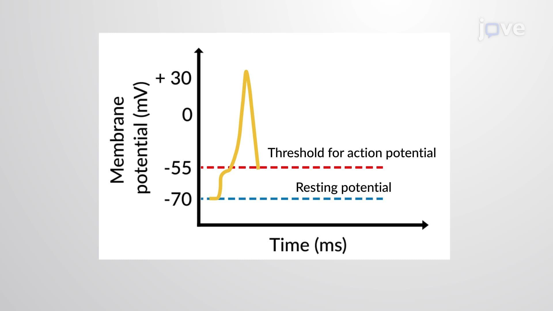
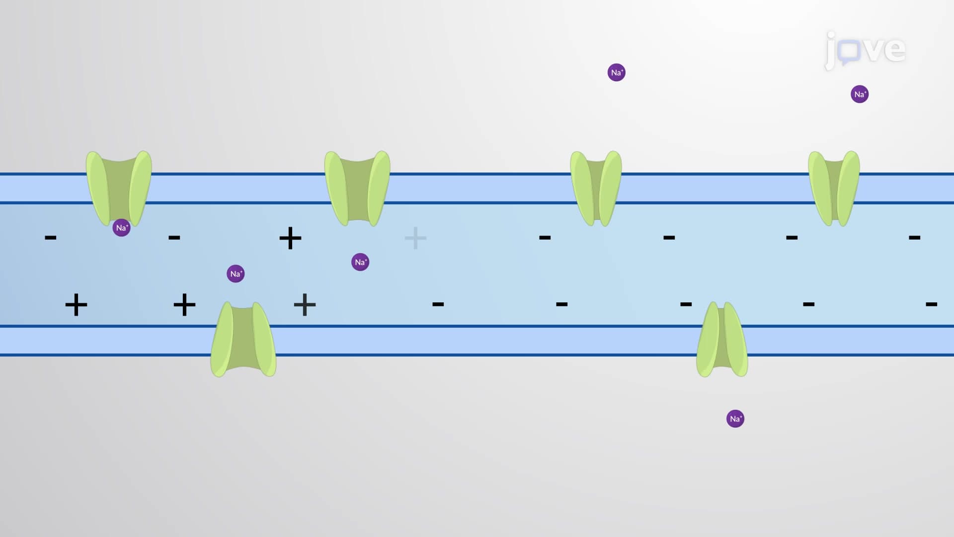
The axon represents the neuron's transmission line, varying dramatically in length and diameter depending on function. The axon hillock integrates all incoming signals and determines whether to generate an action potential. Myelinated axons, wrapped in fatty sheaths produced by oligodendrocytes (CNS) or Schwann cells (PNS), conduct signals up to 100 times faster than unmyelinated fibers. Nodes of Ranvier, gaps between myelin segments, enable saltatory conduction-the rapid "jumping" of electrical signals. This mechanism explains why multiple sclerosis, which destroys myelin, causes severe motor and sensory deficits.
Axon terminals house synaptic vesicles containing neurotransmitters like dopamine, serotonin, and acetylcholine. When action potentials arrive, calcium influx triggers vesicle fusion and neurotransmitter release into the synaptic cleft. This chemical signaling allows precise control over target cells, producing either excitatory or inhibitory effects. Understanding this process helps nursing students on the NCLEX explain how psychiatric medications like SSRIs modify synaptic transmission to treat depression.
Related Micro-courses