Loading video…


 17 Concepts
 102.1K views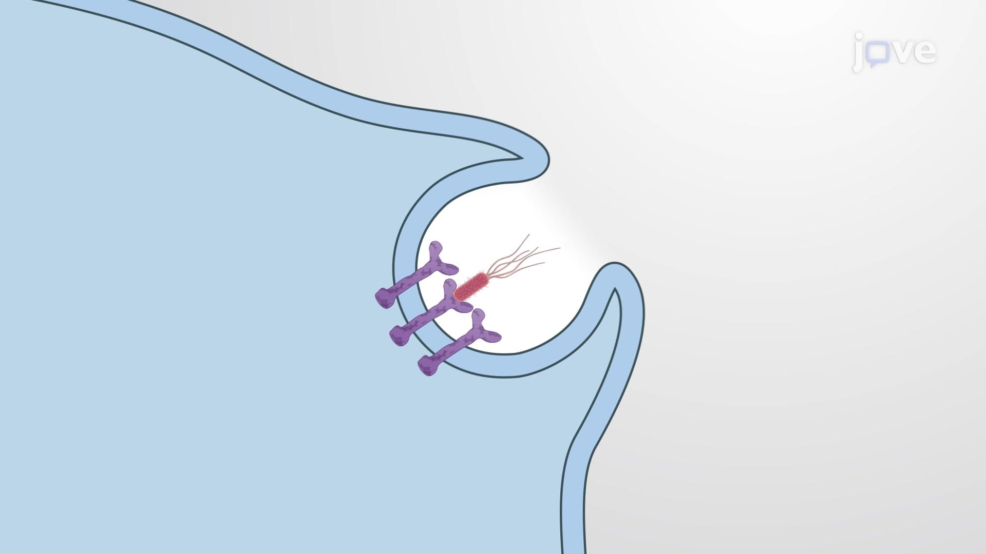

Video Summary: What Is Phagocytosis
Did you know your white blood cells literally "eat" invading bacteria to keep you healthy? Phagocytosis is this fascinating cellular process where immune cells engulf and destroy harmful particles. When a macrophage encounters E. coli bacteria in your bloodstream, it extends projections called pseudopods to surround and digest the threat. Watch the full video on JoVE Coach to master this concept with expert-led visuals and step-by-step explanations.
Phagocytosis represents one of nature's most elegant defense strategies, allowing cells to literally consume threats to maintain organism health. This specialized form of endocytosis enables immune cells to identify, capture, and destroy harmful particles ranging from bacteria to cellular debris. Unlike passive transport mechanisms, phagocytosis requires active cellular participation and energy investment, making it a highly regulated and purposeful process.
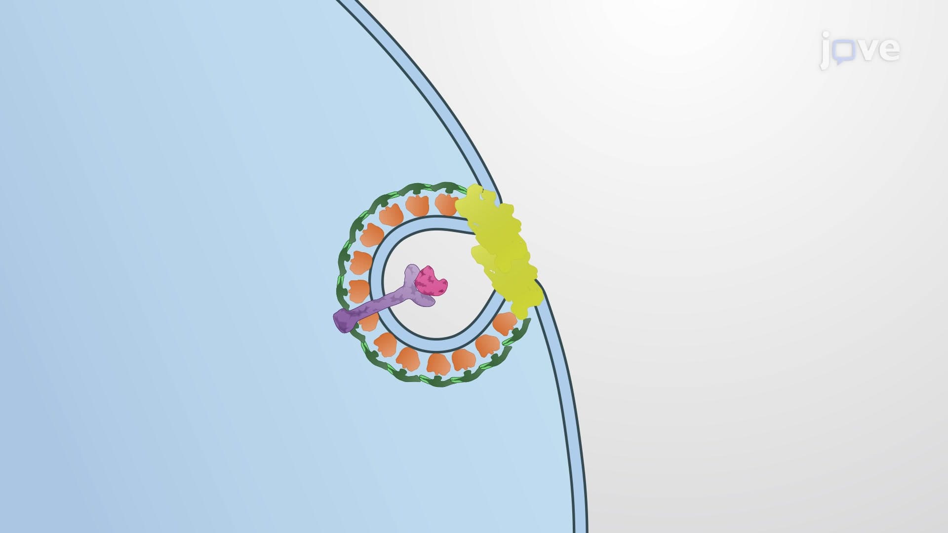
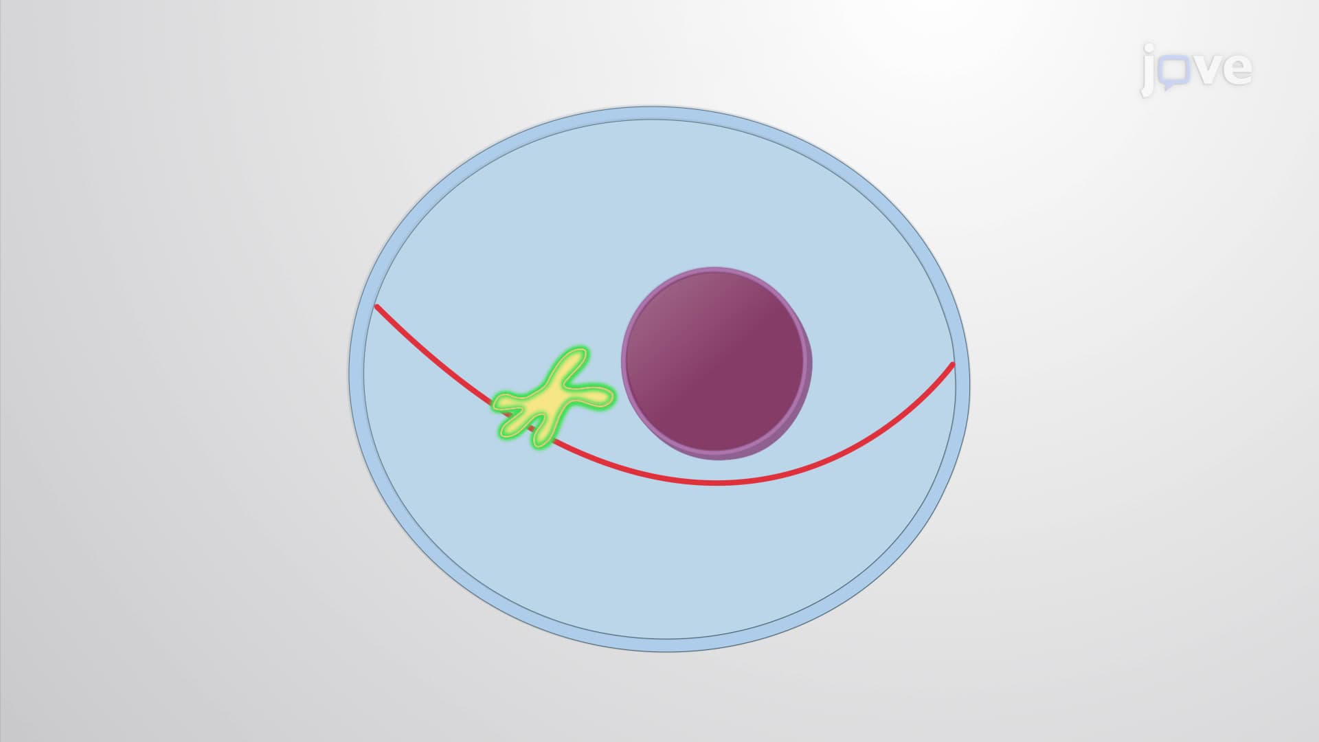
The phagocytosis process begins when specialized receptors on the cell surface recognize molecular patterns associated with foreign particles or damaged tissue. These pattern recognition receptors (PRRs) act like cellular security guards, distinguishing between "self" and "non-self" materials. Once recognition occurs, the cell initiates dramatic membrane reorganization to form pseudopods-finger-like projections that extend outward to surround the target particle.
Professional phagocytes like macrophages and neutrophils excel at this process. For instance, alveolar macrophages in your lungs constantly patrol air sacs, engulfing dust particles, bacteria, and other inhaled contaminants. This continuous surveillance prevents respiratory infections and maintains lung health-a process particularly relevant for understanding diseases like pneumonia covered in AP Biology and college immunology courses.
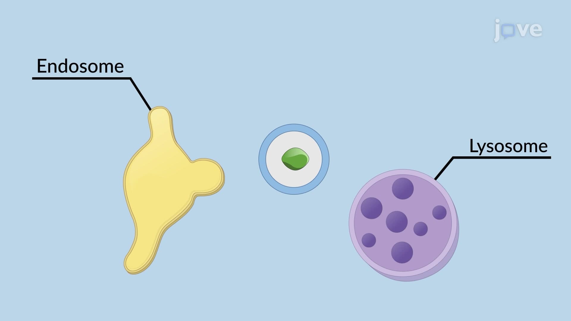
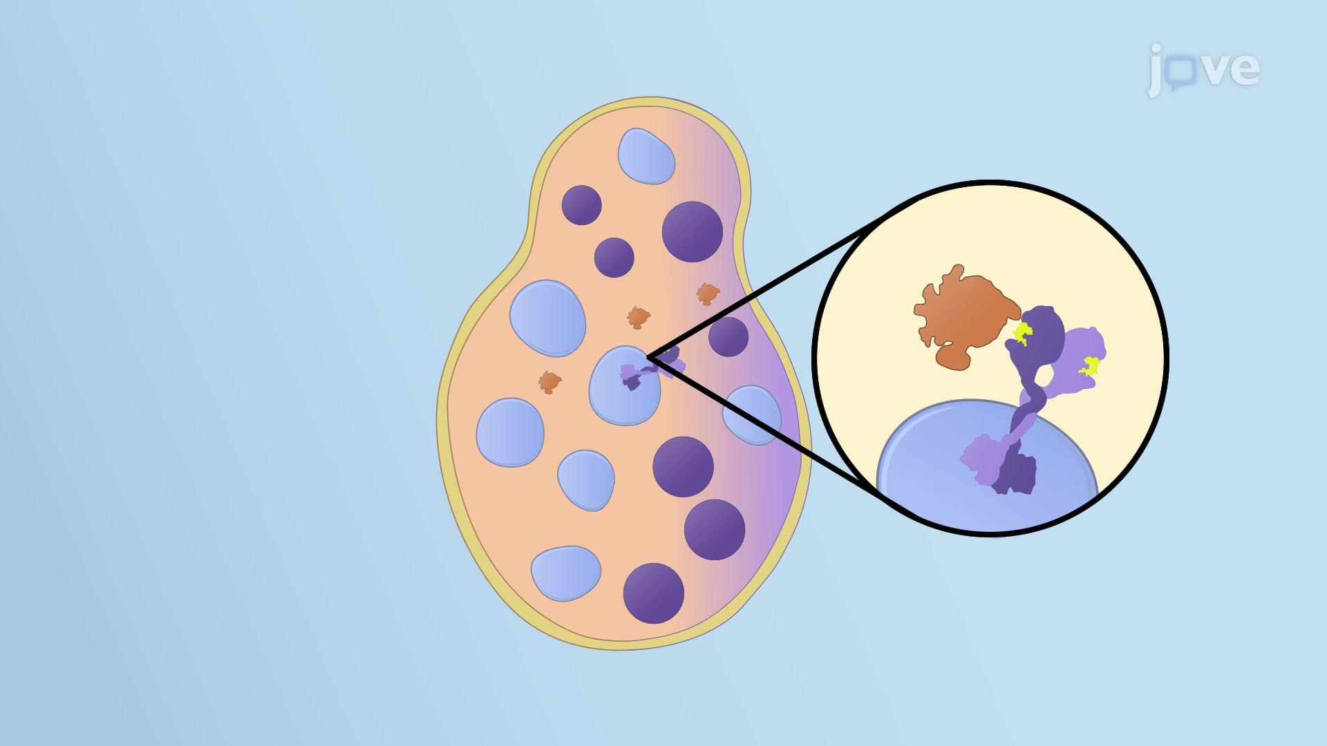
After pseudopods completely surround their target, membrane fusion creates a phagosome-essentially a cellular prison containing the captured particle. However, the story doesn't end there. The phagosome must fuse with lysosomes, organelles packed with digestive enzymes, to form a phagolysosome. This fusion event transforms the cellular prison into an execution chamber where acidic conditions and powerful enzymes break down the engulfed material.
This process proves critical for medical understanding. When studying antibiotic resistance for the MCAT, students learn how some bacteria like Mycobacterium tuberculosis can survive within phagosomes by preventing phagolysosome formation-leading to persistent infections that challenge treatment protocols used in US hospitals.
Phagocytosis knowledge appears frequently on standardized exams, from AP Biology questions about immune system function to MCAT passages on cellular biology. Understanding this process helps explain why immunocompromised patients face increased infection risks and how vaccines work by training phagocytes to recognize specific pathogens. College courses in cell biology, immunology, and microbiology extensively cover phagocytosis as foundational knowledge for future healthcare professionals.
Related Micro-courses