Loading subject page…

- Cell Biology
- Extracellular Matrix in Animals
- Basal Lamina are the Specialized Form of ECM
Micro-course: Extracellular Matrix in Animals
45,311 views
Learn key concepts and earn your certificate.


Basal Lamina are the Specialized Form of ECM

00:00
01:03
Video Summary: Basal Lamina are the Specialized Explained
Did you know that your kidneys filter over 180 liters of blood daily through microscopic barriers? Basal lamina are the specialized extracellular structures that make this filtration possible, acting as molecular gatekeepers throughout your body. At places like Johns Hopkins Hospital, doctors study how these thin protein sheets separate different cell types while allowing selective passage of molecules. Basal lamina are the specialized explained through their unique composition of collagen IV, laminins, and other glycoproteins that create cellular boundaries. Watch the full video on JoVE Coach to master this concept with expert-led visuals and step-by-step explanations.
- Understand how basal lamina are the specialized extracellular matrix structures that separate different cell types and tissues
- Identify the key molecular components including type IV collagen, laminins, entactin, and perlecan that give basal lamina their unique properties
- Analyze the location-specific functions of basal lamina in neuromuscular junctions, epithelial tissues, and kidney glomeruli
- Compare how basal lamina organization varies depending on the attached cell type and tissue requirements
- Evaluate the role of basal lamina as molecular filters that selectively permit or restrict passage of different substances
- Apply knowledge of basal lamina structure to explain disease processes like diabetic nephropathy and muscular dystrophy
- Recognize basal lamina as crucial connectors linking cells to the broader extracellular matrix of connective tissues
- Explore how disruption of basal lamina integrity leads to various pathological conditions
Understanding How Basal Lamina are The Specialized Matrix Structures
Basal lamina are the specialized thin sheets of extracellular matrix that serve as fundamental architectural elements in multicellular organisms. Unlike the thicker basement membranes visible under light microscopy, basal lamina represent the electron microscopy-visible component that directly contacts cell surfaces. These structures demonstrate remarkable adaptability, with their composition and organization precisely tailored to meet the specific functional demands of different tissues.
Molecular Architecture and Specialized Components
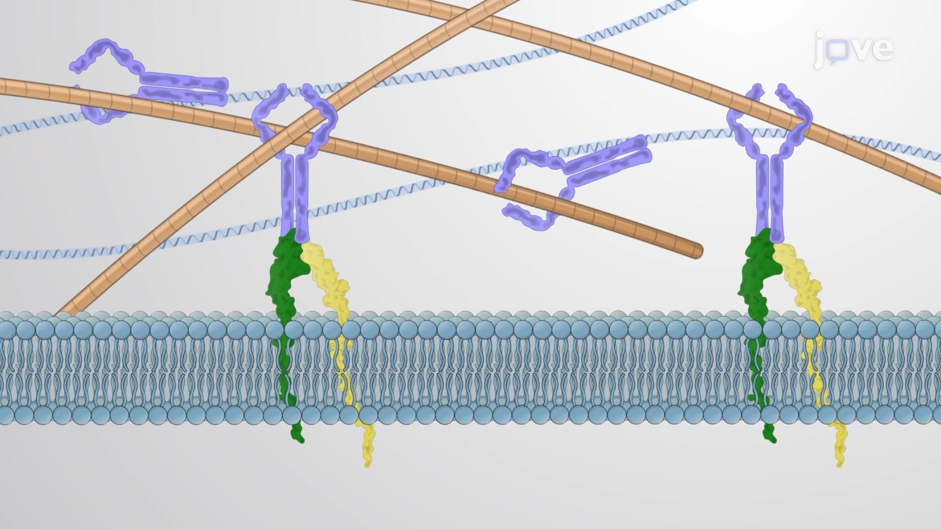
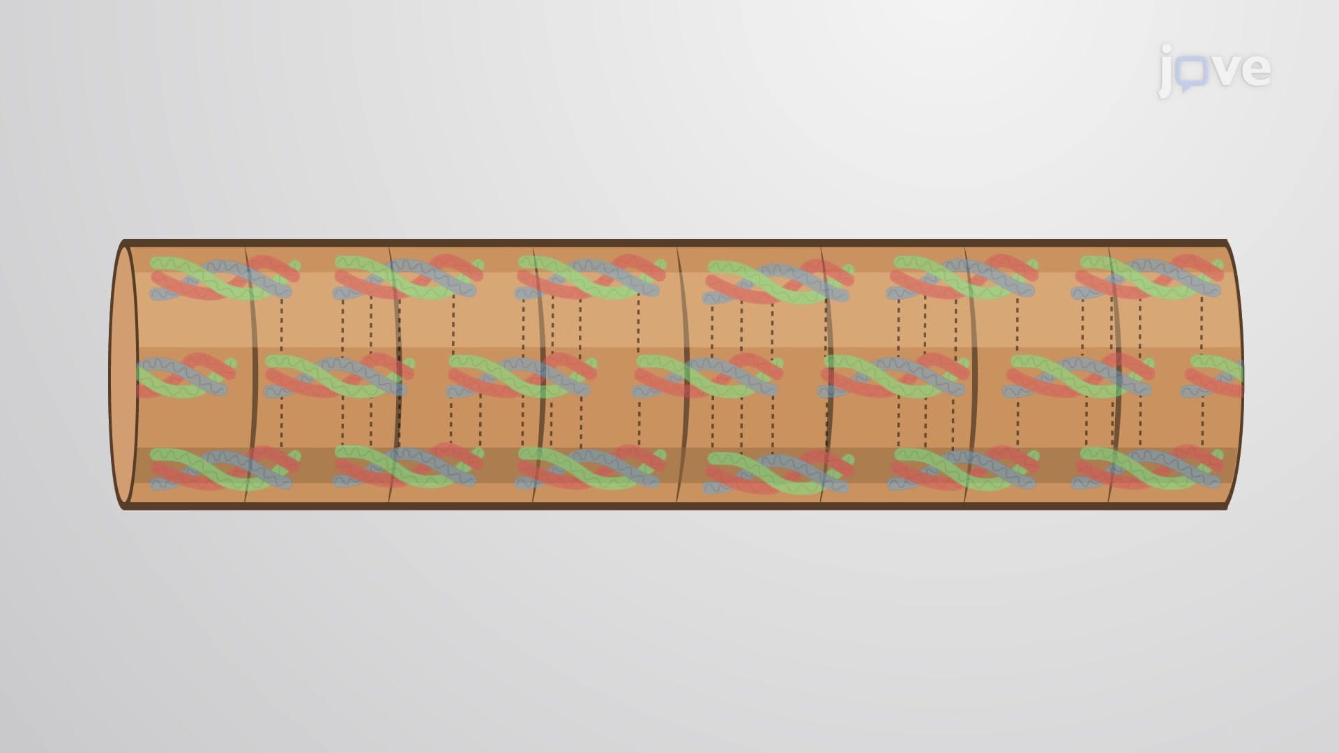
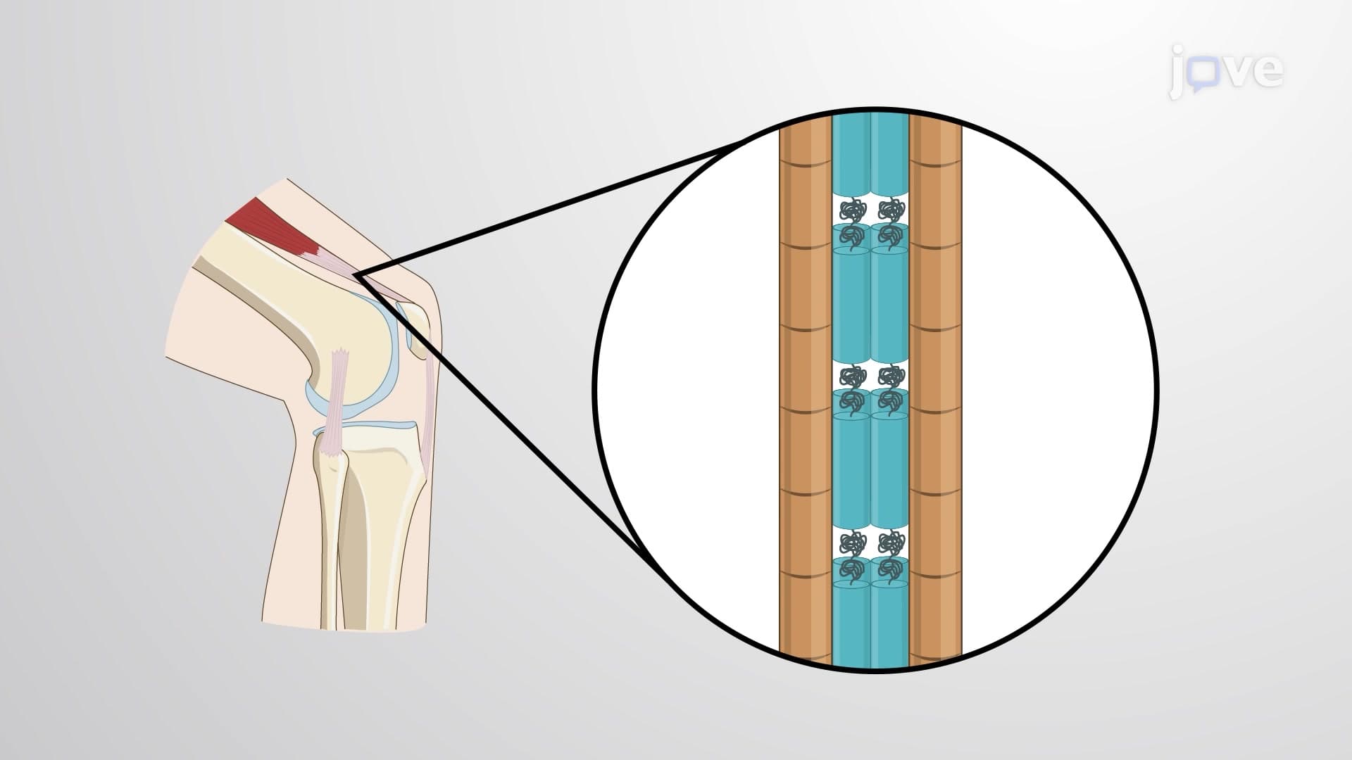
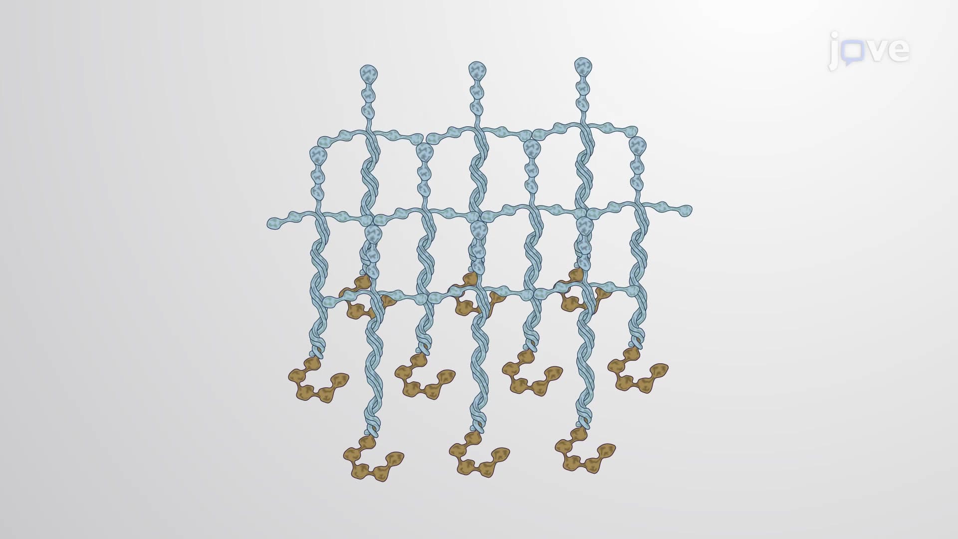
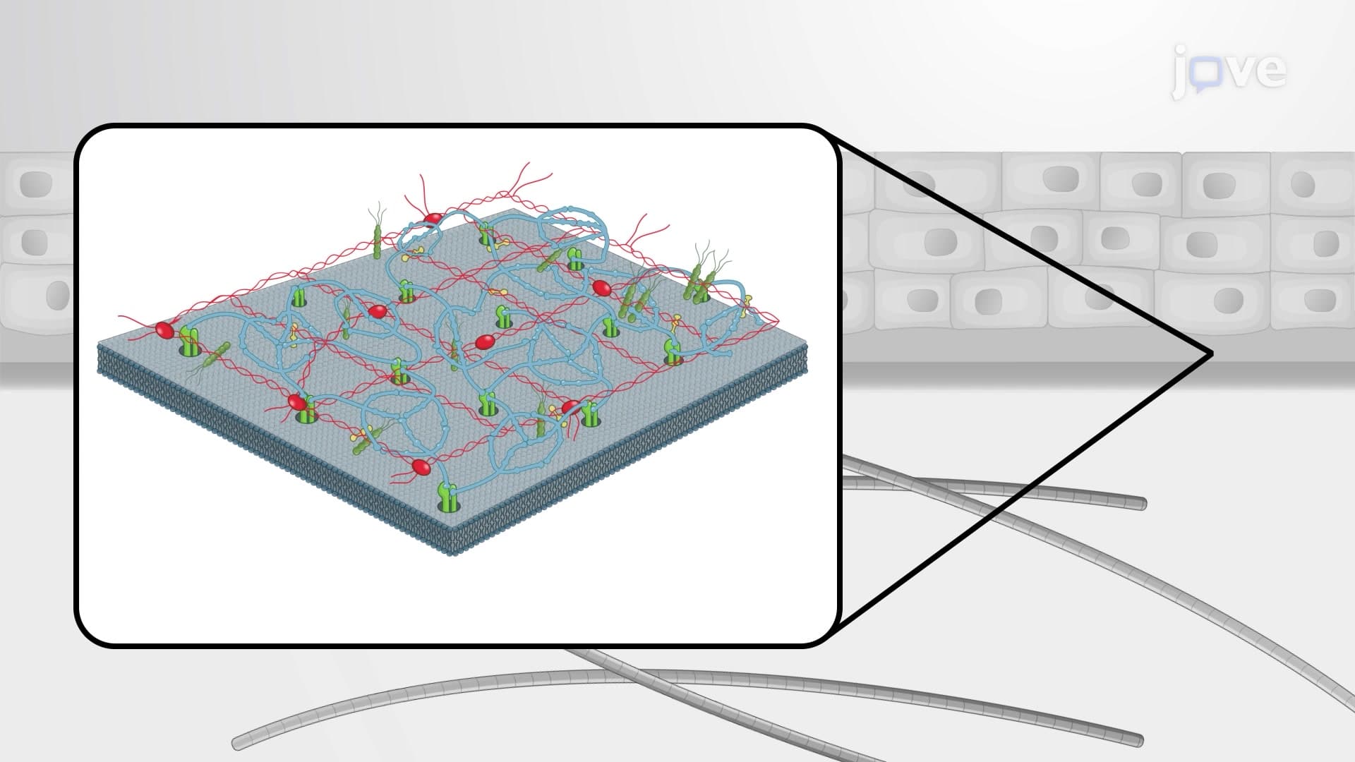
The sophisticated molecular composition of basal lamina reflects their diverse functions. Type IV collagen forms the primary structural network, creating a flexible yet strong foundation. This collagen type differs significantly from the fibrillar collagens found in tendons and bones, featuring a more branched, net-like arrangement. Laminins serve as crucial cell-binding proteins, with different laminin isoforms expressed in tissue-specific patterns. For instance, laminin-211 predominates in muscle tissue, while laminin-511 is characteristic of epithelial basal lamina.
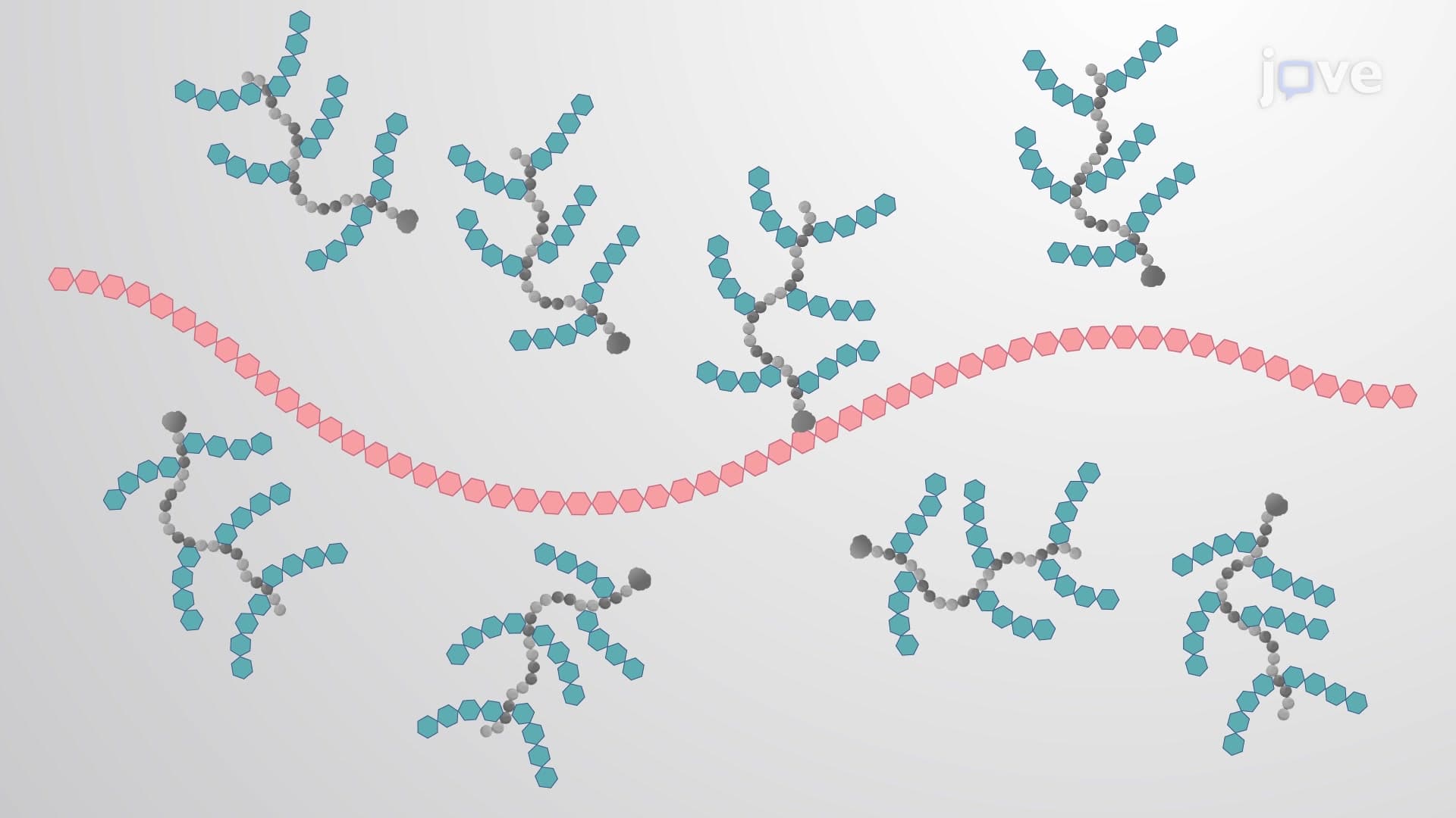
Entactin (also called nidogen) acts as a molecular bridge, connecting laminin to type IV collagen networks. Perlecan, a large heparan sulfate proteoglycan, contributes to the charge-selective properties of basal lamina, particularly important in kidney filtration. These components work synergistically, with their relative concentrations varying based on tissue-specific requirements.
Location-Specific Functions in Human Physiology
At neuromuscular junctions, basal lamina create specialized synaptic clefts essential for proper nerve-muscle communication. Research at institutions like Harvard Medical School has shown how disruption of these structures contributes to conditions like myasthenia gravis. In epithelial tissues, basal lamina provide crucial polarity cues, helping cells distinguish their apical (free) surface from their basal (attached) surface.
The kidney glomerulus presents perhaps the most clinically relevant example of basal lamina function. Here, the glomerular basement membrane acts as a sophisticated molecular sieve, preventing proteins larger than albumin from entering the urine while allowing smaller molecules and water to pass freely. Medical students studying for the MCAT often encounter questions about how diabetes damages this filtration barrier, leading to proteinuria and eventual kidney failure.
Clinical Relevance and Educational Applications
Understanding basal lamina proves essential for students preparing for advanced coursework and professional exams. AP Biology students frequently encounter these structures when studying cell biology and tissue organization. The concept appears regularly in college-level cell biology courses and features prominently in medical school curricula, particularly in histology and pathology courses.
Healthcare professional exams like the NCLEX and HESI A2 often include questions about basement membrane function in various disease states. Students should focus on connecting structural knowledge with functional outcomes, understanding how changes in basal lamina composition lead to observable clinical symptoms.
Frequently Asked Questions
Basal lamina are the specialized thin layers of extracellular matrix that separate different cell types and provide structural support. They consist of unique proteins like type IV collagen and laminins that create flexible, selective barriers. These structures are essential for maintaining tissue organization and enabling selective molecular transport.
MCAT questions often focus on basal lamina function in kidney filtration and disease processes like diabetic nephropathy. AP Biology exams typically test understanding of their role in tissue organization and cell-matrix interactions. Students should know the key molecular components and be able to explain location-specific functions.
Unlike fibrous connective tissue matrices, basal lamina form thin, sheet-like structures directly attached to cell surfaces. They contain specialized proteins like type IV collagen and laminins not found in other ECM types. Their organization creates selective barriers rather than providing mechanical strength like tendons or cartilage.
Diabetic patients at medical centers like Mayo Clinic often develop kidney disease due to basal lamina thickening in glomeruli, impairing filtration. Muscular dystrophy involves defective muscle basal lamina, leading to progressive weakness. Cancer metastasis requires tumor cells to break through basal lamina barriers.
Basic understanding of proteins and carbohydrates is sufficient for most educational levels. High school students can grasp the concept by focusing on function rather than detailed molecular interactions. College students should understand protein-protein interactions, but complex biochemistry isn't required for foundational knowledge.
Create comparison charts showing basal lamina composition and function in different tissues like muscle, kidney, and epithelium. Practice drawing simplified diagrams showing molecular organization. Focus on connecting structure to function, especially for clinical scenarios involving filtration and cell adhesion.
Study integrin-mediated cell adhesion to understand how cells interact with basal lamina. Explore extracellular matrix remodeling in wound healing and development. Investigate how matrix metalloproteinases regulate basal lamina turnover in normal physiology and disease states.
Expect exam questions comparing basal lamina composition across different tissues and explaining functional consequences of component deficiencies. Lab practicals often include electron micrographs requiring identification of basal lamina structures. Problem-solving questions may involve predicting effects of specific protein mutations.
Related Micro-courses
6 Concepts
12 Concepts

11 Concepts