Loading subject page…


45,326 views

Video Summary: Laminins are the Adhesive Proteins Explained
Ever wonder how your skin stays attached to underlying tissues during intense physical activity? Laminins are the adhesive proteins that act like molecular "glue" in your body's basement membranes, preventing tissue layers from separating. These specialized glycoproteins are critical in conditions like muscular dystrophy, where laminin defects cause muscle weakness in thousands of Americans annually. Laminins are The Adhesive Proteins Explained reveals how these three-chain molecules create strong cellular connections while enabling vital cell-to-cell communication. Watch the full video on JoVE Coach to master this concept with expert-led visuals and step-by-step explanations.
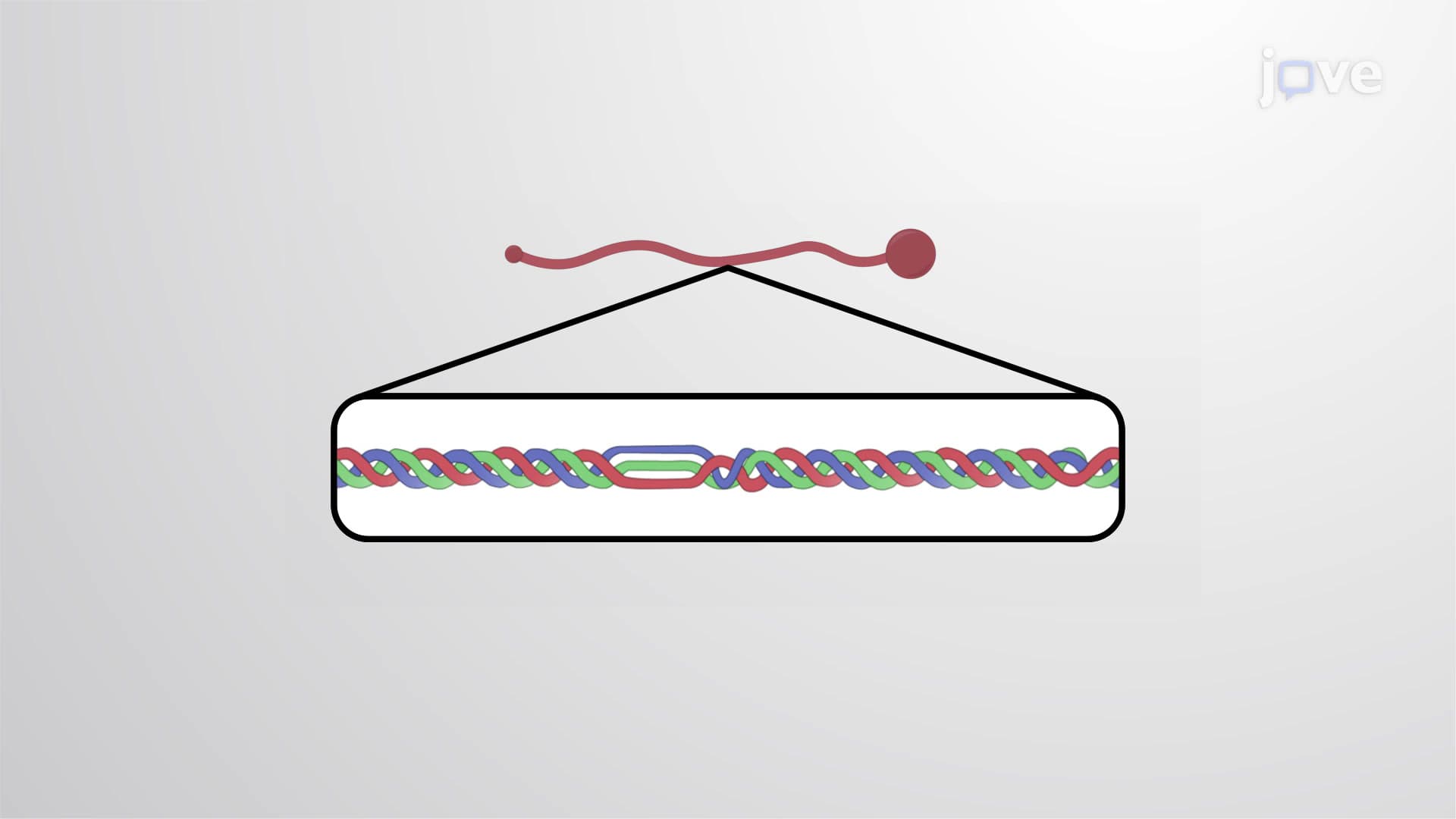
Laminins are The Adhesive Proteins represent one of the most crucial protein families in human biology, serving as the primary adhesive components of basement membranes. These large, complex glycoproteins possess a distinctive three-chain structure that resembles a cross when viewed under electron microscopy. The alpha, beta, and gamma chains each contribute unique properties, with the alpha chain being the largest and most variable among different laminin types.
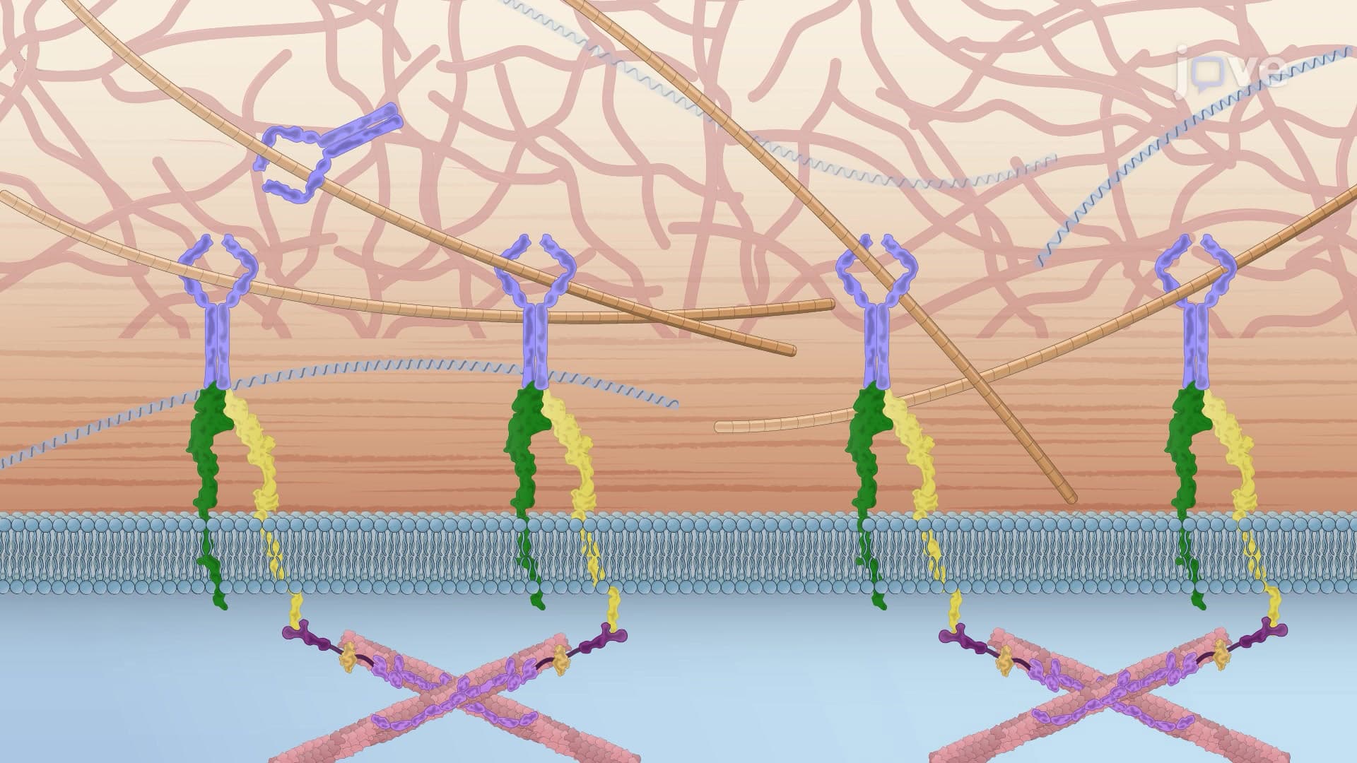
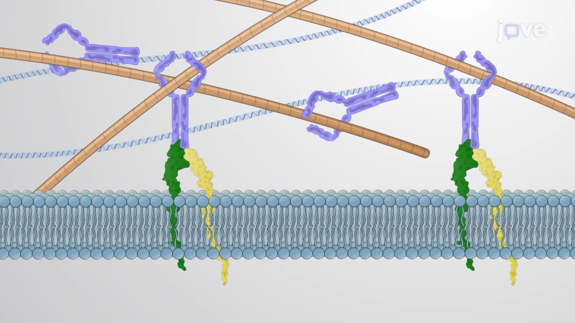
The molecular organization of laminins reflects their dual function as both structural anchors and communication facilitators. At the C-terminus, the three chains intertwine in a coiled-coil formation, creating a stable central domain. This region contains the laminin G-like domains, particularly on the alpha chain, which serve as primary binding sites for cell surface receptors. These domains interact specifically with integrins (especially alpha6beta1 and alpha3beta1) and dystroglycan, establishing firm connections between cells and their surrounding matrix.
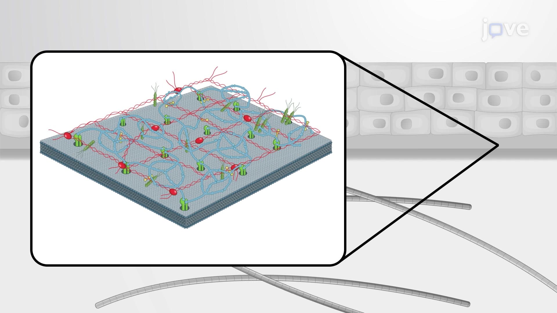
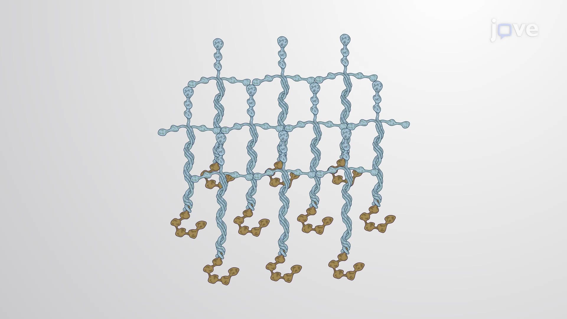
The N-terminal regions of laminin chains remain separate, forming globular domains that enable laminin-to-laminin interactions. This self-assembly capability allows laminins to create mesh-like networks that provide both flexibility and strength to basement membranes. In tissues like kidney glomeruli and lung alveoli, this network must withstand constant mechanical stress while maintaining selective permeability.
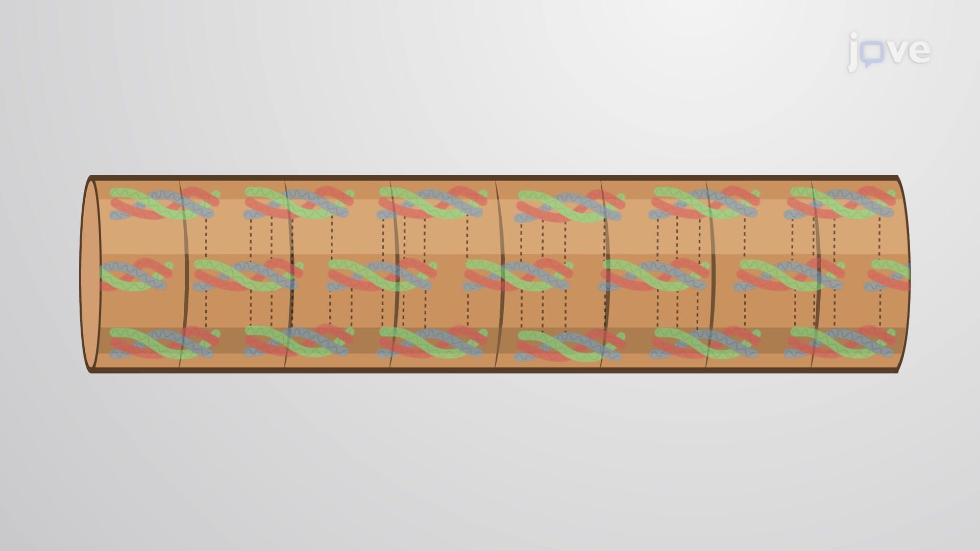
The integration of laminins with other basement membrane components creates a sophisticated biological composite material. Type IV collagen provides tensile strength, while perlecan contributes compressive resistance and growth factor binding. Entactin (also called nidogen) acts as a bridging molecule, linking laminin networks to collagen IV networks. This collaboration is essential for proper organ development and function.
Laminin dysfunction underlies several genetic disorders that affect thousands of Americans. Congenital muscular dystrophies often result from mutations in laminin alpha2 chains, leading to muscle weakness and developmental delays. Epidermolysis bullosa involves defects in laminin-332 (formerly laminin-5), causing severe skin fragility. Understanding these connections helps explain why students studying for the MCAT or AP Biology exams encounter laminins in contexts ranging from embryology to pathology.
For college biochemistry courses and pre-medical studies, laminins exemplify how protein structure determines function. Their modular design allows different laminin isoforms to serve specialized roles in various tissues, from the blood-brain barrier to the dermal-epidermal junction. This concept frequently appears on USMLE Step 1 examinations, particularly in questions about basement membrane disorders and tissue repair mechanisms.
Related Micro-courses