Loading subject page…


45,326 views

Video Summary: What Is Fibril Associated Collagen
Ever wonder why your Achilles tendon can withstand forces equivalent to supporting a 1,200-pound weight? The secret lies in fibril associated collagen, specialized proteins that act like molecular bridges, connecting and strengthening collagen fibers throughout your body. These remarkable molecules transform ordinary collagen fibrils into robust, interconnected networks that give tendons, cartilage, and other tissues their incredible durability. Watch the full video on JoVE Coach to master this concept with expert-led visuals and step-by-step explanations.
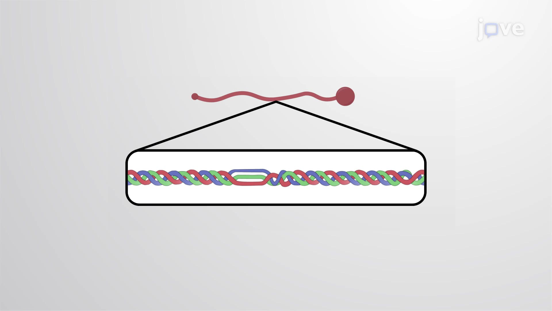
Fibril associated collagen represents a specialized class of structural proteins that function as molecular architects within our connective tissues. Unlike the long, continuous triple helices found in fibrillar collagens such as Type I, fibril associated collagens feature unique structural adaptations that enable them to serve as cross-linking agents. These proteins contain shorter triple-helical regions interrupted by flexible domains, creating molecules perfectly designed for bridging and reinforcement roles.
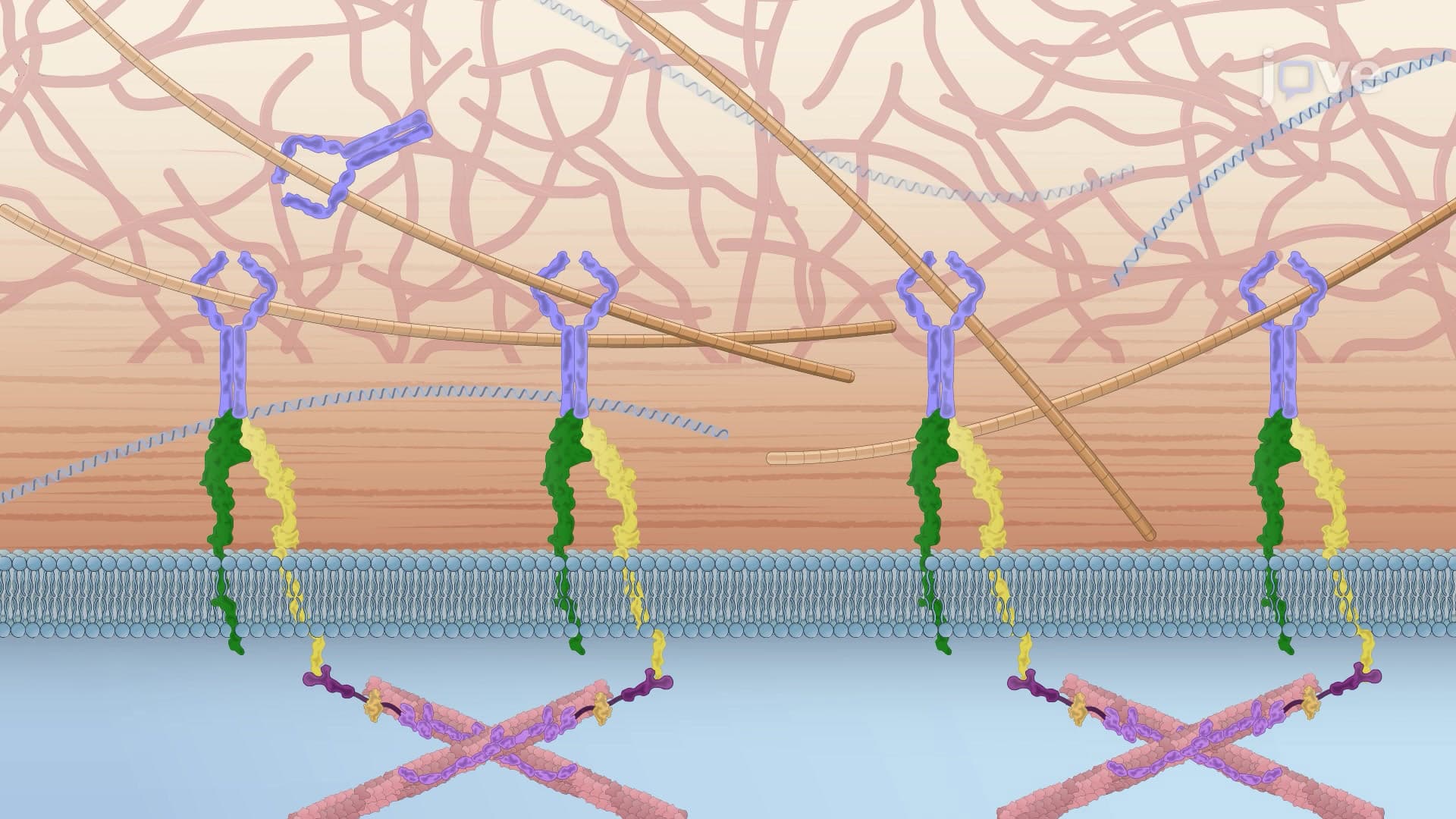
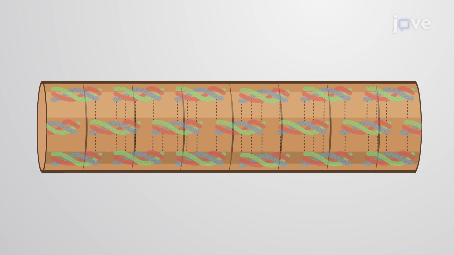
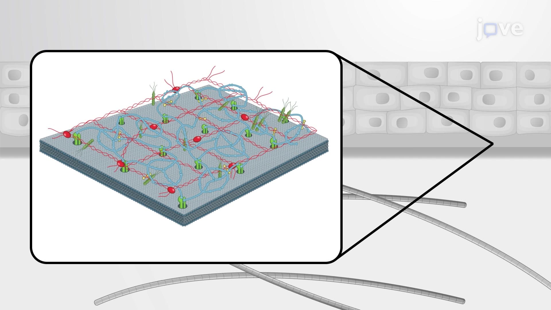
The architectural brilliance of these collagens becomes apparent when examining their strategic positioning within tissue matrices. Rather than forming the primary structural scaffold like fibrillar collagens, fibril associated collagens occupy the spaces between existing collagen fibrils, where they create a three-dimensional network of interconnections. This positioning allows them to transmit mechanical forces across multiple fibrils, dramatically increasing the overall strength and resilience of the tissue.
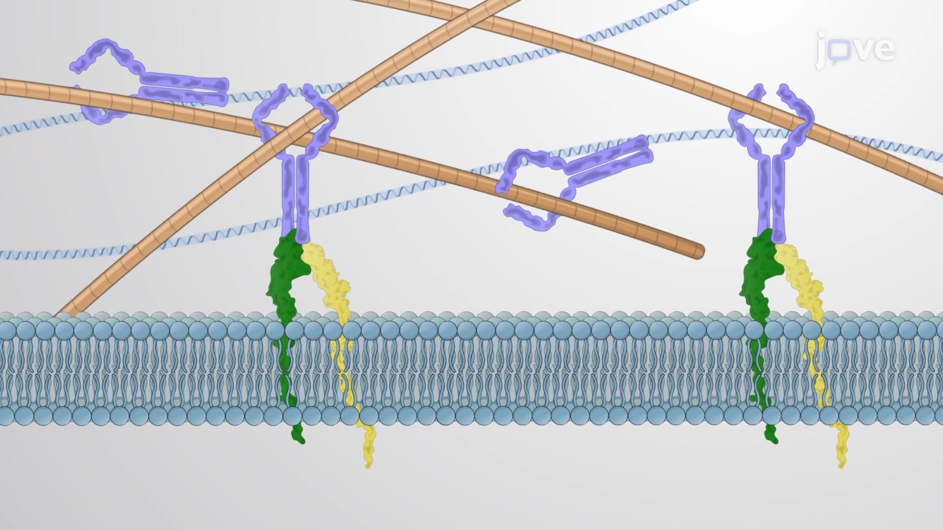
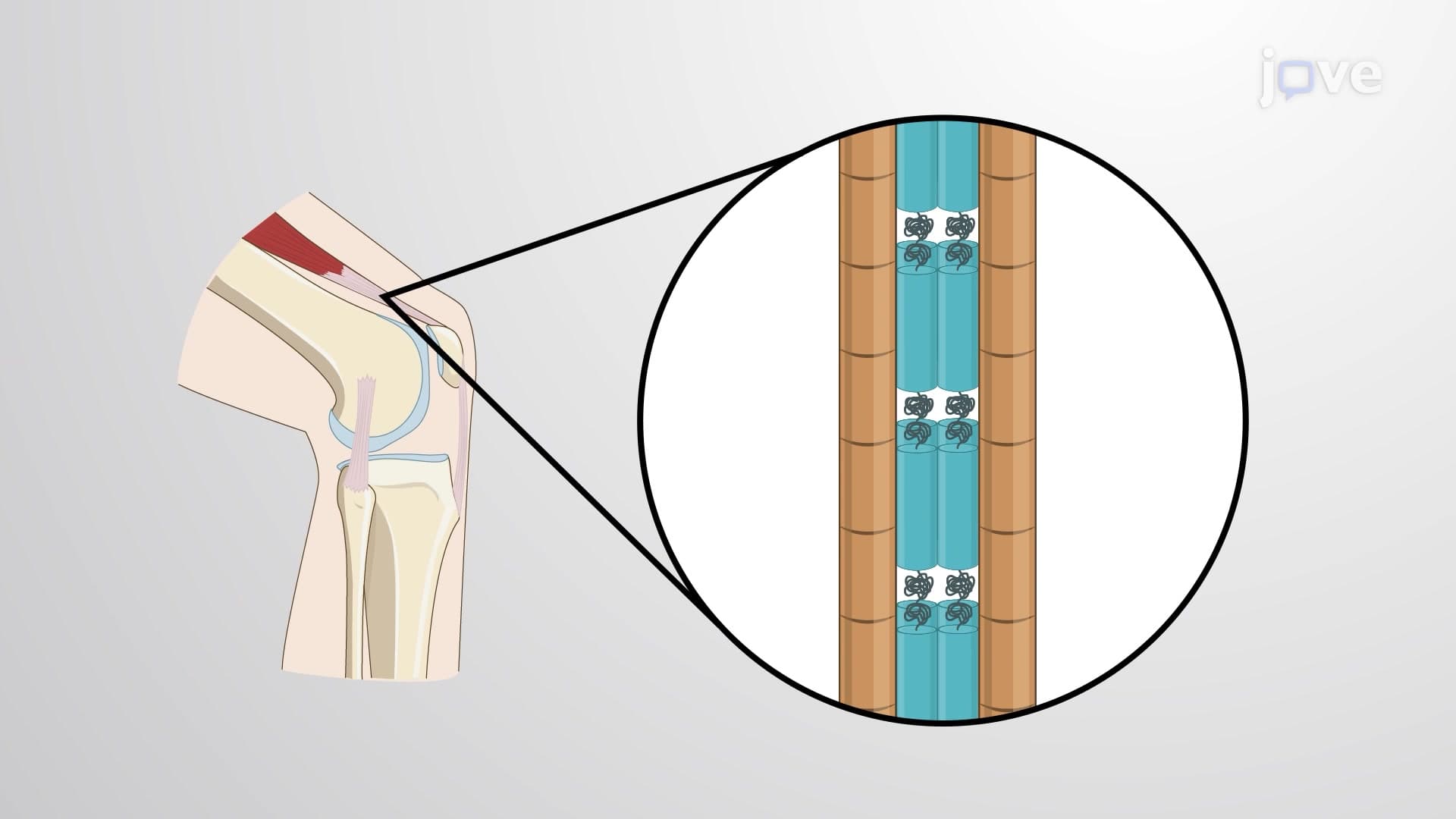
Type VI fibril associated collagen exemplifies precision molecular engineering in biological systems. Its structure features a relatively short triple helix flanked by large globular domains at both termini. These globular regions contain binding sites that enable Type VI molecules to associate with one another, forming microfibrils that wrap around the surface of Type I collagen fibrils like molecular cables.
In major weight-bearing tendons such as the patellar tendon (crucial for knee extension in athletes), Type VI collagen microfibrils create non-covalent associations with the underlying Type I collagen framework. This interaction increases fibril diameter and enhances the tendon's ability to withstand the repetitive loading forces experienced during activities like jumping or running. The clinical importance becomes evident in conditions like Ullrich congenital muscular dystrophy, where Type VI collagen mutations lead to muscle weakness and joint contractures.
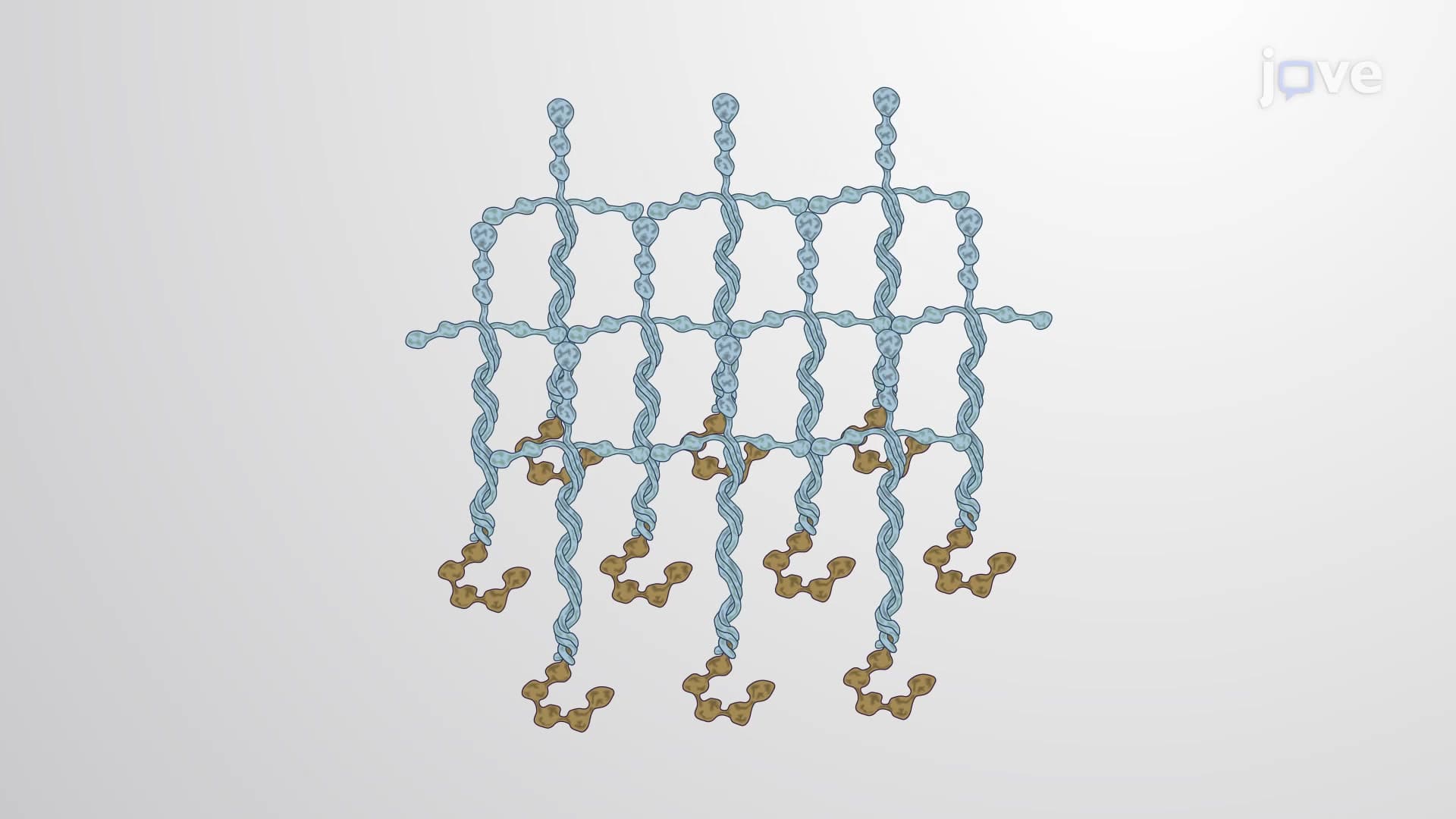
Type IX fibril associated collagen showcases even greater structural complexity, featuring multiple triple-helical domains connected by flexible kink regions. This modular design allows the molecule to bend and adapt while maintaining strong associations with Type II collagen fibrils in cartilaginous tissues. The N-terminal globular domain projects outward from the fibril surface, creating binding sites for other matrix components.
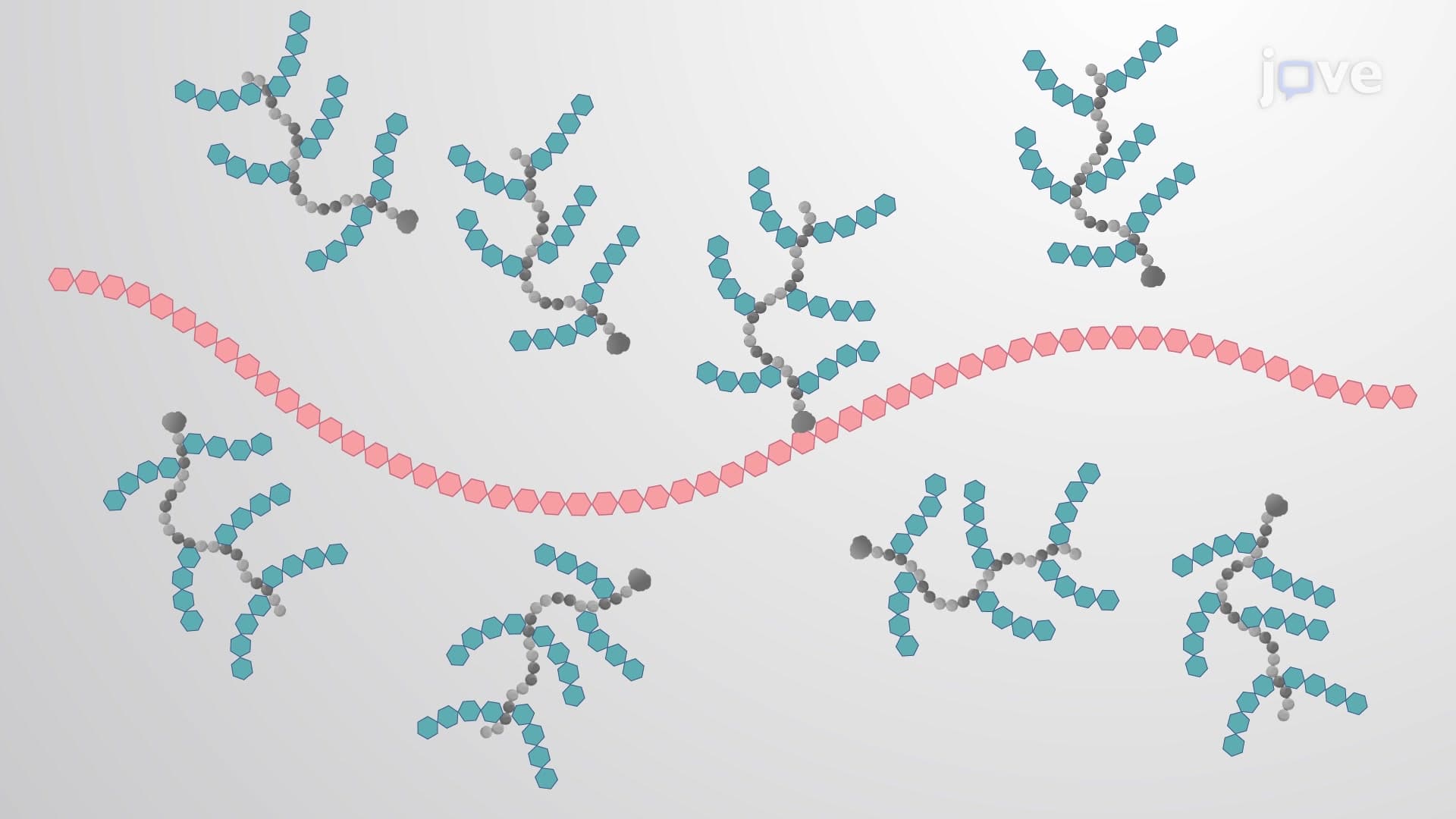
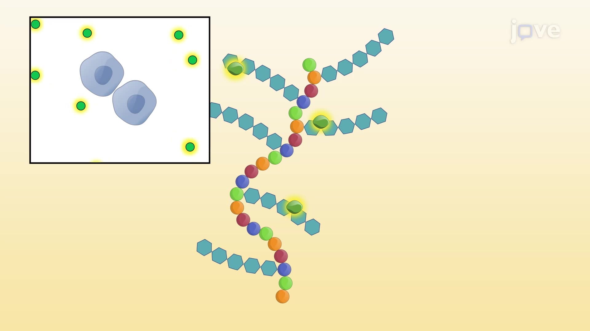
Perhaps most remarkably, Type IX collagen carries covalently attached chondroitin sulfate chains at its flexible kink regions. These glycosaminoglycan side chains extend into the surrounding matrix space, where they interact with proteoglycans, hyaluronic acid, and other matrix molecules. This creates a integrated network that gives cartilage its unique ability to resist compression while maintaining flexibility, essential properties for joint surfaces that must withstand decades of repetitive loading in structures like the knee meniscus.
For students preparing for advanced placement biology or college-level anatomy courses, understanding fibril associated collagen provides crucial insights into structure-function relationships in biological materials. MCAT preparation particularly benefits from grasping how these molecular interactions contribute to tissue mechanics, as questions frequently explore the connections between protein structure and physiological function.
Related Micro-courses