Loading video…


 8 Concepts
 40.5K views


Video Summary: Satellite Stem Cells and Muscular Dystrophy Explained
Did you know that your muscles contain dormant "repair crews" that spring into action whenever you work out or get injured? Satellite stem cells and muscular dystrophy are intrinsically connected through the body's muscle repair mechanisms. When these specialized stem cells fail to function properly, as seen in patients at Johns Hopkins Hospital with Duchenne muscular dystrophy, progressive muscle weakness occurs. Understanding Satellite Stem Cells And Muscular Dystrophy Explained reveals how these cellular repair systems work and why their dysfunction leads to devastating muscle diseases. Watch the full video on JoVE Coach to master this concept with expert-led visuals and step-by-step explanations.
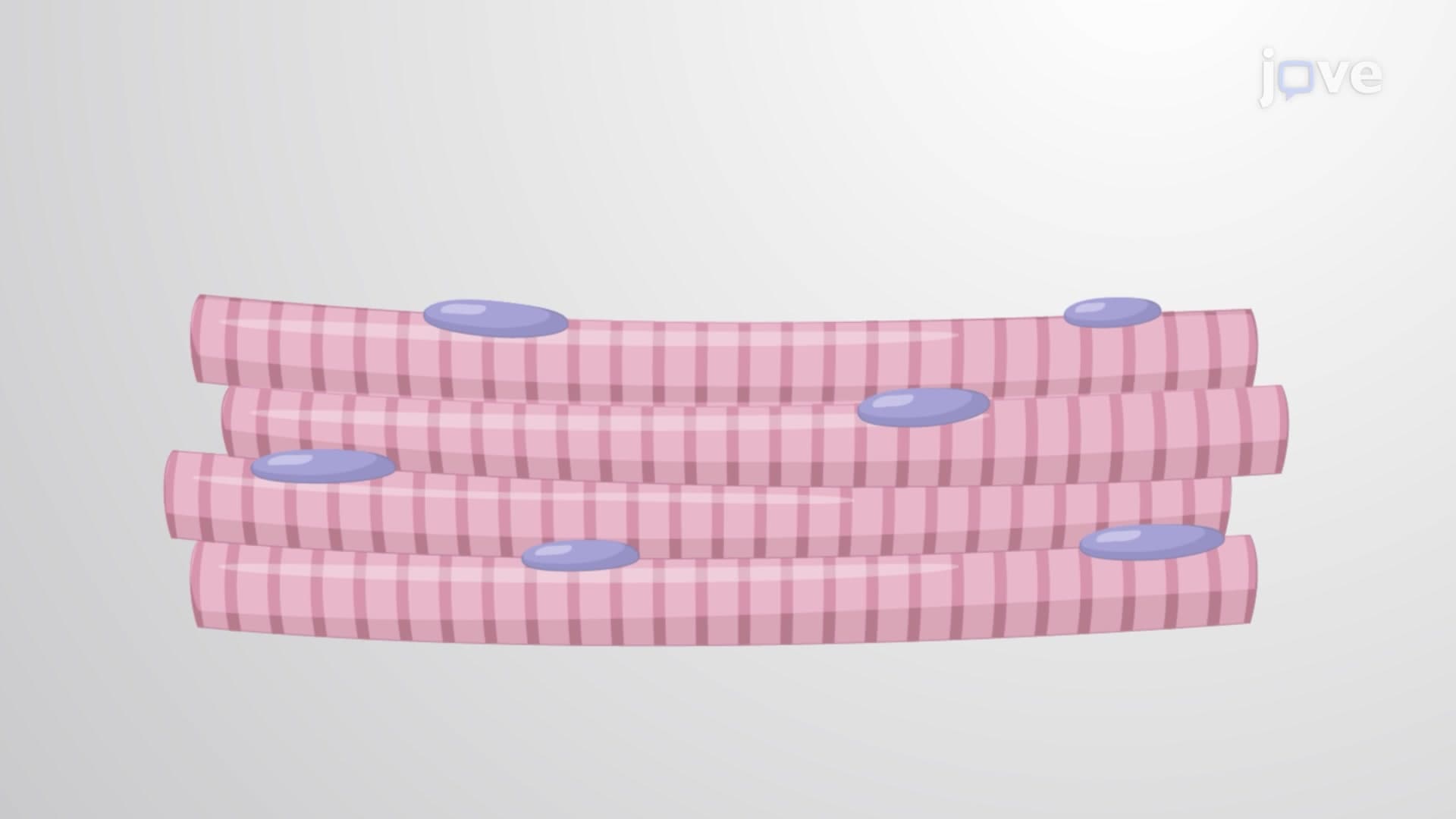
Satellite stem cells represent one of the most fascinating examples of adult stem cell biology in action. These multipotent cells serve as the body's muscle repair system, remaining dormant until called into action by injury, exercise, or disease. Located in the space between the muscle fiber and its surrounding basement membrane, satellite cells account for approximately 2-7% of all muscle nuclei in healthy adults.
When you perform resistance training at your local gym or sustain a muscle injury during sports, satellite cells receive biochemical signals that trigger their activation. This process involves several key transcription factors, including Pax7 and MyoD, which also play crucial roles during embryonic muscle development. Once activated, satellite cells undergo asymmetric division-one daughter cell maintains the stem cell pool (self-renewal), while the other differentiates into myoblasts.
The regeneration process follows a precise sequence: activated satellite cells proliferate, differentiate into myoblasts, and then fuse with existing damaged muscle fibers or form new myotubes. This mechanism explains why strength training leads to muscle growth and why minor muscle injuries heal effectively in healthy individuals. Students preparing for the MCAT or AP Biology exams should understand this as a prime example of adult stem cell plasticity and tissue homeostasis.
Duchenne muscular dystrophy (DMD) affects approximately 1 in 3,500 male births in the United States, making it the most common form of childhood muscular dystrophy. The disease results from mutations in the dystrophin gene, located on the X chromosome. Dystrophin normally acts as a structural protein that connects the muscle fiber's internal cytoskeleton to the surrounding extracellular matrix, providing mechanical stability during muscle contraction.
In DMD patients, the absence of functional dystrophin creates a cascade of cellular problems. While satellite cells initially attempt to repair the constantly damaged muscle fibers, they eventually become exhausted and lose their regenerative capacity. Research conducted at institutions like the University of California, San Francisco, has shown that DMD satellite cells exhibit altered gene expression patterns and reduced proliferative potential compared to healthy controls.
The progressive nature of DMD reflects the gradual failure of satellite cell-mediated repair mechanisms. Patients typically show symptoms by age 3-5, require wheelchairs by adolescence, and face life-threatening complications by their twenties. Current treatment strategies, including corticosteroids and emerging gene therapies like Eteplirsen (approved by the FDA), aim to preserve remaining muscle function and potentially enhance satellite cell activity.
Understanding this relationship between satellite stem cells and muscular dystrophy is essential for students pursuing careers in medicine, physical therapy, or biomedical research. The concept frequently appears in college-level cell biology courses and serves as an excellent example of how basic stem cell research translates to clinical applications.
Related Micro-courses