Loading subject page…


157,177 views

Video Summary: Pinching Off of Coated Vesicles Explained
Every second, millions of tiny cellular packages called vesicles transport critical cargo throughout your cells-but how do they actually detach from their origin? The pinching off of coated vesicles involves fascinating molecular machinery that works like microscopic scissors, with different mechanisms for various vesicle types. For instance, when neurons at Johns Hopkins medical research labs study synaptic transmission, they observe clathrin-coated vesicles using dynamin proteins to literally twist off from cell membranes. Watch the full video on JoVE Coach to master this concept with expert-led visuals and step-by-step explanations.
The pinching off of coated vesicles represents one of cell biology's most elegant transport mechanisms, enabling cells to package and deliver cargo between organelles with remarkable precision. This process varies significantly depending on the vesicle type, creating distinct pathways that students encounter frequently in AP Biology and college biochemistry courses.
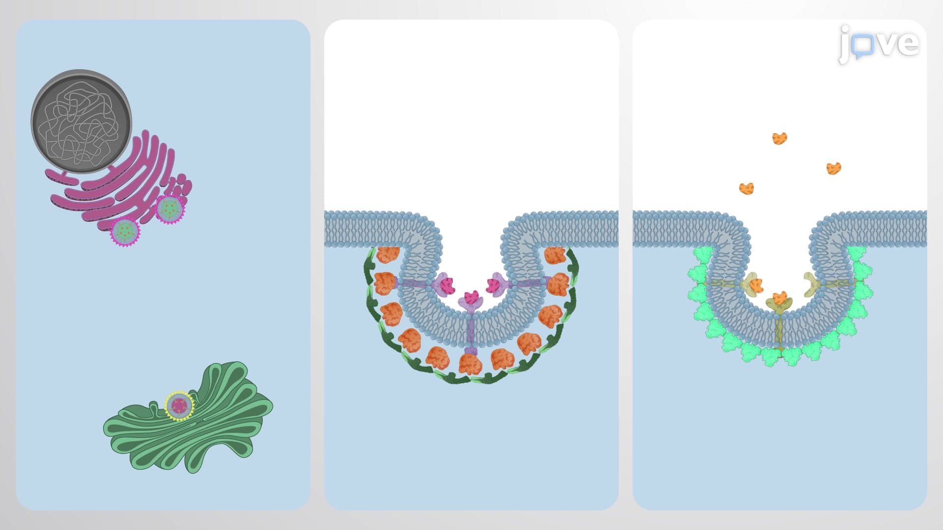
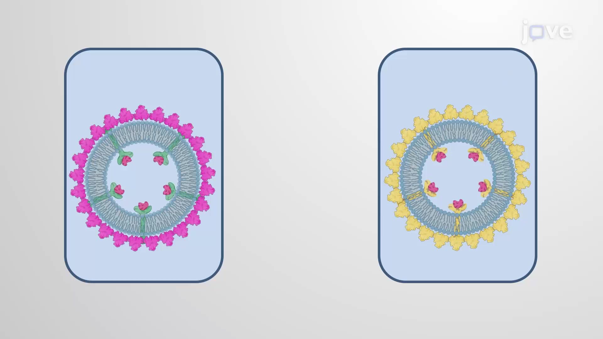
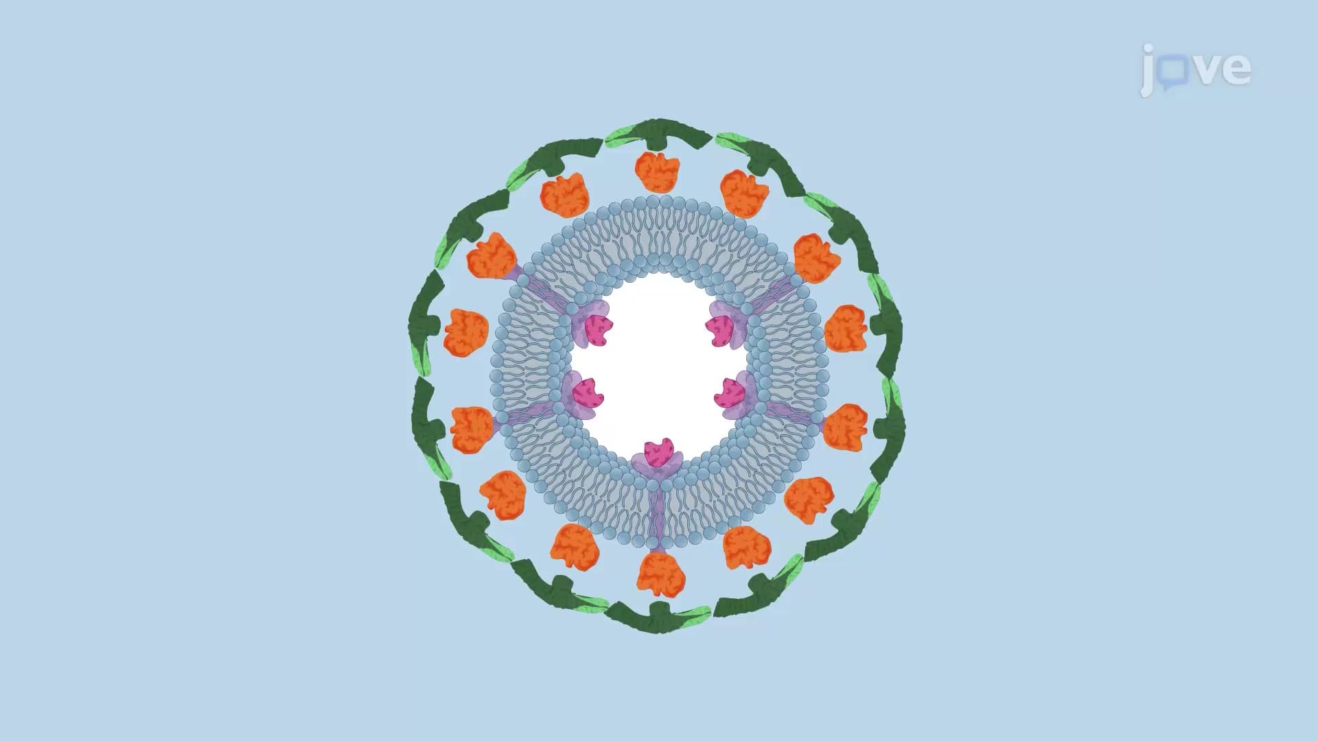
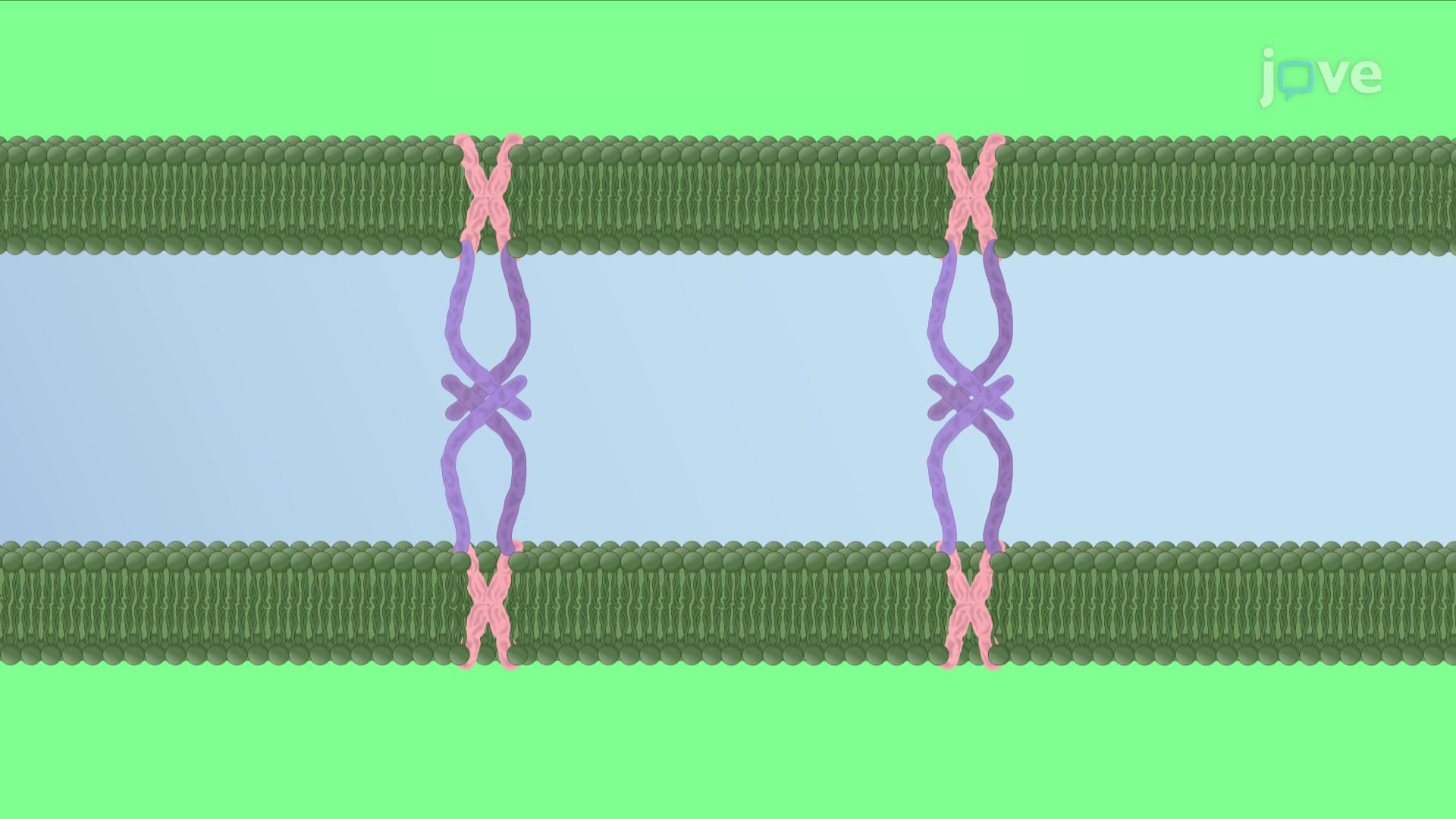
COPI and COPII vesicles demonstrate the simpler end of the spectrum, achieving separation through spontaneous membrane fusion events. These vesicles bud from the endoplasmic reticulum and Golgi apparatus when membrane leaflets on opposite sides of the budding region naturally fuse together. This process resembles how soap bubbles naturally separate when their connecting neck becomes too thin to maintain stability.
Research at Stanford University's cell biology laboratories has shown that this spontaneous mechanism relies on membrane curvature and lipid composition changes that favor energetically favorable separation. The beauty of this system lies in its self-sufficiency-no additional proteins or energy sources are required beyond the natural membrane dynamics.
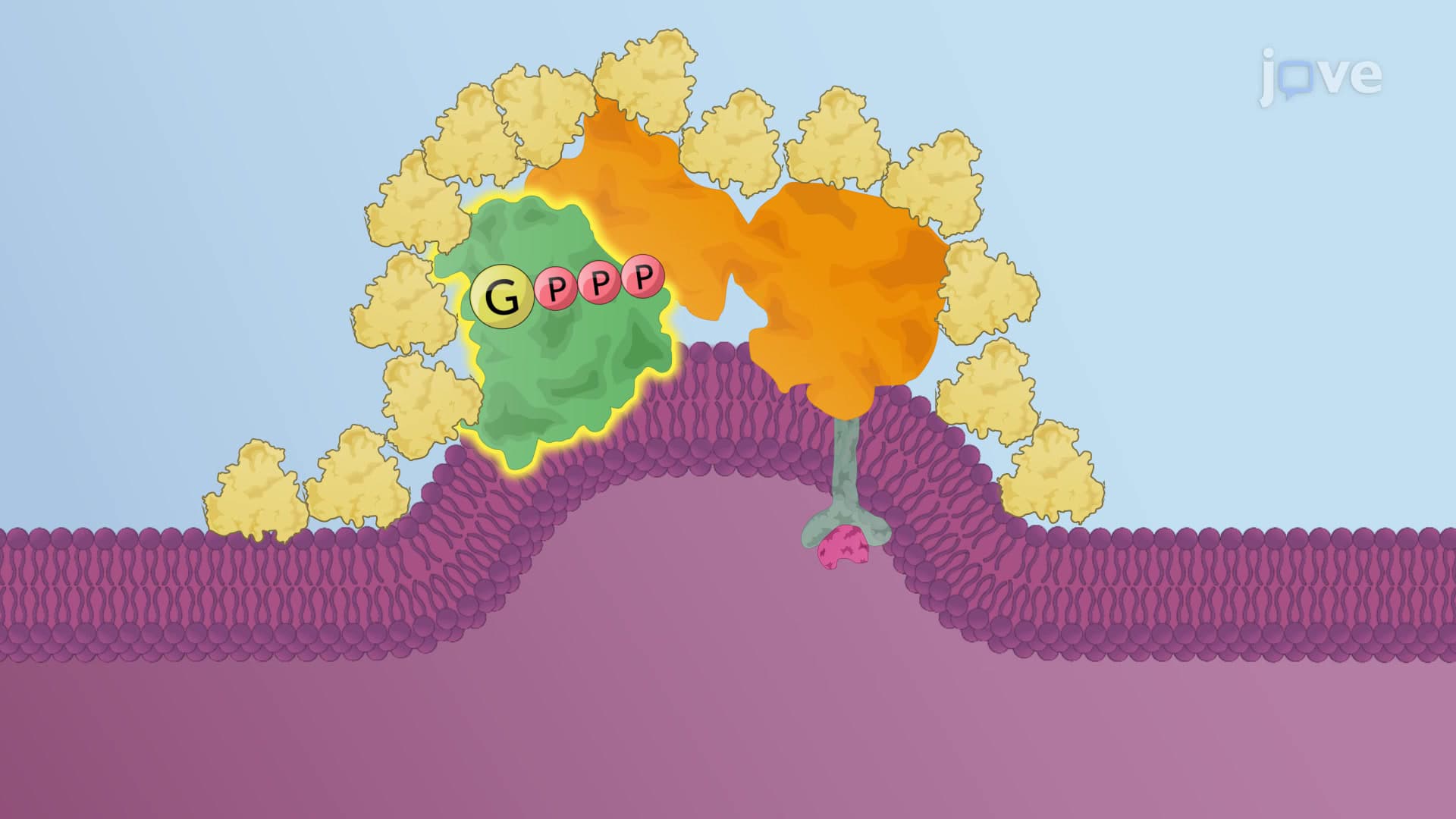
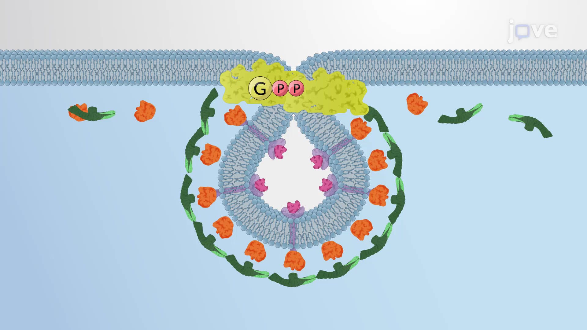
Clathrin-coated vesicles require sophisticated molecular machinery centered around dynamin, a GTP-binding protein that functions like a molecular motor. Students preparing for the MCAT often struggle with this concept until they visualize dynamin as a spring-loaded collar that tightens around the vesicle neck.
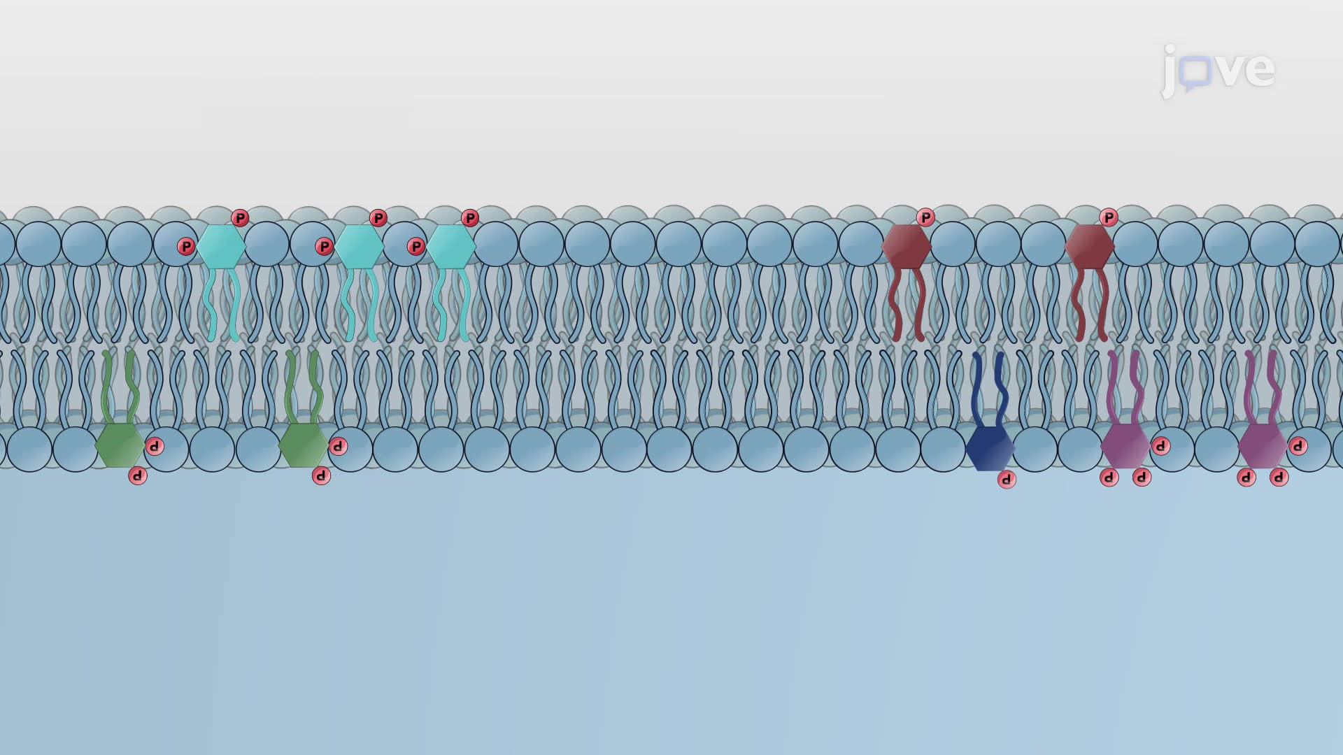
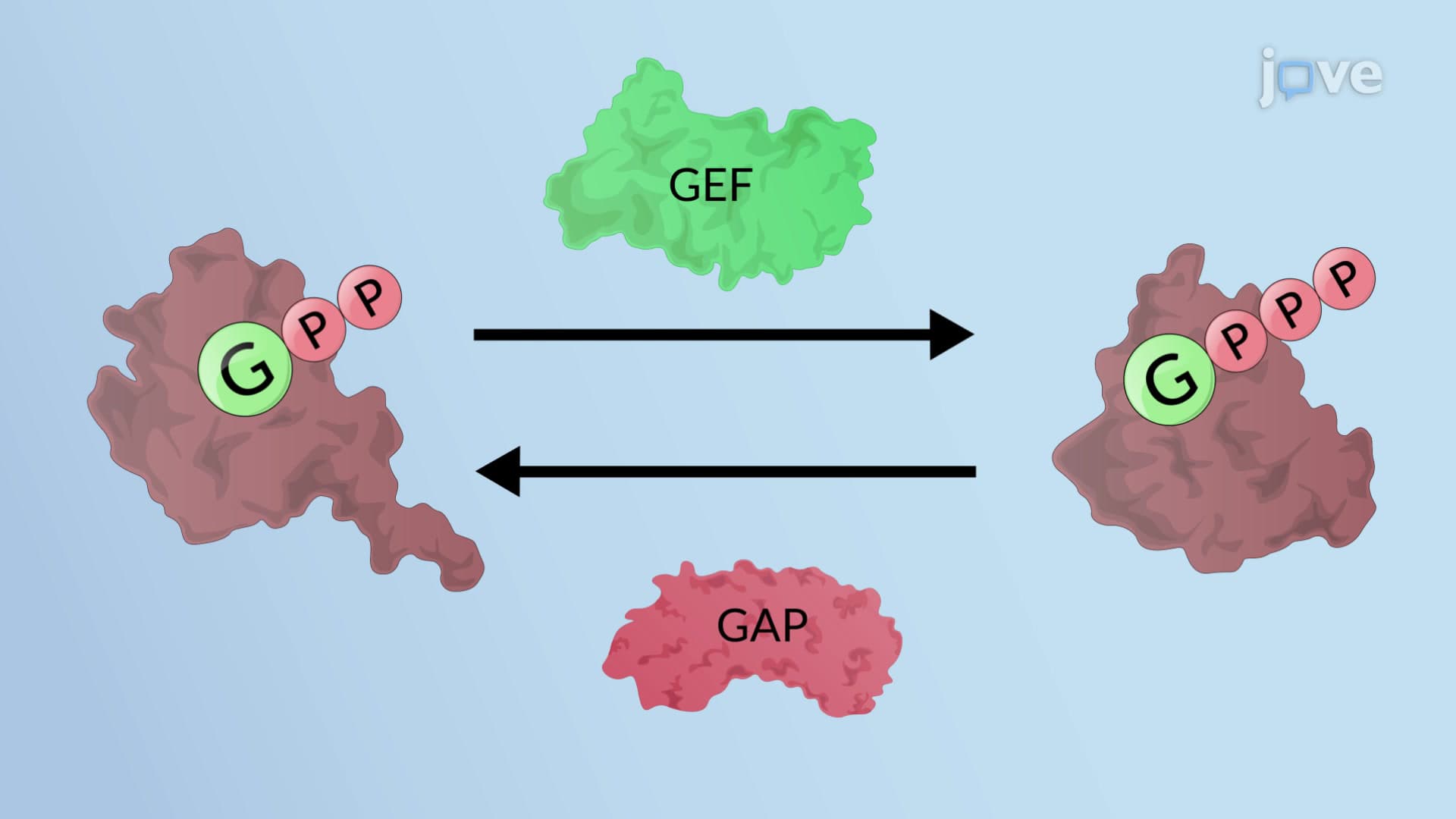
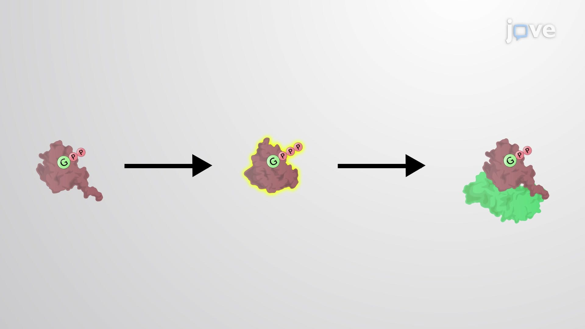
The process begins when dynamin assembles into a helical structure around the narrow neck connecting the budding vesicle to its parent membrane. This protein contains specialized domains: a PIP-binding region that anchors it to the membrane and GTPase domains that harness energy from GTP hydrolysis. When GTP breaks down to GDP, the released energy drives a dramatic conformational change in dynamin's structure.
The conformational change causes dynamin's helical structure to contract and twist, creating a scissor-like action that literally pinches the vesicle free from the membrane. Harvard Medical School researchers have documented this process using advanced microscopy techniques, revealing that the twisting motion generates enough mechanical force to sever the lipid bilayer connections.
Following successful pinching, a cascade of coat disassembly events begins. PIP phosphatase enzymes rapidly deplete phosphatidylinositol 4,5-bisphosphate from the newly formed vesicle membrane, weakening the attachment between adaptor proteins and the membrane surface. This destabilization allows chaperone proteins Hsp70 and auxilin to bind and systematically dismantle the clathrin coat structure, preparing the vesicle for cargo delivery to its target organelle.
Related Micro-courses