Loading subject page…


157,134 views

Video Summary: What Is Autophagy
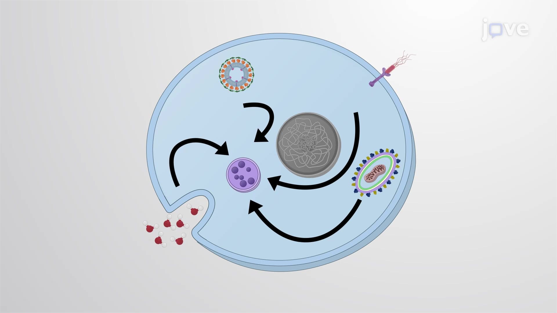
Did you know your cells literally eat themselves to survive? Autophagy is a vital cellular recycling process where cells break down damaged components under stress conditions like starvation or infection. When patients at Johns Hopkins Hospital undergo fasting before surgery, their cells activate autophagy pathways to maintain energy homeostasis. This self-cleaning mechanism removes worn-out organelles and protein aggregates that normal disposal systems can't handle. Watch the full video on JoVE Coach to master this concept with expert-led visuals and step-by-step explanations.
Autophagy represents one of biology's most elegant solutions to cellular maintenance. Unlike the proteasome system that degrades individual proteins, autophagy tackles larger cellular components-entire organelles, protein aggregates, and even invading pathogens. This process becomes critical when cells face metabolic stress, making it a cornerstone topic in AP Biology and college-level cell biology courses.
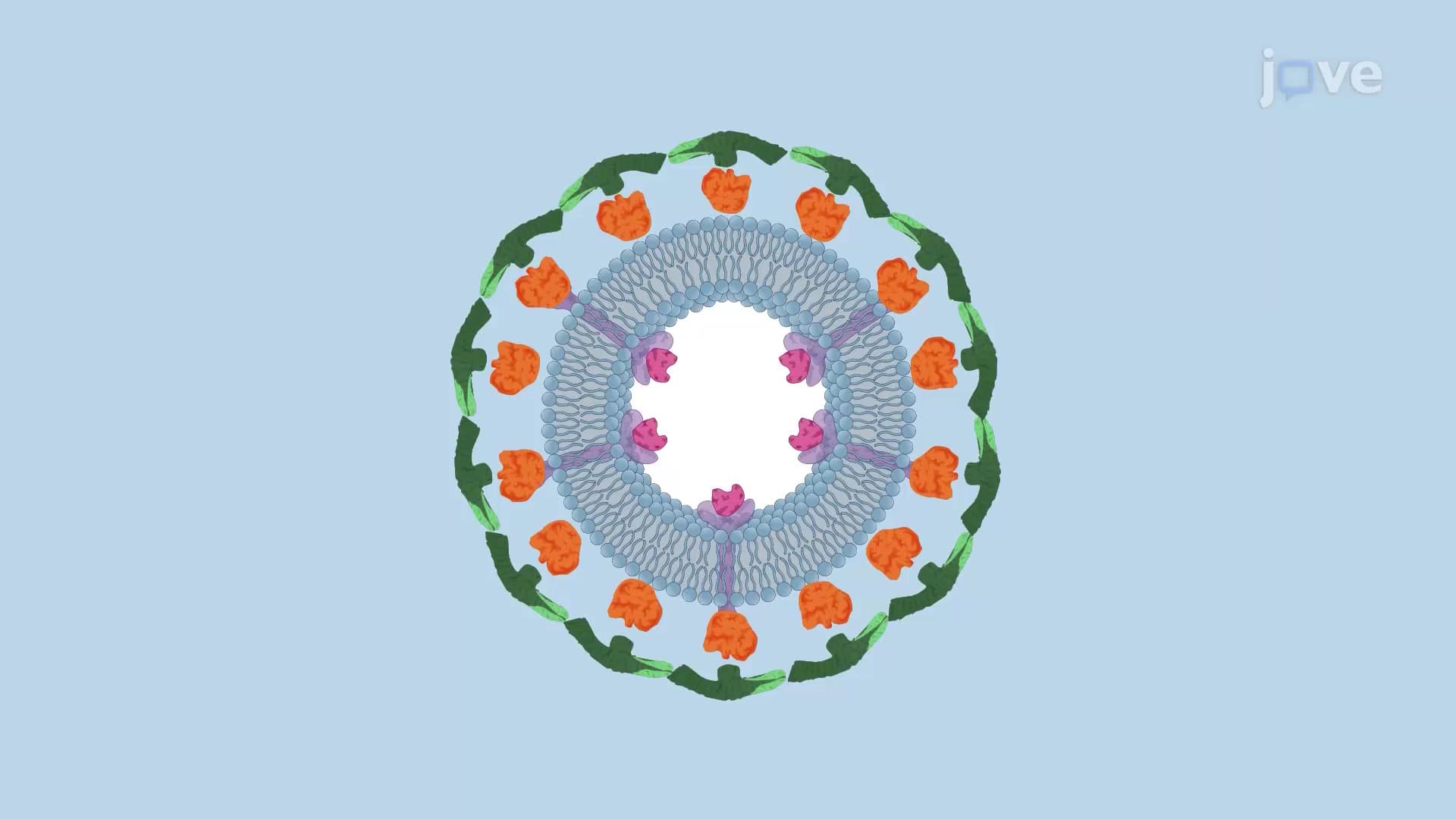
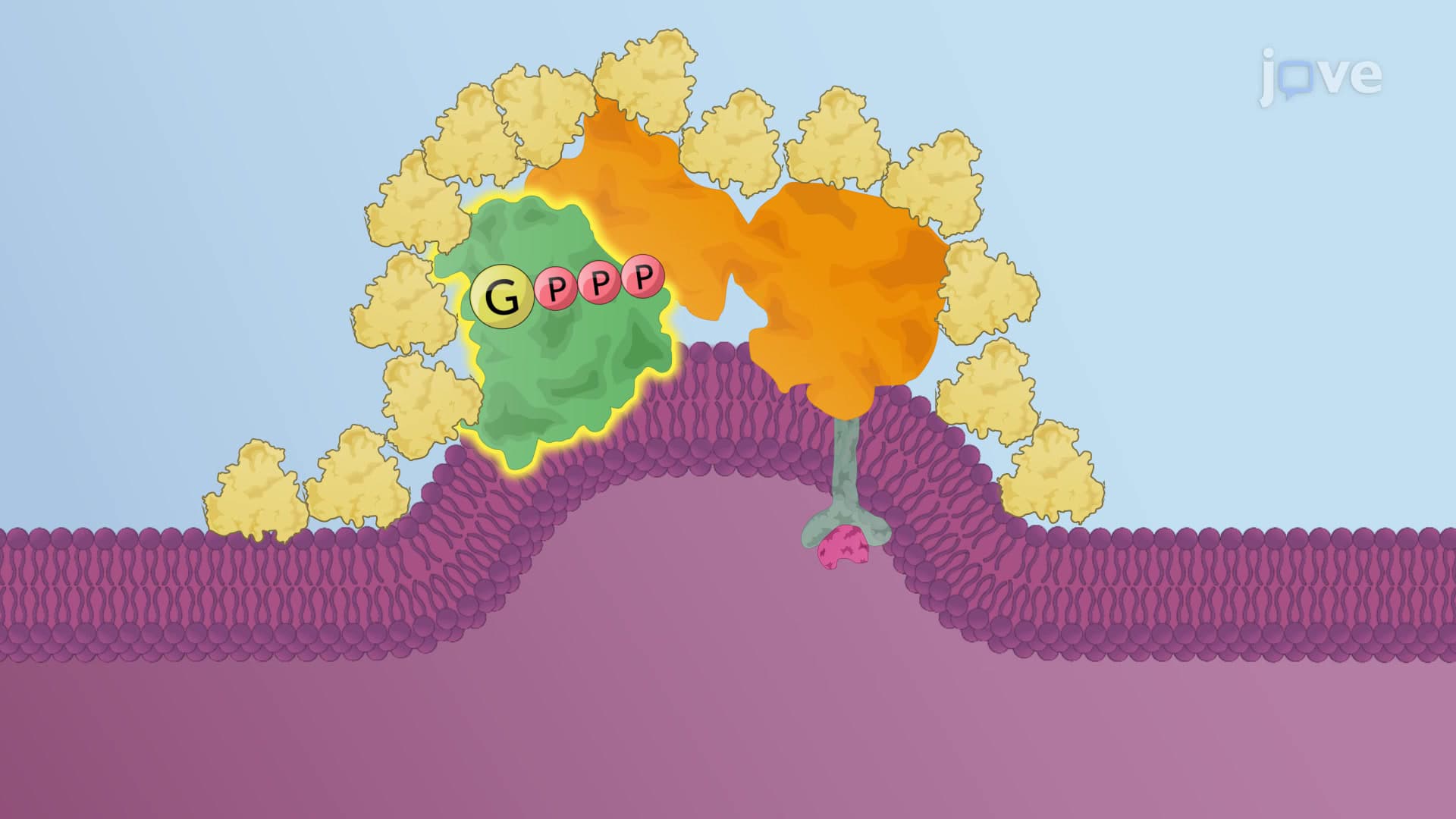
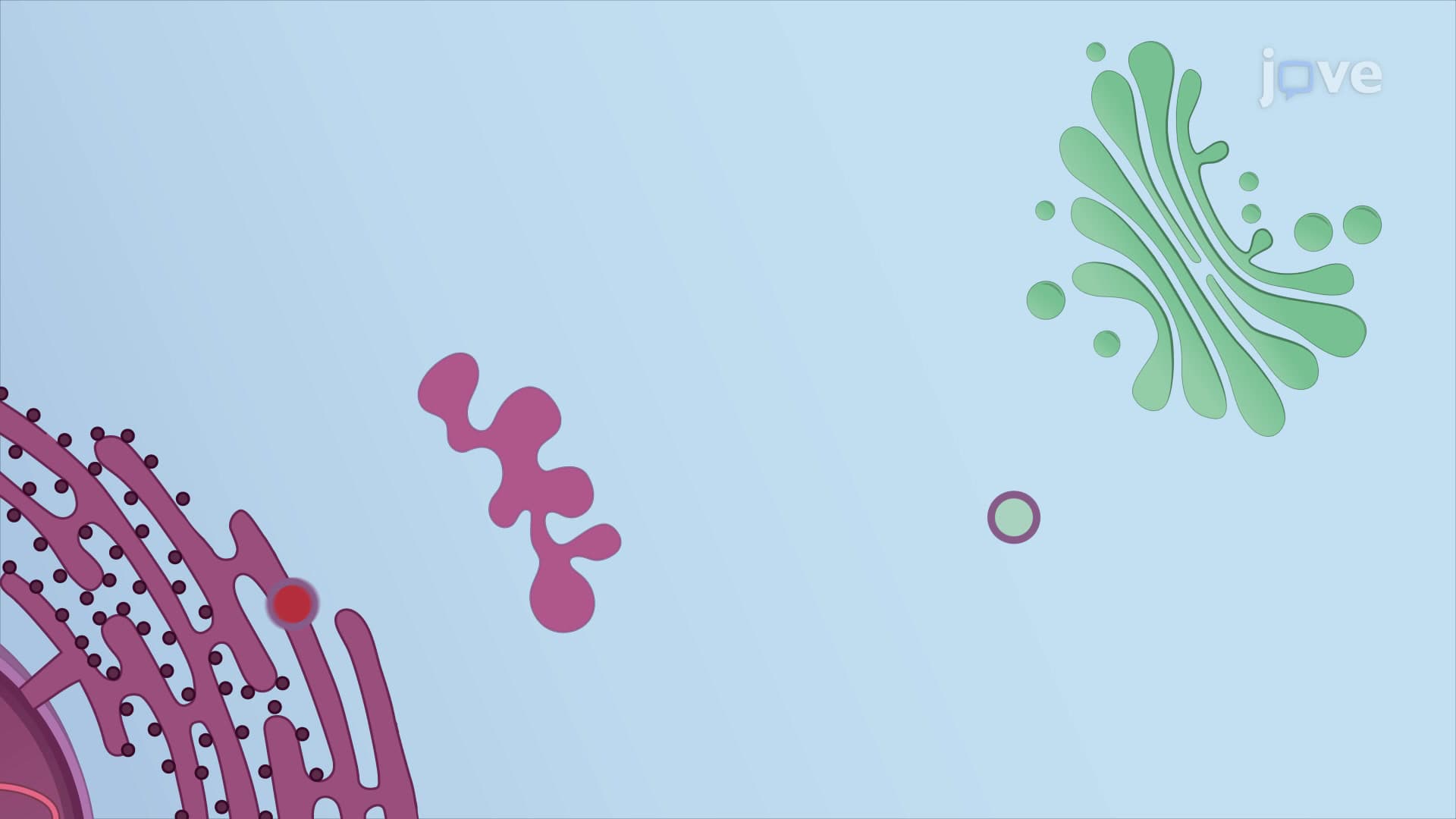
The autophagy process unfolds through distinct phases that students must master for exams like the MCAT. Initially, stressed cells form a crescent-shaped membrane structure called a phagophore. This structure expands around cellular cargo, creating a double-membrane vesicle termed an autophagosome. The outer membrane contains specific protein markers, particularly ATG9, which acts like a postal code directing the autophagosome to its destination.
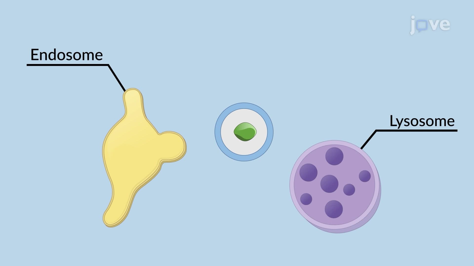
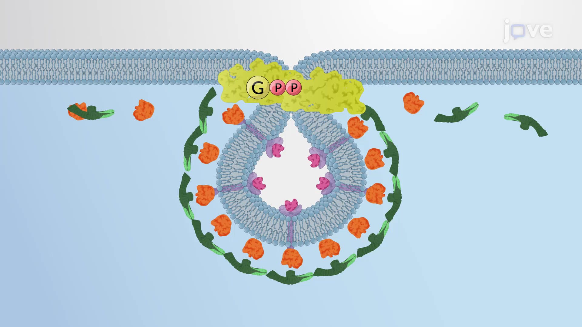
SNARE proteins facilitate the crucial fusion step between autophagosomes and lysosomes, forming autolysosomes. This mechanism mirrors the neurotransmitter release process studied in AP Biology, where SNARE proteins mediate membrane fusion at synapses. Understanding this parallel helps students grasp both concepts more effectively.
Autophagy dysfunction contributes to numerous diseases studied in medical schools across the United States. Huntington's disease, researched extensively at institutions like Harvard Medical School, involves protein aggregates that overwhelm normal autophagy mechanisms. Similarly, cancer cells often manipulate autophagy-sometimes promoting it for survival during chemotherapy, other times suppressing it to avoid cell death.
Alzheimer's disease provides another compelling example. Researchers at Stanford University have demonstrated that impaired autophagy leads to tau protein accumulation, contributing to neurodegeneration. These connections make autophagy a high-yield topic for pre-med students preparing for the MCAT's biological foundations section.
The metabolic aspects of autophagy frequently appear on college exams and standardized tests. During fasting states, autophagy breaks down cellular components to provide amino acids for essential protein synthesis. This process explains why intermittent fasting has gained attention in American healthcare discussions, though students should understand the complex regulation involved rather than oversimplifying the benefits.
Related Micro-courses