Loading subject page…


157,156 views

Video Summary: What Is Protein Glycosylation
Did you know that nearly every protein on your cell surface is decorated with sugar molecules that determine whether your immune system recognizes it as "self" or "foreign"? Protein glycosylation is the process where cells attach carbohydrate chains called glycans to proteins, fundamentally altering their structure and function. This critical cellular mechanism occurs in two main types: O-linked glycosylation (attaching sugars to serine or threonine) and N-linked glycosylation (attaching to asparagine), with the latter beginning in the endoplasmic reticulum using a 14-sugar precursor. Blood type determination in humans directly depends on specific glycosylation patterns on red blood cell proteins. Watch the full video on JoVE Coach to master this concept with expert-led visuals and step-by-step explanations.
Protein glycosylation represents one of the most prevalent post-translational modifications in eukaryotic cells, affecting over 50% of all human proteins. This sophisticated biochemical process involves the covalent attachment of carbohydrate structures to specific amino acid residues, dramatically expanding protein diversity and functionality. Unlike genetic mutations that alter primary sequence, glycosylation creates a dynamic layer of protein modification that cells can regulate in response to developmental cues, environmental conditions, and disease states.
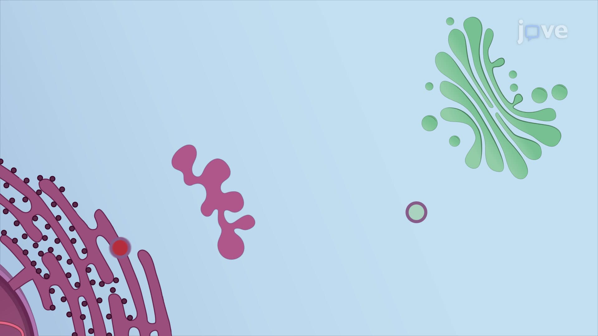
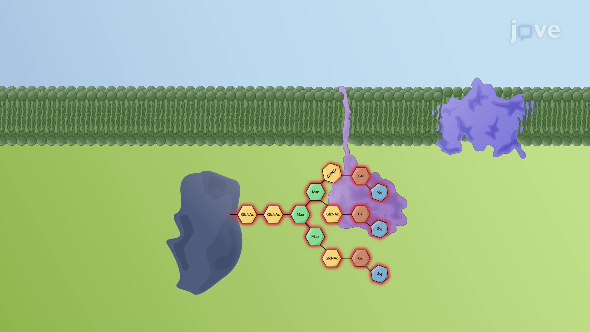
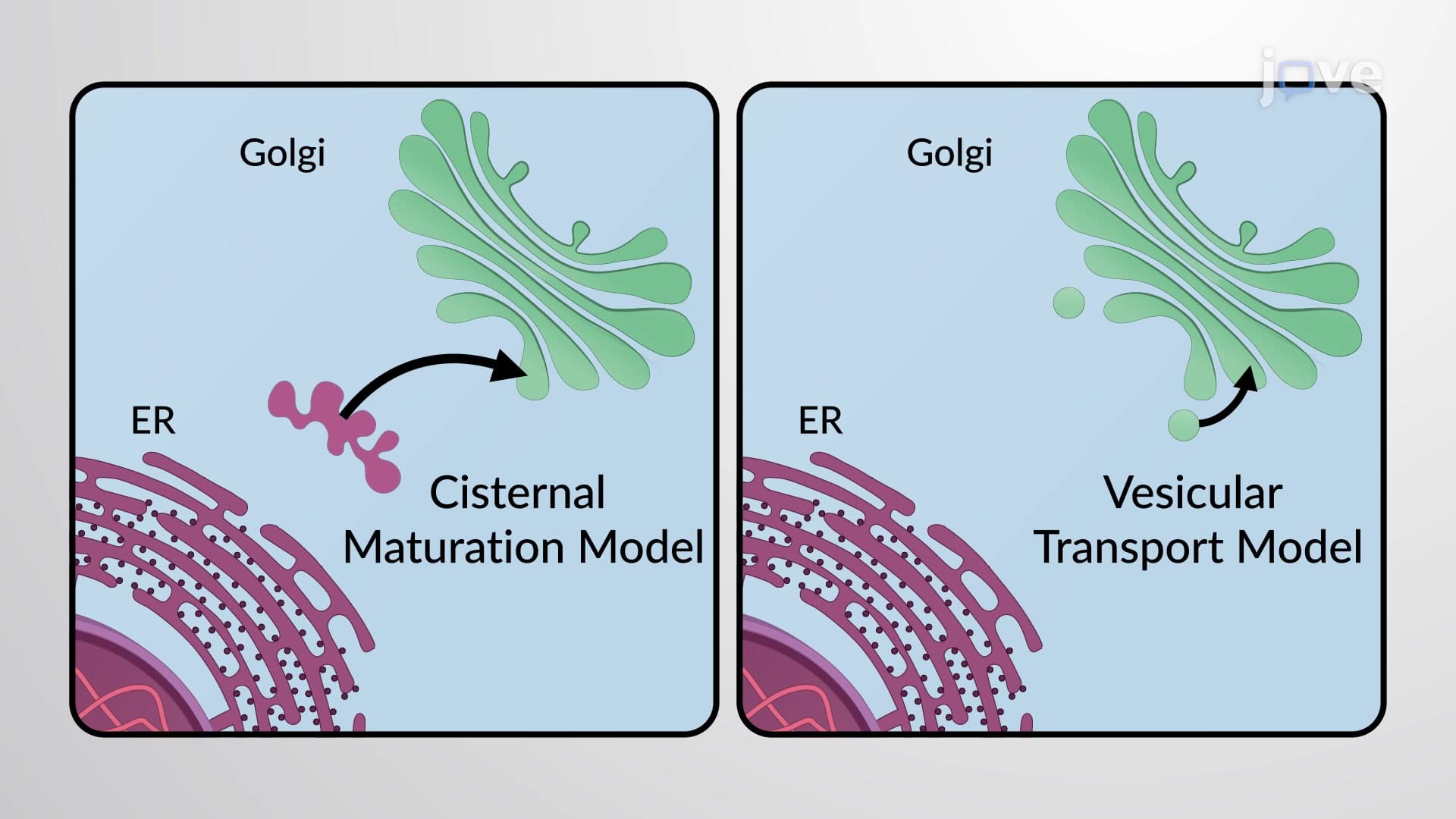
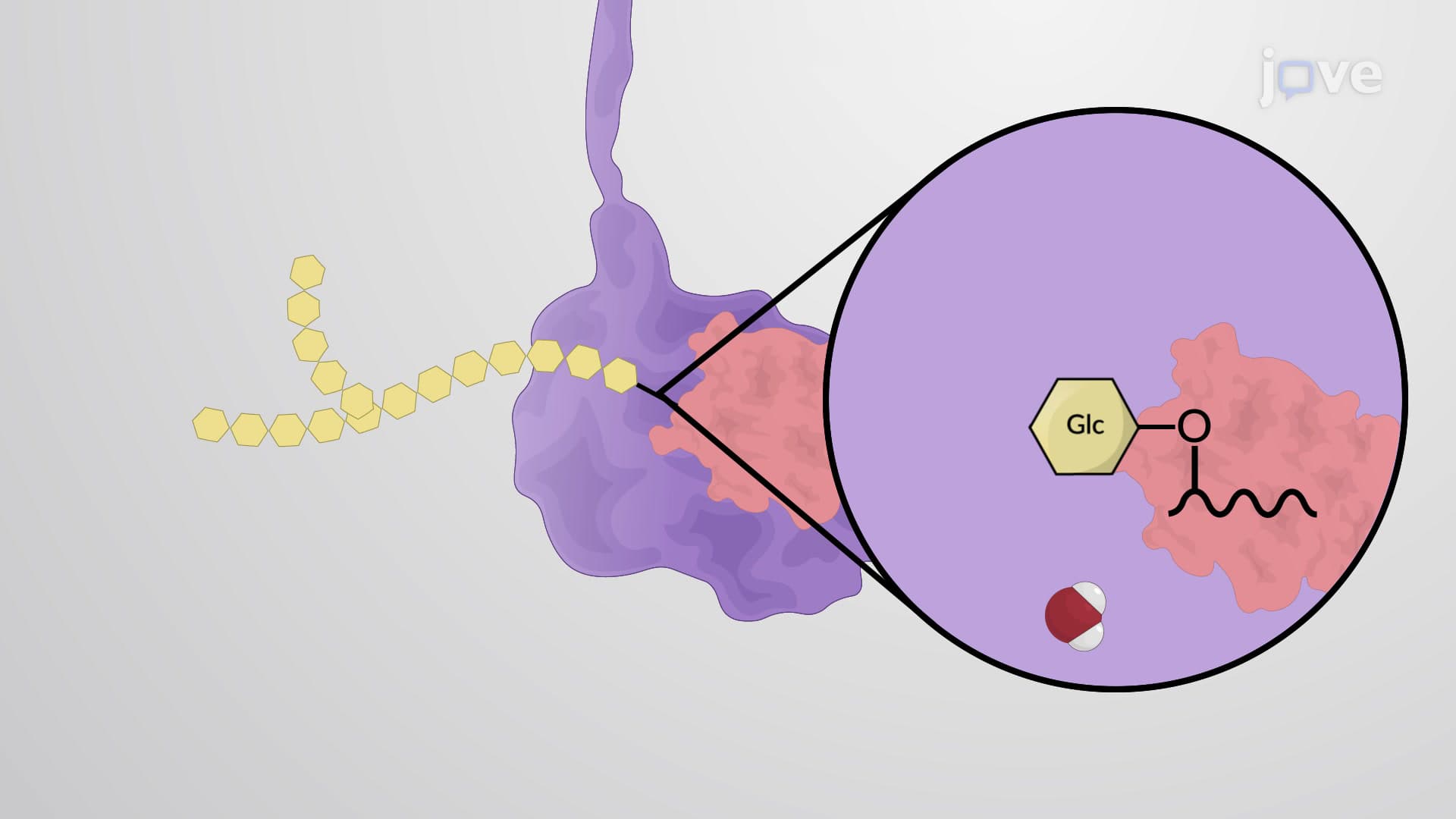
The cellular machinery employs two distinct mechanisms for protein glycosylation. O-linked glycosylation occurs primarily in the Golgi apparatus, where glycosyltransferases attach sugar residues to hydroxyl groups of serine and threonine residues. This process typically begins with N-acetylgalactosamine addition and can result in complex branched structures. In contrast, N-linked glycosylation initiates in the endoplasmic reticulum through a more elaborate process involving a lipid-linked oligosaccharide precursor.
The N-linked pathway begins with the assembly of a 14-sugar precursor on dolichol phosphate, a specialized lipid carrier embedded in the ER membrane. This precursor contains a precise arrangement: three glucose molecules, nine mannose units, and two N-acetylglucosamine residues. The enzyme oligosaccharyltransferase recognizes the consensus sequence Asn-X-Ser/Thr (where X is any amino acid except proline) and transfers the entire glycan en bloc to the asparagine residue.
Glycosylation patterns serve as molecular signatures that influence protein folding, stability, and cellular trafficking. In the immune system, glycoproteins on cell surfaces act as identification markers-consider how ABO blood group antigens result from specific glycosylation patterns on red blood cell proteins. The A and B antigens differ by a single sugar residue, yet this minor variation can trigger life-threatening transfusion reactions.
For students preparing for advanced coursework, understanding glycosylation proves essential across multiple disciplines. AP Biology students encounter this concept when studying cell membranes and protein structure, while pre-medical students face detailed glycosylation questions on the MCAT biochemistry section. Medical students must grasp how glycosylation defects contribute to congenital disorders of glycosylation (CDGs), a group of genetic diseases affecting approximately 1 in 20,000 births in the United States.
The endoplasmic reticulum maintains strict quality control over glycoprotein folding through the calnexin/calreticulin cycle. Improperly folded glycoproteins are retained in the ER and ultimately degraded, preventing potentially harmful proteins from reaching their cellular destinations. Dysregulation of this process contributes to various diseases, including certain cancers where altered glycosylation patterns promote metastasis and immune evasion.
Related Micro-courses