Loading subject page…


157,151 views

Video Summary: What Is Vesicular Tubular Clusters
Ever wondered how your cells sort and deliver thousands of proteins without mixing up their destinations? Vesicular tubular clusters form when transport vesicles from the endoplasmic reticulum fuse together, creating highways for protein trafficking-similar to how FedEx sorts packages at distribution centers before final delivery. These cellular structures are crucial for understanding diseases like cystic fibrosis, where protein transport goes wrong. Watch the full video on JoVE Coach to master this concept with expert-led visuals and step-by-step explanations.
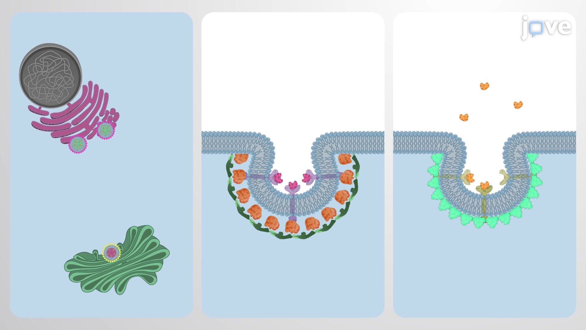
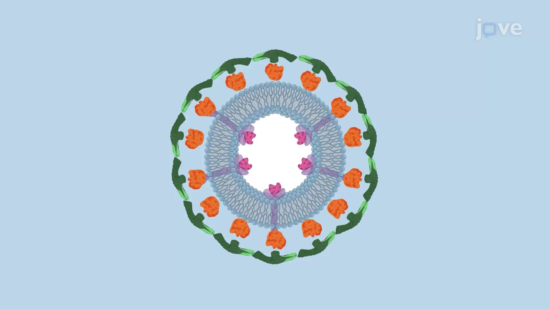
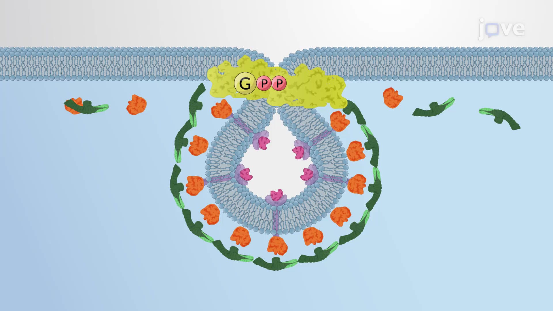
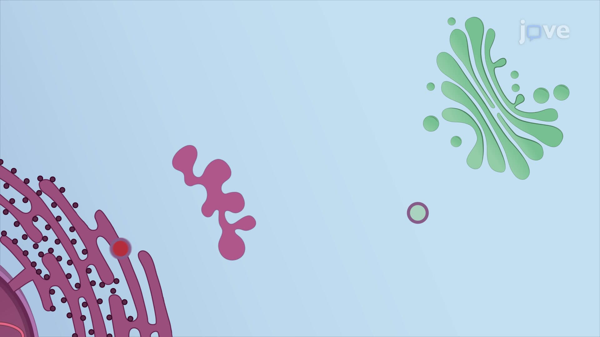
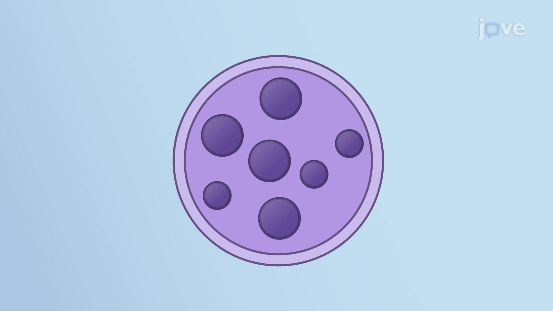
Vesicular tubular clusters represent a critical intermediate structure in the cellular secretory pathway, forming when COPII-coated transport vesicles from the endoplasmic reticulum undergo homotypic fusion. This process begins at ER exit sites-specialized ribosome-free regions where cargo proteins destined for secretion are packaged into transport vesicles.
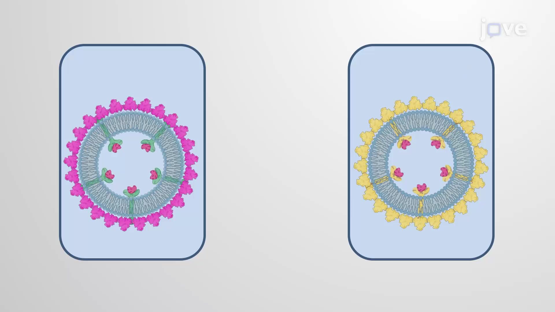
The fusion mechanism involves sophisticated molecular machinery. NSF (N-ethylmaleimide sensitive factor) proteins act as cellular "crowbars," prying apart spent SNARE protein complexes to reset them for new rounds of fusion. Fresh v-SNAREs and t-SNAREs then zipper together between adjacent vesicles, pulling their membranes close enough to merge-a process essential for maintaining proper cellular organization.
VTCs exist within the ERGIC, a dynamic region that serves as a quality control checkpoint between protein synthesis and final processing. Here, incompletely folded proteins receive additional time for proper maturation, while cellular machinery sorts cargo based on destination signals-much like how mail sorting facilities separate packages by ZIP codes.
For students preparing for the MCAT or AP Biology exams, understanding ERGIC function is crucial. This compartment frequently appears in questions about protein trafficking diseases, including Alzheimer's disease, where amyloid precursor protein processing goes awry in this very region.
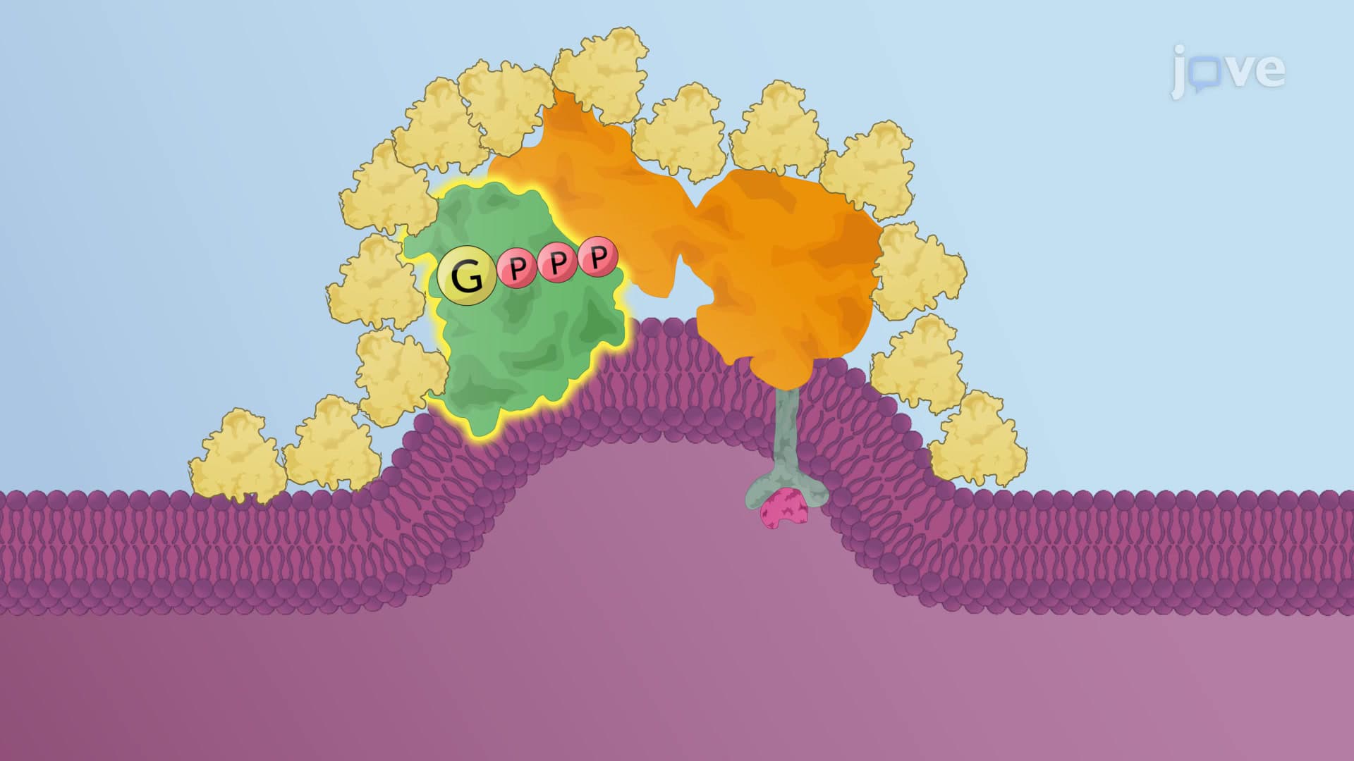
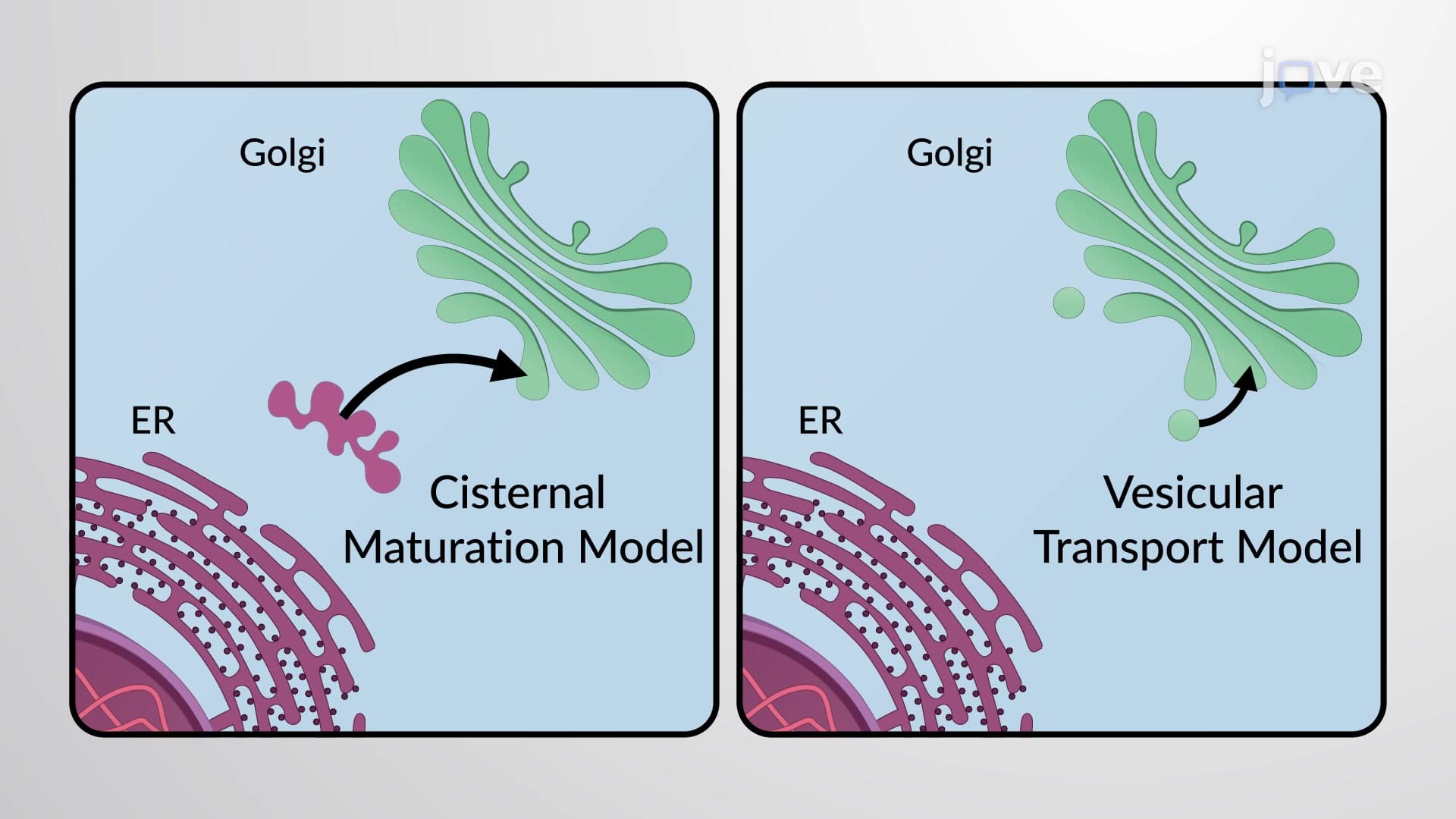
VTCs don't remain stationary-they move along microtubule highways toward the Golgi apparatus, powered by motor proteins. During transit, COPI vesicles spontaneously bud from the tubular clusters, executing the ER retrieval pathway. This quality control mechanism ensures that ER-resident proteins and cargo receptors don't accidentally reach inappropriate cellular destinations.
The coat protein switching mechanism-from COPII to COPI-remains an active research area. College biochemistry courses often explore this mystery, as understanding coat protein dynamics could unlock new therapeutic approaches for trafficking disorders like cystic fibrosis, where defective CFTR protein never reaches the cell surface.
Pharmaceutical companies like Pfizer and Johnson & Johnson invest heavily in understanding VTC biology for drug development. Many genetic diseases result from trafficking defects: Tay-Sachs disease involves lysosomal enzyme misrouting, while some forms of diabetes stem from insulin processing problems in the secretory pathway. These conditions highlight why mastering vesicular transport concepts proves essential for future healthcare professionals.
Related Micro-courses