Loading subject page…

4,623 views

Video Summary: Imaging Studies I Kidney Ureter Explained
Did you know that kidney stones affect 1 in 10 Americans at some point in their lives, making imaging studies i kidney ureter one of the most commonly ordered diagnostic procedures in US emergency departments? The imaging studies i kidney ureter tutorial demonstrates how KUB (kidney, ureter, and bladder) X-rays help detect everything from kidney stones to structural abnormalities. At hospitals like Johns Hopkins, this 15-20 minute procedure guides critical treatment decisions for thousands of patients annually. Watch the full video on JoVE Coach to master this concept with expert-led visuals and step-by-step explanations.
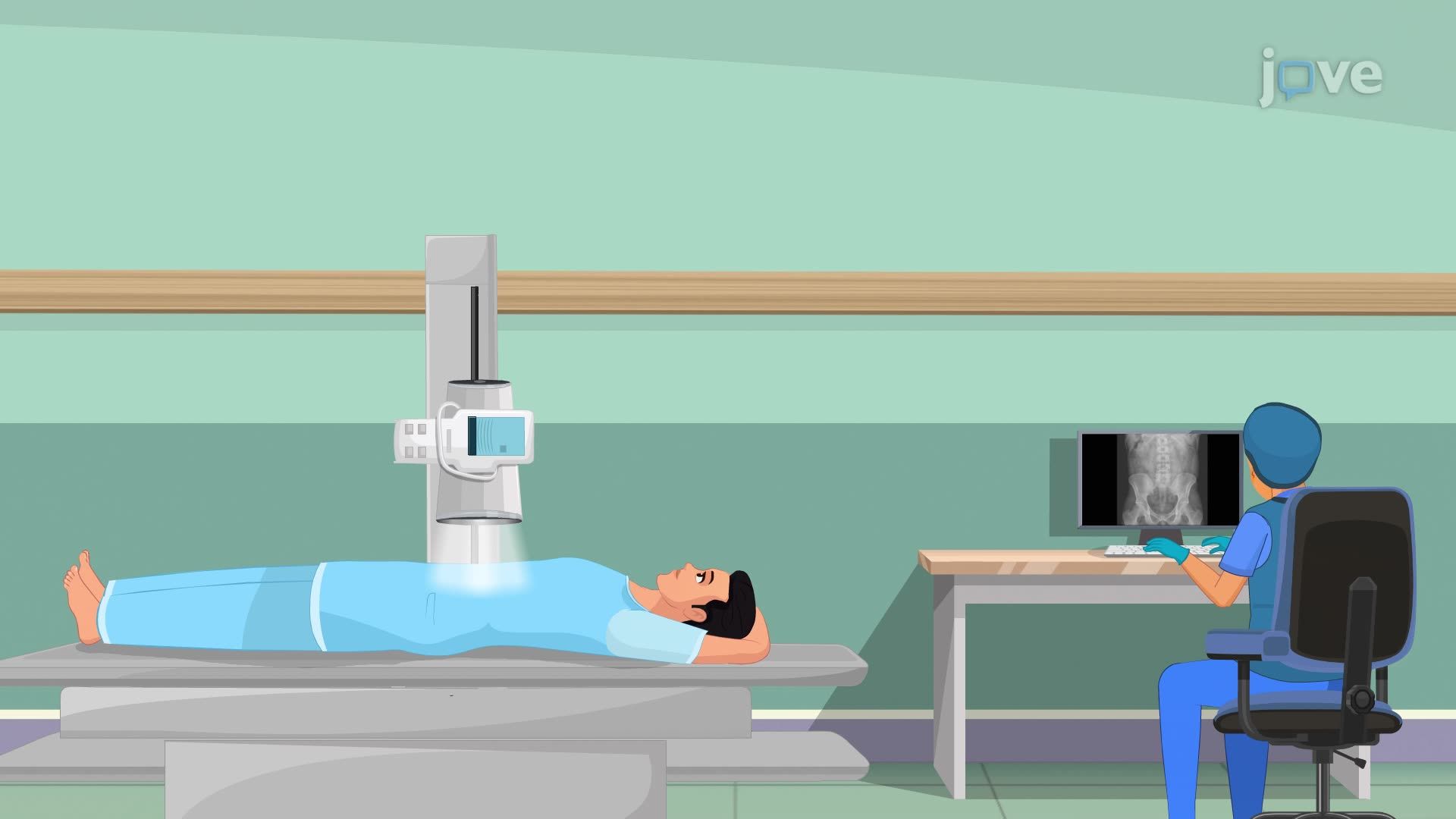
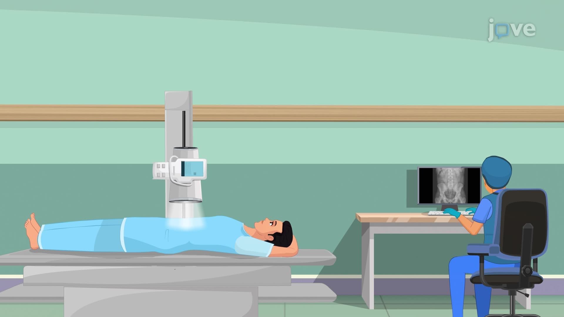
Imaging studies i kidney ureter represent a cornerstone of urological diagnostics, providing healthcare professionals with critical insights into urinary tract anatomy and pathology. The KUB (kidney, ureter, bladder) study serves as a first-line imaging technique that captures the entire urinary system in a single, comprehensive view. This non-invasive procedure utilizes plain radiography to create detailed images of the retroperitoneal space, where the kidneys and ureters reside, extending down to the pelvis where the bladder is located.
The diagnostic power of how imaging studies i kidney ureter works extends far beyond simple anatomical visualization. These studies excel at detecting radiopaque kidney stones, which appear as bright white spots on X-ray images due to their calcium content. Major medical centers like Mayo Clinic and Cleveland Clinic rely heavily on KUB imaging to evaluate patients presenting with flank pain, hematuria, or suspected urinary obstruction. The procedure can identify hydronephrosis (kidney swelling), bladder distension, and even certain tumors that alter normal organ contours.
For students preparing for healthcare careers, understanding the imaging studies i kidney ureter concept is essential for MCAT success, particularly in the biological and biochemical foundations section. The procedure's simplicity masks its diagnostic complexity, while the X-ray itself takes mere seconds, proper interpretation requires deep anatomical knowledge and clinical correlation.
The imaging studies i kidney ureter overview encompasses critical pre-procedure preparations that ensure optimal image quality and patient safety. Nurses play a vital role in patient education, explaining the procedure's purpose and addressing concerns about radiation exposure. Metal removal is crucial because metallic objects create artifacts that can obscure pathology, a lesson emphasized in NCLEX-RN examinations.
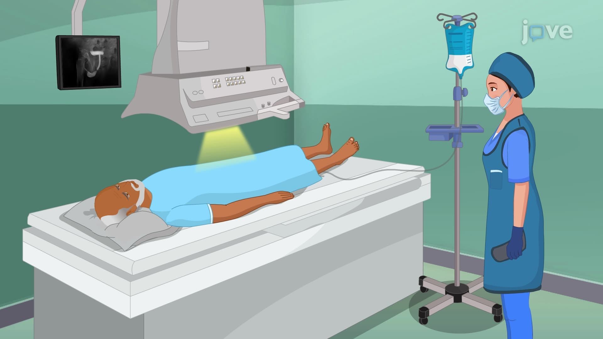
Patient positioning significantly impacts diagnostic accuracy. The standard supine position provides excellent visualization of most urinary tract structures, while upright positioning can demonstrate kidney mobility and help differentiate fixed from mobile masses. Oblique views may reveal overlapping structures more clearly, particularly in patients with complex anatomy or suspected retroperitoneal pathology.
The imaging studies i kidney ureter study guide emphasizes post-procedure hydration as both a therapeutic and preventive measure. Increased fluid intake promotes kidney function and may help flush small debris or contrast material from the urinary system. This recommendation reflects evidence-based practice standards taught in nursing programs across institutions like Johns Hopkins School of Nursing and UCSF School of Nursing.
Documentation requirements align with Joint Commission standards and state nursing board regulations. Accurate recording of patient tolerance, positioning used, and any complications ensures continuity of care and legal compliance. These documentation skills are regularly tested on the HESI A2 and TEAS examinations required for nursing school admission.
Related Micro-courses