Loading subject page…

4,623 views

Video Summary: Imaging Studies Iv Magnetic Resonance Imaging Explained
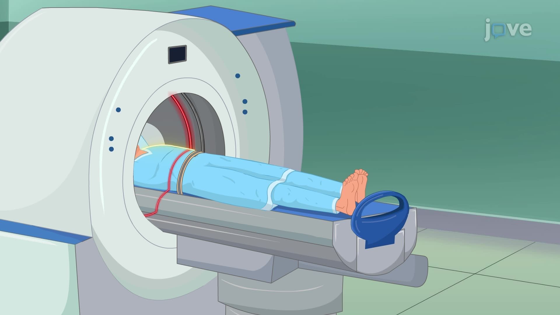
Did you know that MRI machines can detect kidney tumors as small as 1 centimeter without exposing patients to any radiation? Imaging studies IV magnetic resonance imaging uses powerful magnets and radio waves to create incredibly detailed images of internal organs. At Johns Hopkins Hospital, radiologists rely on this advanced technique to diagnose conditions like endometriosis and ovarian cysts with remarkable precision. Watch the full video on JoVE Coach to master this concept with expert-led visuals and step-by-step explanations.
Imaging Studies IV Magnetic Resonance Imaging Explained represents one of medicine's most sophisticated diagnostic tools, combining physics principles with advanced computer technology. Unlike X-rays or CT scans, MRI produces detailed anatomical images without ionizing radiation, making it particularly valuable for repeated imaging in chronic conditions. The technology relies on the magnetic properties of hydrogen atoms, which are abundant in human tissue due to our body's high water content.
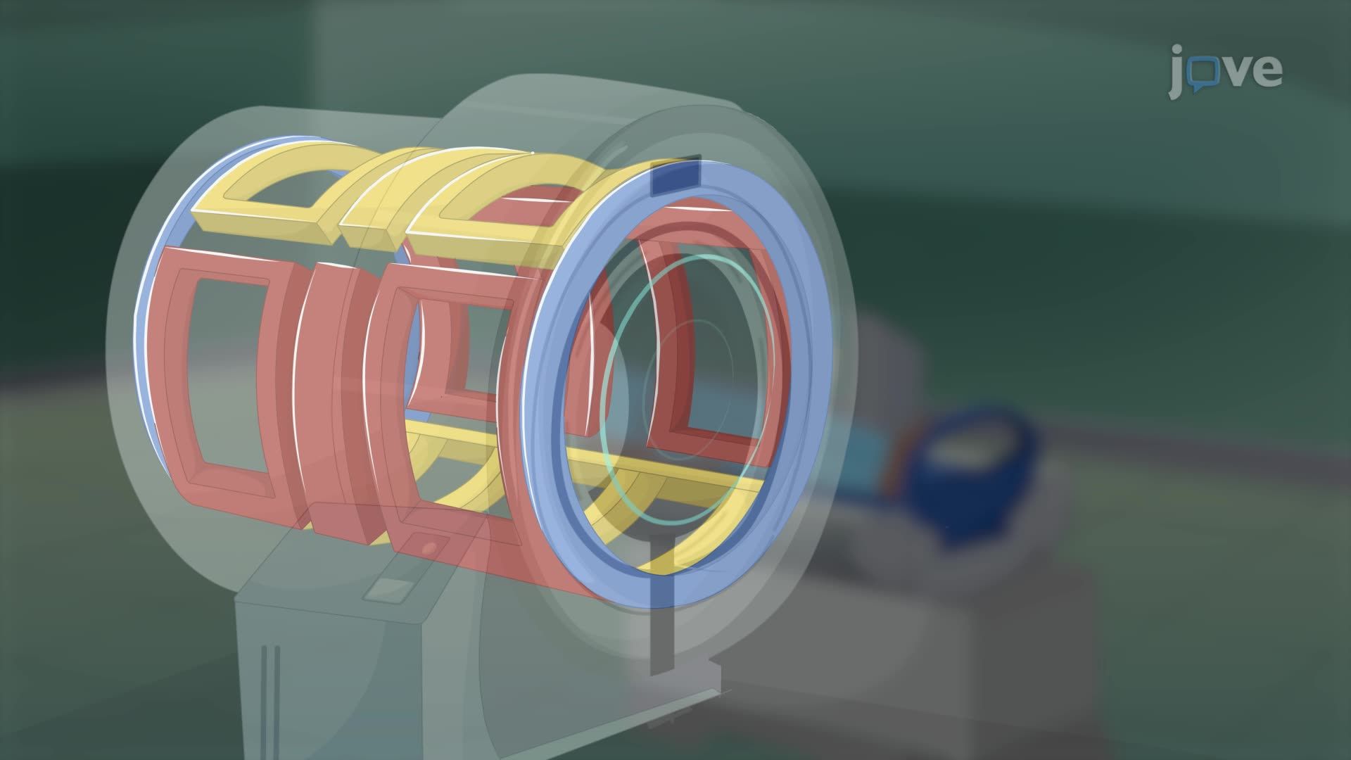
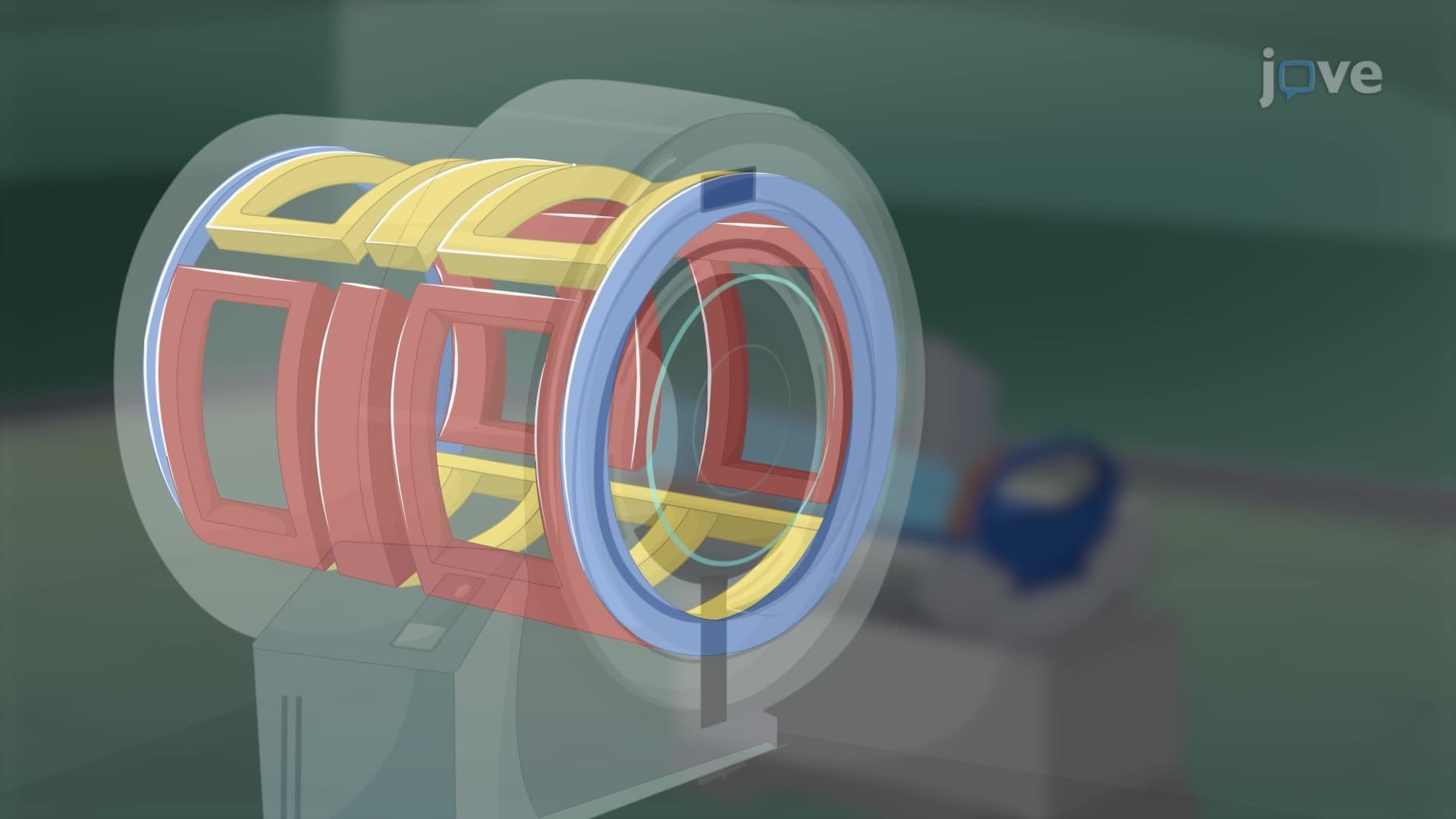
The MRI process begins when patients enter a powerful magnetic field, typically ranging from 1.5 to 3 Tesla-about 30,000 times stronger than Earth's magnetic field. This intense magnetism forces hydrogen protons in body tissues to align like tiny compass needles. When radiofrequency pulses are applied, these aligned protons absorb energy and temporarily flip their orientation. As the radio waves cease, protons return to their original alignment, releasing energy that MRI detectors capture and convert into electrical signals.
Major medical centers like Mayo Clinic and Cleveland Clinic utilize MRI extensively for urological and gynecological diagnoses. The technology excels at differentiating between various tissue types, enabling radiologists to distinguish between benign cysts and malignant tumors with exceptional accuracy. For students preparing for the MCAT or pursuing pre-med tracks, understanding MRI's diagnostic capabilities is crucial, as questions frequently appear on standardized exams regarding imaging modality selection for specific clinical scenarios.
Healthcare professionals must carefully screen patients for metallic implants, pacemakers, or foreign bodies before MRI procedures. The powerful magnetic field can cause ferromagnetic objects to move dangerously or malfunction. Pre-procedure protocols include obtaining detailed medical histories, assessing kidney function (especially when contrast agents are planned), and ensuring patients fast appropriately. Post-procedure care focuses on monitoring for contrast reactions and maintaining adequate hydration to facilitate dye elimination through the kidneys.
Related Micro-courses