Loading subject page…

4,623 views

Video Summary: Imaging Studies V Intravenous Urography Guide
When a patient arrives at Johns Hopkins Emergency Department with severe flank pain, doctors need to quickly visualize their urinary system to diagnose potential kidney stones. Imaging studies v intravenous urography provides this critical diagnostic capability by using contrast dye to highlight the kidneys, ureters, and bladder on X-ray images. This technique helps medical professionals at major US hospitals identify blockages, tumors, and structural abnormalities throughout the urinary tract. Watch the full video on JoVE Coach to master this concept with expert-led visuals and step-by-step explanations.
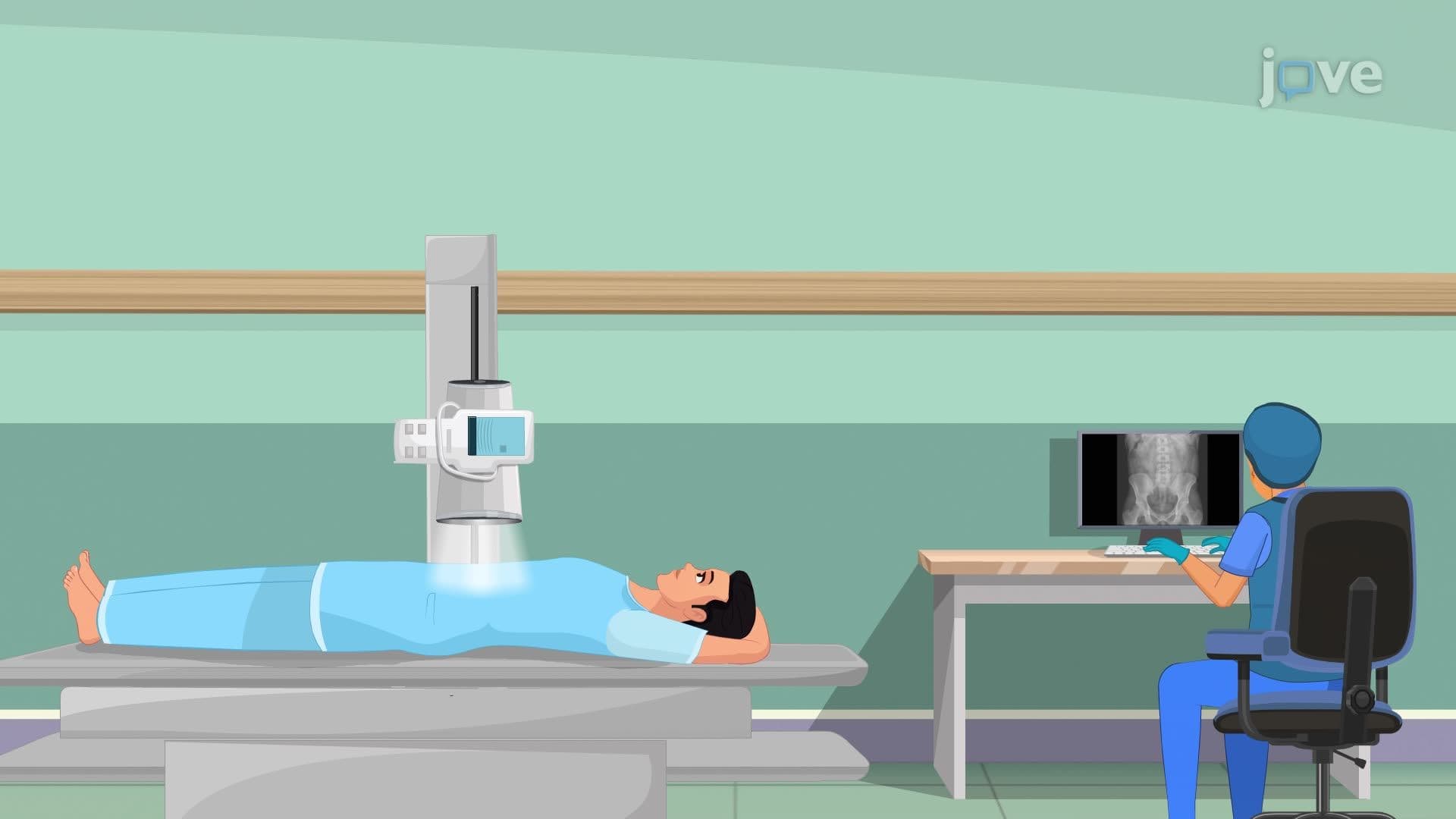
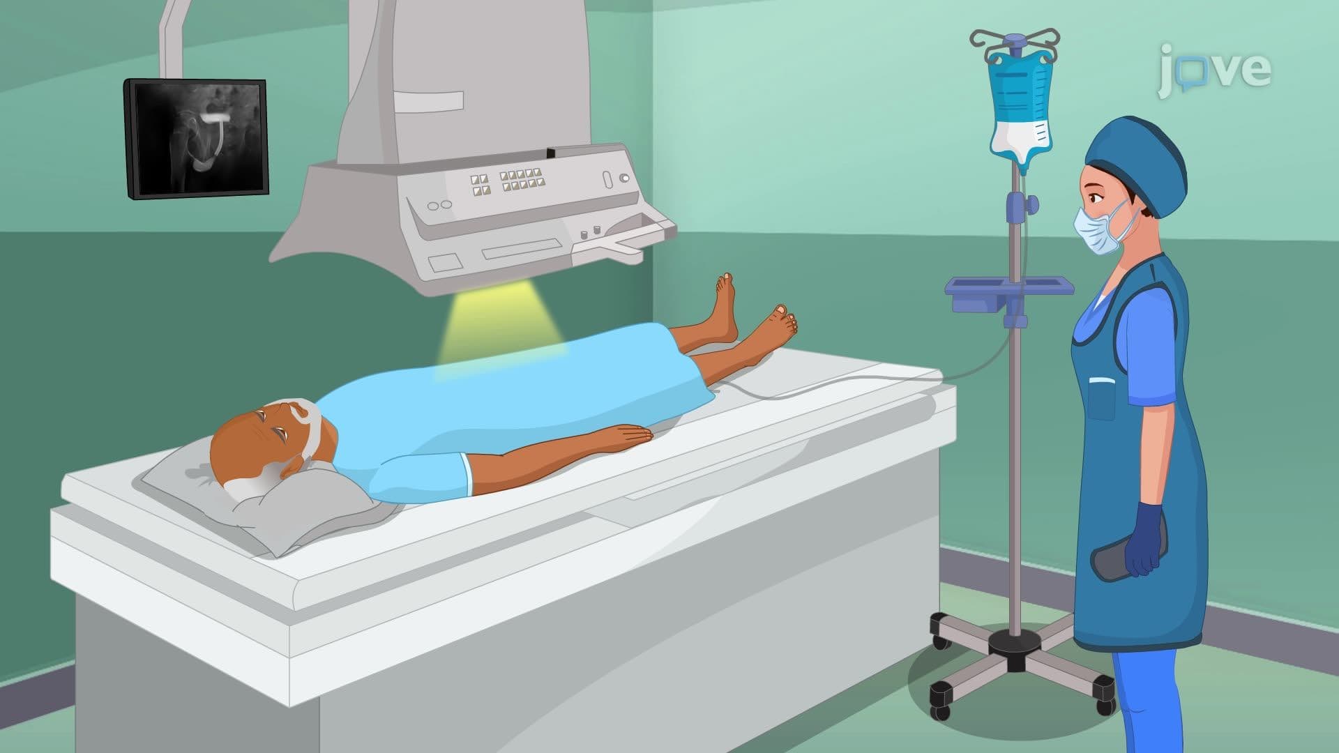
Imaging studies v intravenous urography represents a cornerstone diagnostic technique in modern urology and emergency medicine. This procedure leverages the kidney's natural filtration function to create detailed images of the entire urinary tract. When contrast dye enters the bloodstream through a peripheral vein, typically in the arm, the kidneys immediately begin filtering it from the blood. As this dye travels through the nephrons, collecting ducts, renal pelvis, ureters, bladder, and urethra, sequential X-ray images capture its movement, creating a roadmap of urinary tract anatomy and function.
US medical centers like Mayo Clinic and Cleveland Clinic routinely use intravenous urography to diagnose various conditions. Kidney stones appear as filling defects or obstructions in the contrast flow, while tumors create irregular shadows or complete blockages. An enlarged prostate shows characteristic bladder outlet obstruction patterns. The timing of dye appearance and clearance also reveals kidney function, making this technique valuable for assessing both structural and functional abnormalities. Emergency departments particularly rely on this imaging when patients present with acute flank pain, hematuria, or suspected urinary tract infections.
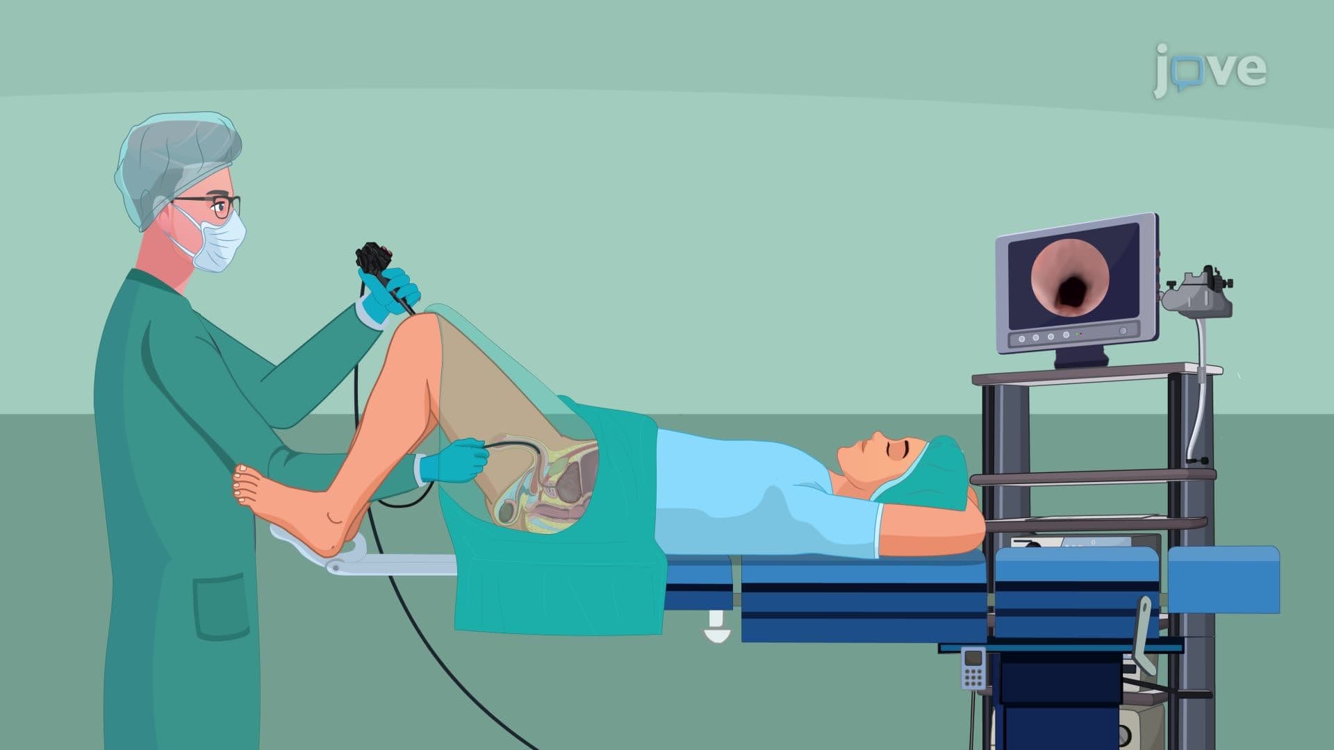
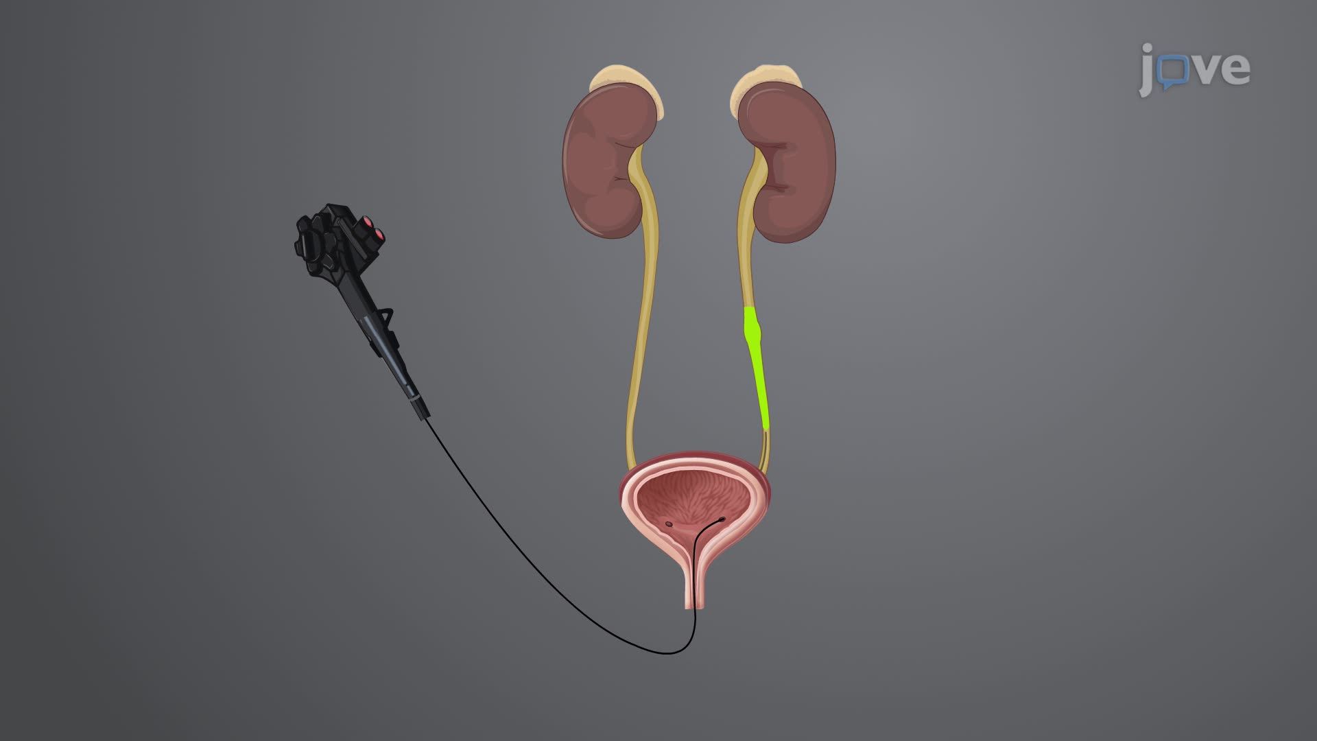
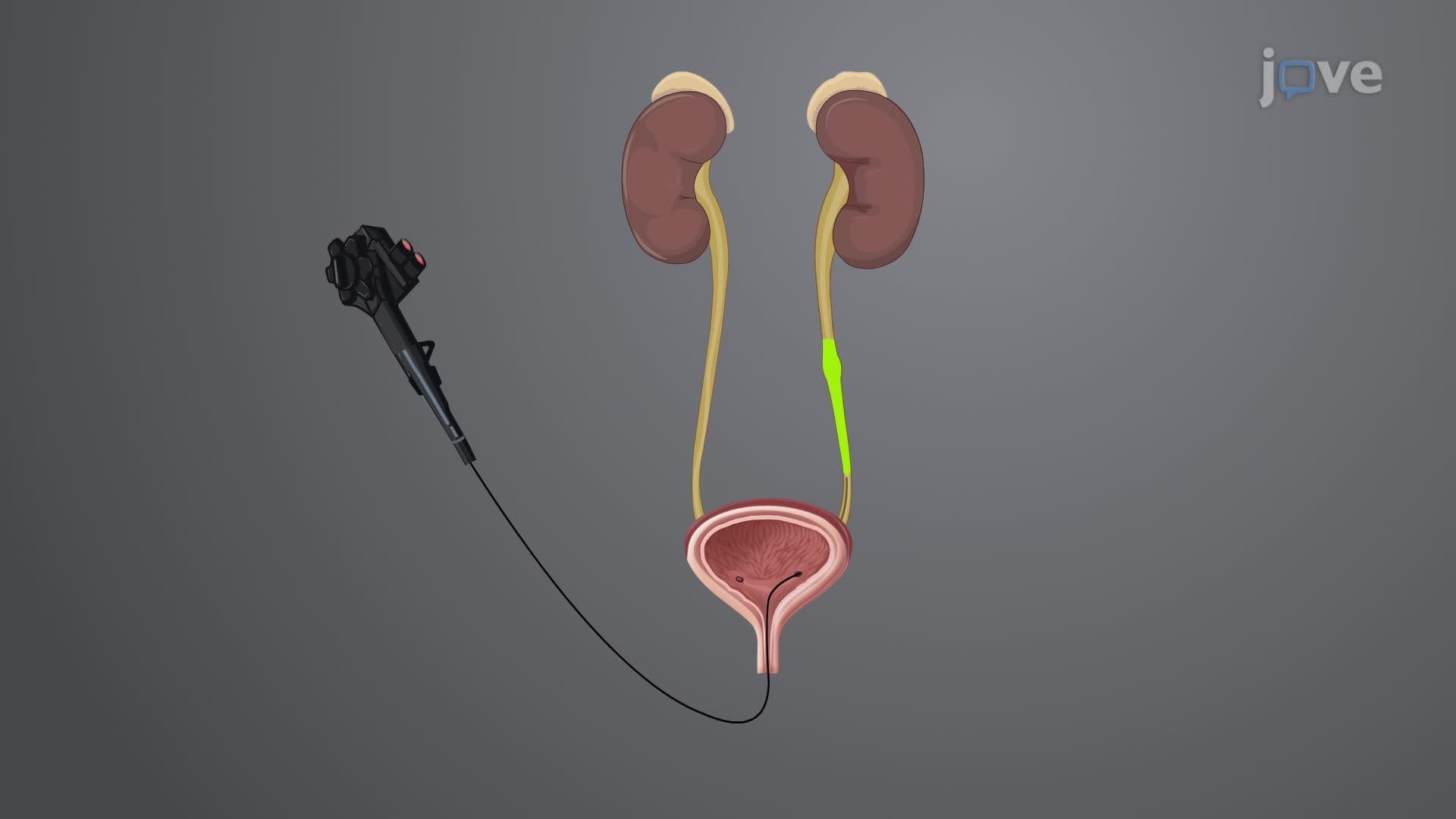
When standard intravenous urography proves insufficient or contraindicated, retrograde pyelography offers an alternative approach. This technique involves inserting a cystoscope through the urethra into the bladder, then advancing specialized catheters directly into the ureters. Contrast dye injected through these catheters provides superior visualization of upper urinary tract structures. US hospitals use this method for patients with severe kidney dysfunction, contrast allergies, or when precise ureteral anatomy assessment is crucial for surgical planning.
Students preparing for the MCAT encounter intravenous urography concepts in organ systems and diagnostic imaging sections. AP Biology courses cover kidney filtration principles that directly apply to understanding contrast dye movement. College anatomy and physiology courses often include this topic when discussing urinary system function. NCLEX exam questions frequently test nursing students' knowledge of pre-procedure patient preparation, including hydration status and allergy screening. Understanding these imaging techniques provides essential foundation knowledge for healthcare careers and demonstrates the practical application of kidney physiology concepts.
Related Micro-courses