Loading subject page…
- Medical Surgical Nursing
- Genitourinary Tract Diagnostic Procedures
- Imaging Studies VI: Voiding Cystourethrography and Cystography
Micro-course: Genitourinary Tract Diagnostic Procedures
4,623 views
Learn key concepts and earn your certificate.


Imaging Studies VI: Voiding Cystourethrography and Cystography

00:00
01:22
Video Summary: Imaging Studies Vi Voiding Cystourethrography Guide
Every year, over 150,000 children in the US undergo imaging studies VI voiding cystourethrography procedures to diagnose urinary tract conditions that could lead to kidney damage if left untreated. This specialized imaging technique combines real-time fluoroscopy with contrast agents to evaluate bladder function, detect vesicoureteral reflux, and assess urethral abnormalities. At major pediatric hospitals like Boston Children's Hospital, this procedure helps diagnose conditions in young patients experiencing recurrent UTIs. Watch the full video on JoVE Coach to master this concept with expert-led visuals and step-by-step explanations.
- Understand the fundamental principles of imaging studies VI voiding cystourethrography and its role in urological diagnosis
- Identify the key anatomical structures evaluated during VCUG procedures, including the bladder, urethra, and ureters
- Analyze the step-by-step procedural components from catheter insertion through post-void imaging
- Compare voiding cystourethrography with standard cystography techniques and their respective diagnostic applications
- Evaluate the clinical indications for VCUG, including vesicoureteral reflux detection and bladder neck obstruction assessment
- Apply knowledge of contrast agent usage and fluoroscopic imaging principles in urological diagnostics
- Recognize the importance of real-time imaging during the voiding phase for comprehensive urinary tract evaluation
- Explore the clinical significance of post-void residual measurements and their implications for patient care
Understanding Imaging Studies VI Voiding Cystourethrography Fundamentals
Imaging studies VI voiding cystourethrography represents a cornerstone diagnostic tool in modern urology, particularly valuable for evaluating pediatric and adult urinary tract disorders. This sophisticated imaging technique combines the precision of fluoroscopic technology with contrast enhancement to provide comprehensive real-time visualization of the entire lower urinary tract system.
The procedure's clinical significance extends far beyond simple anatomical imaging. In US medical practice, VCUG studies are frequently ordered by pediatric urologists at institutions like Children's Hospital of Philadelphia when patients present with recurrent urinary tract infections, suspected vesicoureteral reflux, or congenital urological abnormalities. The technique's ability to capture dynamic bladder function during both filling and emptying phases makes it irreplaceable for certain diagnostic scenarios.
Procedural Components and Technical Methodology
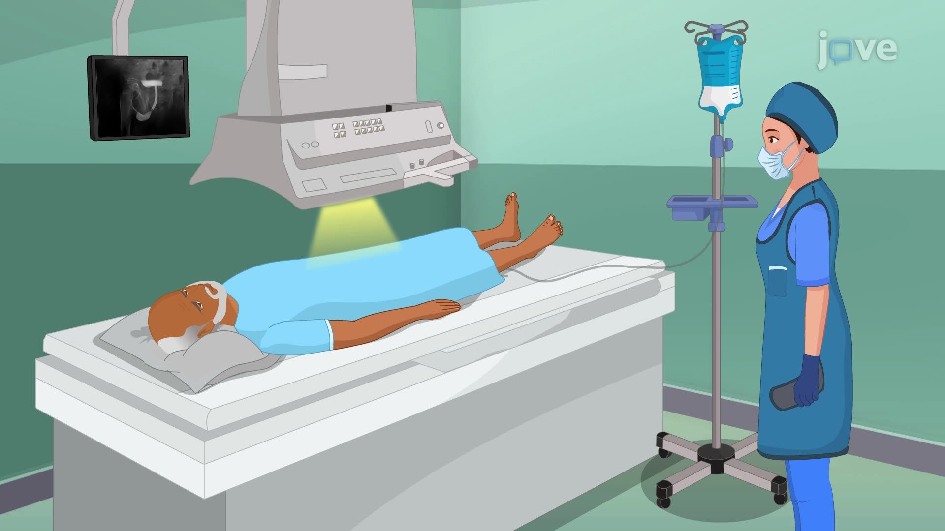
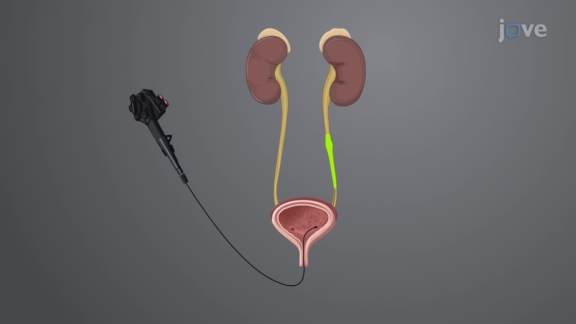
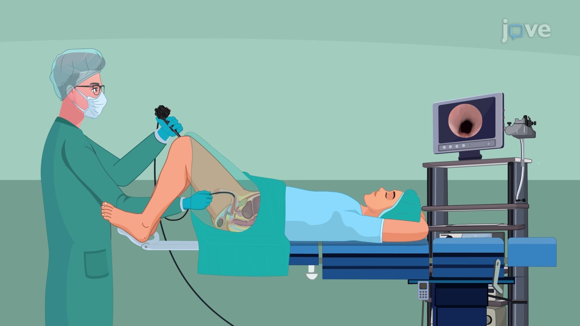
The VCUG procedure follows a systematic approach that maximizes diagnostic yield while minimizing patient discomfort. Initial preparation involves sterile catheter insertion through the urethra, a technique requiring precise anatomical knowledge that students encounter in advanced anatomy courses and clinical rotations. The contrast agent-typically iodinated solution-is carefully introduced to achieve optimal bladder distention while avoiding overfilling that could compromise natural voiding patterns.
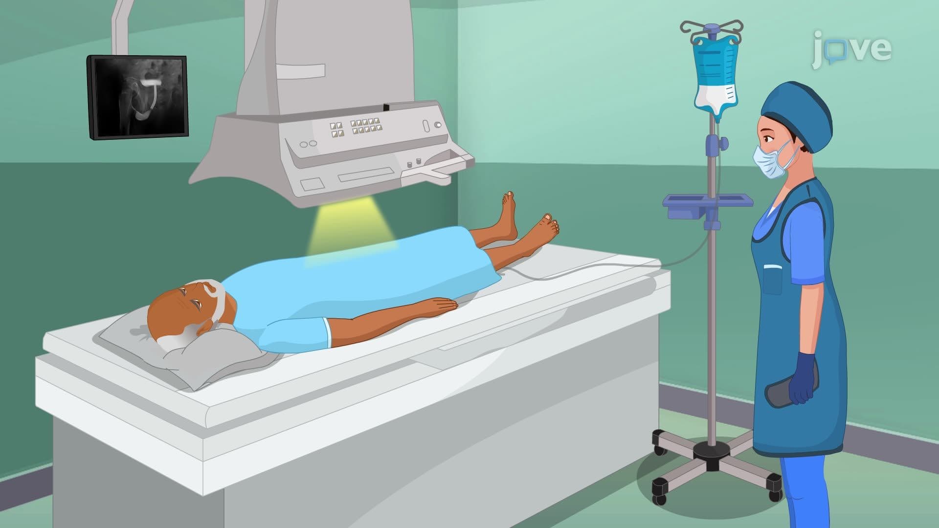
Fluoroscopic imaging during the filling phase captures crucial structural information about bladder wall integrity, capacity, and shape. This real-time visualization allows radiologists to identify abnormalities such as bladder diverticula, trabeculation, or anatomical variants that static imaging might miss. For students preparing for the MCAT or advanced placement biology exams, understanding this dynamic imaging concept reinforces broader principles of structure-function relationships in biological systems.
Clinical Applications and Diagnostic Capabilities
The voiding phase represents the procedure's most diagnostically valuable component, as it reveals functional abnormalities that may not be apparent during static imaging. Vesicoureteral reflux-the abnormal backflow of urine from bladder to kidneys-can only be definitively diagnosed during this active voiding phase. This condition affects approximately 1% of children in the US and can lead to serious complications including kidney scarring and hypertension if undetected.
Post-void residual assessment provides critical information about bladder emptying efficiency and potential neurological or anatomical causes of incomplete voiding. In clinical practice at major US medical centers like Mayo Clinic, these measurements guide treatment decisions and help monitor therapeutic interventions over time.
Comparative Analysis with Standard Cystography
While both techniques utilize contrast enhancement and catheter-based delivery systems, the fundamental difference lies in timing and imaging approach. Standard cystography focuses on static anatomical assessment, making it suitable for evaluating structural abnormalities like bladder rupture following trauma or identifying congenital malformations. However, its inability to capture functional dynamics limits its utility in diagnosing reflux or assessing voiding dysfunction, conditions that require the real-time capabilities unique to VCUG procedures.
Frequently Asked Questions
Voiding cystourethrography is a specialized imaging technique that uses contrast agents and real-time fluoroscopy to evaluate bladder function during both filling and emptying phases. Unlike regular bladder X-rays, VCUG captures dynamic urinary tract function, allowing diagnosis of conditions like vesicoureteral reflux that require observing active urine flow patterns.
MCAT questions often test understanding of imaging modalities within broader urological or diagnostic contexts. Students should focus on the functional aspects of bladder physiology, contrast agent principles, and the relationship between anatomical structure and diagnostic capability. USMLE Step 1 may include VCUG in pediatric nephrology scenarios or imaging interpretation questions.
Advanced anatomy and physiology courses, diagnostic imaging classes, and clinical laboratory science programs frequently include VCUG principles. Pre-nursing students encounter this material in pathophysiology courses, while radiology technology programs provide hands-on procedural training. Medical school curricula integrate VCUG within urology and pediatric nephrology rotations.
Pediatric hospitals like Seattle Children's Hospital use VCUG primarily for evaluating children with recurrent UTIs or suspected kidney reflux. Adult applications include assessing bladder dysfunction following spinal cord injuries or evaluating treatment outcomes after urological surgeries. The procedure helps prevent kidney damage by identifying reflux early in childhood.
Not at all - the basic concepts build on fundamental anatomy and physics principles taught in high school biology and chemistry. Students who understand basic urinary system anatomy and can grasp the concept of contrast agents enhancing X-ray visibility have the foundation needed. The procedural steps follow logical medical reasoning that reinforces critical thinking skills.
Create concept maps connecting anatomical structures, procedural steps, and clinical applications. Practice with imaging interpretation exercises and focus on understanding why real-time imaging is essential for certain diagnoses. Review case studies showing normal versus abnormal findings, and connect VCUG indications to broader urological pathophysiology concepts.
Focus on patient care aspects including pre-procedure preparation, patient positioning, and post-procedure monitoring. Understand the nursing implications of catheter insertion, contrast administration, and patient education requirements. NCLEX questions often emphasize safety considerations and appropriate patient communication during urological procedures.
Consider exploring CT urography, MR urography, and nuclear medicine renal scans to understand the complete spectrum of urological imaging. Study how different imaging modalities complement each other in clinical diagnosis, and learn about emerging technologies like contrast-enhanced ultrasound that may supplement or replace traditional VCUG in certain clinical scenarios.
Related Micro-courses