Loading subject page…

4,623 views

Video Summary: Imaging Studies Vii Vascular Imaging Explained
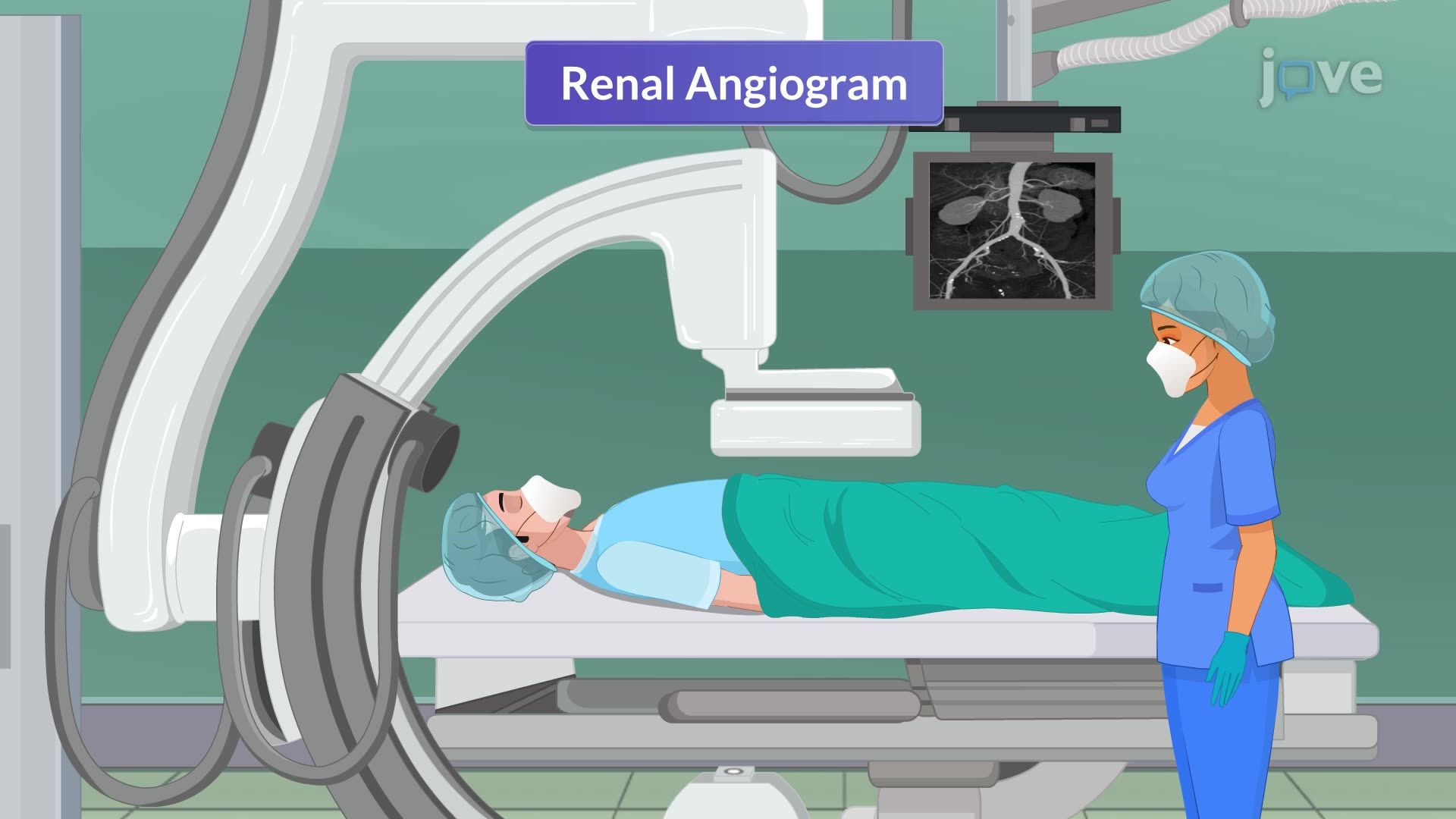
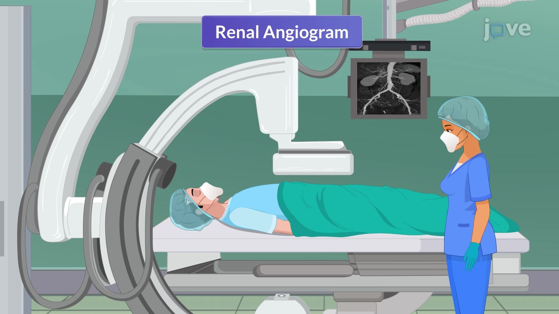
Every year, over 200,000 Americans undergo renal angiograms to diagnose kidney blood vessel disorders that could lead to hypertension or kidney failure. Imaging Studies VII Vascular Imaging reveals how contrast dye and real-time X-ray technology allow radiologists at hospitals like Mayo Clinic to visualize blood flow through renal arteries, identifying blockages and aneurysms before they become life-threatening. Watch the full video on JoVE Coach to master this concept with expert-led visuals and step-by-step explanations.
Imaging Studies VII Vascular Imaging represents a sophisticated diagnostic approach that combines interventional radiology with real-time imaging to visualize blood vessel structures and function. Renal angiography exemplifies this technology, using iodinated contrast agents and fluoroscopic guidance to create detailed maps of kidney vasculature. This technique has revolutionized nephrology and vascular surgery by providing precise anatomical information that guides treatment decisions for conditions ranging from renal artery stenosis to complex aneurysms.
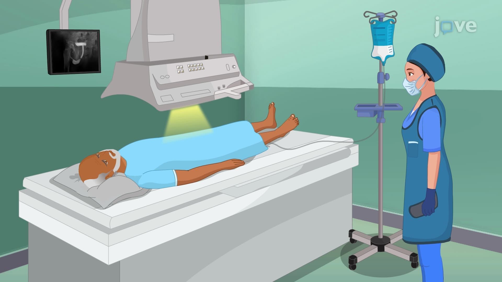
Patient preparation for vascular imaging procedures follows strict medical protocols developed by organizations like the American College of Radiology. The 6-8 hour fasting requirement prevents aspiration complications during sedation, while comprehensive allergy screening identifies patients at risk for contrast-induced reactions. Healthcare providers at institutions like Johns Hopkins emphasize the importance of creatinine level assessment, as contrast agents can potentially cause nephrotoxicity in patients with pre-existing kidney dysfunction. This preparation phase directly correlates with concepts tested in nursing examinations like the NCLEX-RN and medical school assessments.
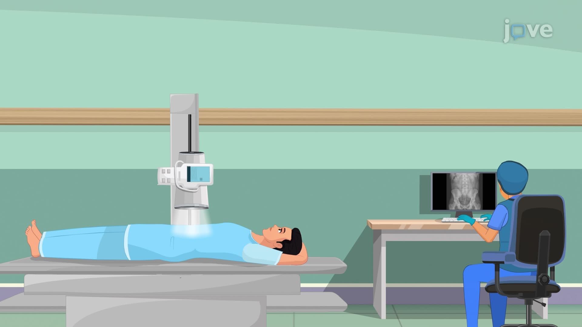
The catheterization process demonstrates advanced interventional techniques where a guidewire and catheter system navigates through the vascular tree under continuous X-ray visualization. Fluoroscopy provides real-time imaging that allows radiologists to precisely position catheters within target vessels while minimizing radiation exposure. The femoral artery approach, preferred at major medical centers like Cleveland Clinic, offers optimal access to the renal circulation while maintaining patient safety. This procedural knowledge appears frequently in MCAT questions related to cardiovascular physiology and medical imaging.
During contrast injection, the imaging sequence captures arterial, capillary, and venous phases of renal blood flow, creating a comprehensive vascular roadmap. Digital subtraction angiography (DSA) technology removes bone and soft tissue shadows, highlighting only contrast-enhanced vessels. Radiologists analyze these images for stenotic lesions, aneurysmal dilatations, arteriovenous malformations, and tumor vascularity. This diagnostic process requires understanding of normal renal anatomy and pathophysiology concepts covered in AP Biology and undergraduate anatomy courses.
Related Micro-courses