Loading subject page…

4,623 views

Video Summary: What Is Imaging Studies Ii Ultrasonography
Every year, over 100 million ultrasound exams are performed in US hospitals, making imaging studies ii ultrasonography one of the most widely used diagnostic tools in modern medicine. This non-invasive technique uses high-frequency sound waves to create real-time images of internal organs, particularly excelling in urinary system evaluation at major medical centers like Johns Hopkins and Mayo Clinic. What is Imaging Studies II Ultrasonography encompasses the comprehensive study of how ultrasound technology diagnoses conditions from kidney stones to bladder disorders through safe, radiation-free imaging. Watch the full video on JoVE Coach to master this concept with expert-led visuals and step-by-step explanations.
Imaging studies ii ultrasonography represents a cornerstone of modern diagnostic medicine, utilizing high-frequency sound waves (typically 2-15 MHz) to create detailed, real-time images of internal body structures. Unlike X-rays or CT scans, ultrasonography operates without ionizing radiation, making it exceptionally safe for repeated use and ideal for monitoring chronic conditions. The technology works through the piezoelectric effect, where electrical energy converts to mechanical sound waves that penetrate tissues and reflect back to create visual representations.
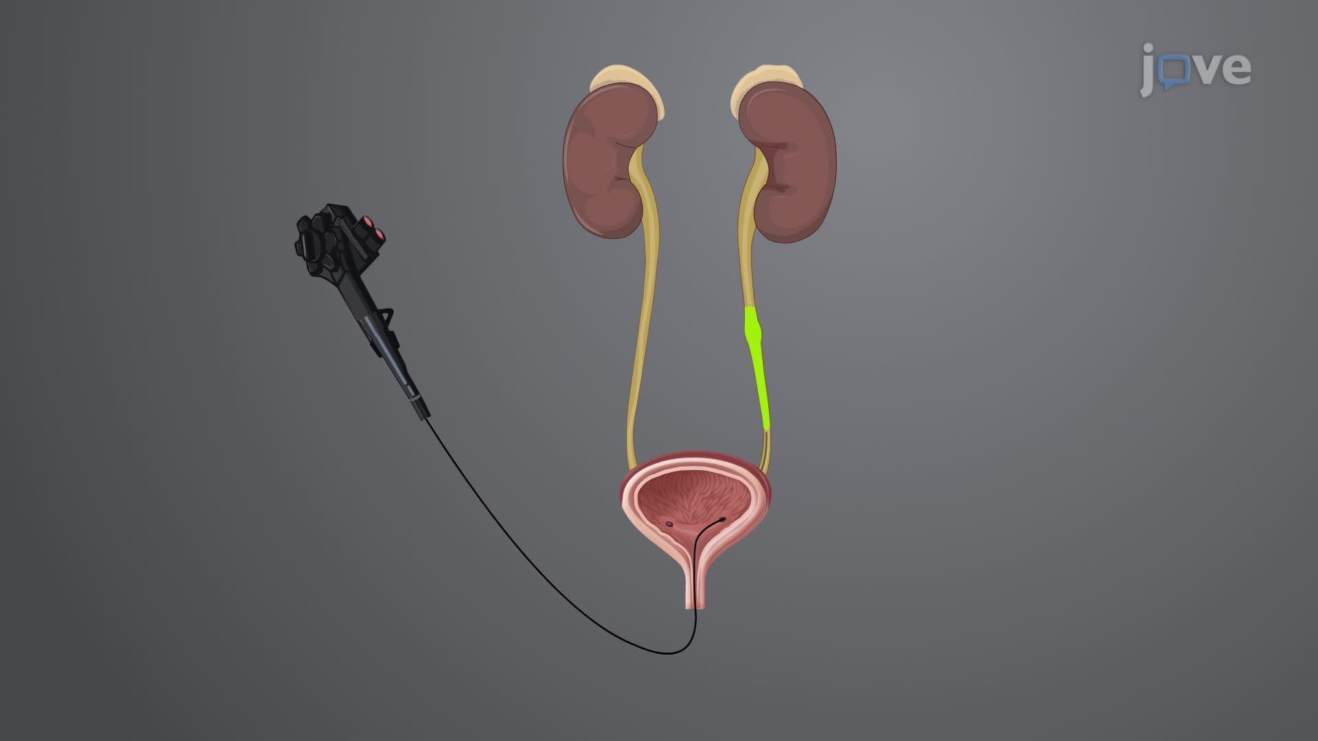
The imaging studies ii ultrasonography overview encompasses extensive diagnostic applications, particularly in urological assessments. At leading US medical institutions like Cleveland Clinic and Massachusetts General Hospital, ultrasound routinely diagnoses kidney stones (urolithiasis), fluid accumulation in kidneys (hydronephrosis), and bladder abnormalities. The technique excels in differentiating between solid masses and fluid-filled cysts, measuring organ dimensions, and assessing blood flow through Doppler applications. For students preparing for healthcare careers, understanding these diagnostic capabilities proves essential for MCAT preparation and nursing school prerequisites.
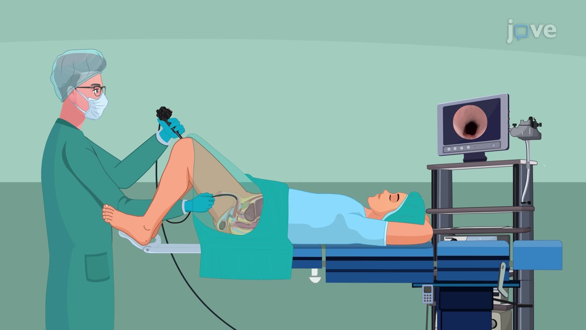
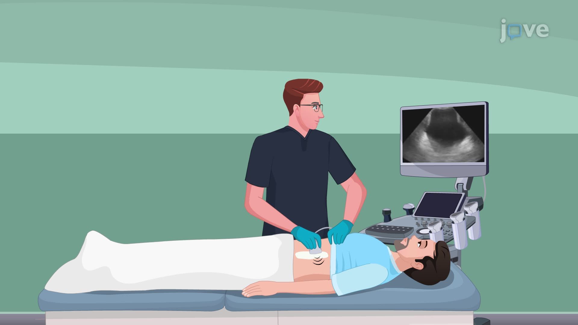
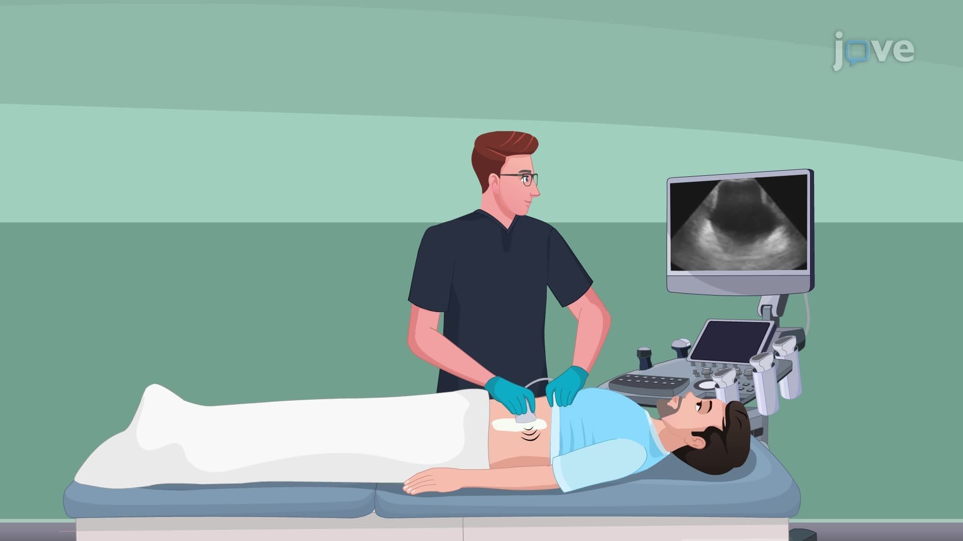
The imaging studies ii ultrasonography concept extends beyond technical operation to encompass comprehensive patient care protocols. Healthcare providers must ensure proper patient preparation, including bladder filling for pelvic ultrasounds and appropriate positioning for optimal image acquisition. The 20-25 minute procedure duration requires patient education about expectations and comfort measures. Post-procedure care involves immediate result documentation and coordination with referring physicians for treatment planning.
Modern ultrasonography continues evolving with 3D/4D imaging capabilities, contrast-enhanced studies, and artificial intelligence integration. Students studying imaging studies ii basics should recognize how technological advancement impacts diagnostic accuracy and patient outcomes. Understanding these developments proves valuable for AP Biology students exploring medical technology applications and college pre-med students preparing for advanced coursework in medical imaging and diagnostic techniques.
Related Micro-courses