Loading video…


 10 Concepts
 2.3K views

Video Summary: Acute Kidney Injury Iv Diagnostic Explained
Did you know that kidney function can decline by 50% before patients show obvious symptoms? Understanding acute kidney injury iv diagnostic techniques is crucial for healthcare professionals, as AKI affects over 13 million Americans annually. At Massachusetts General Hospital, emergency physicians use systematic diagnostic approaches to differentiate between prerenal, intrarenal, and postrenal causes of kidney dysfunction. The Acute Kidney Injury IV Diagnostic Explained framework combines serum creatinine monitoring, urinalysis interpretation, and imaging studies to prevent irreversible kidney damage. Watch the full video on JoVE Coach to master this concept with expert-led visuals and step-by-explanations.
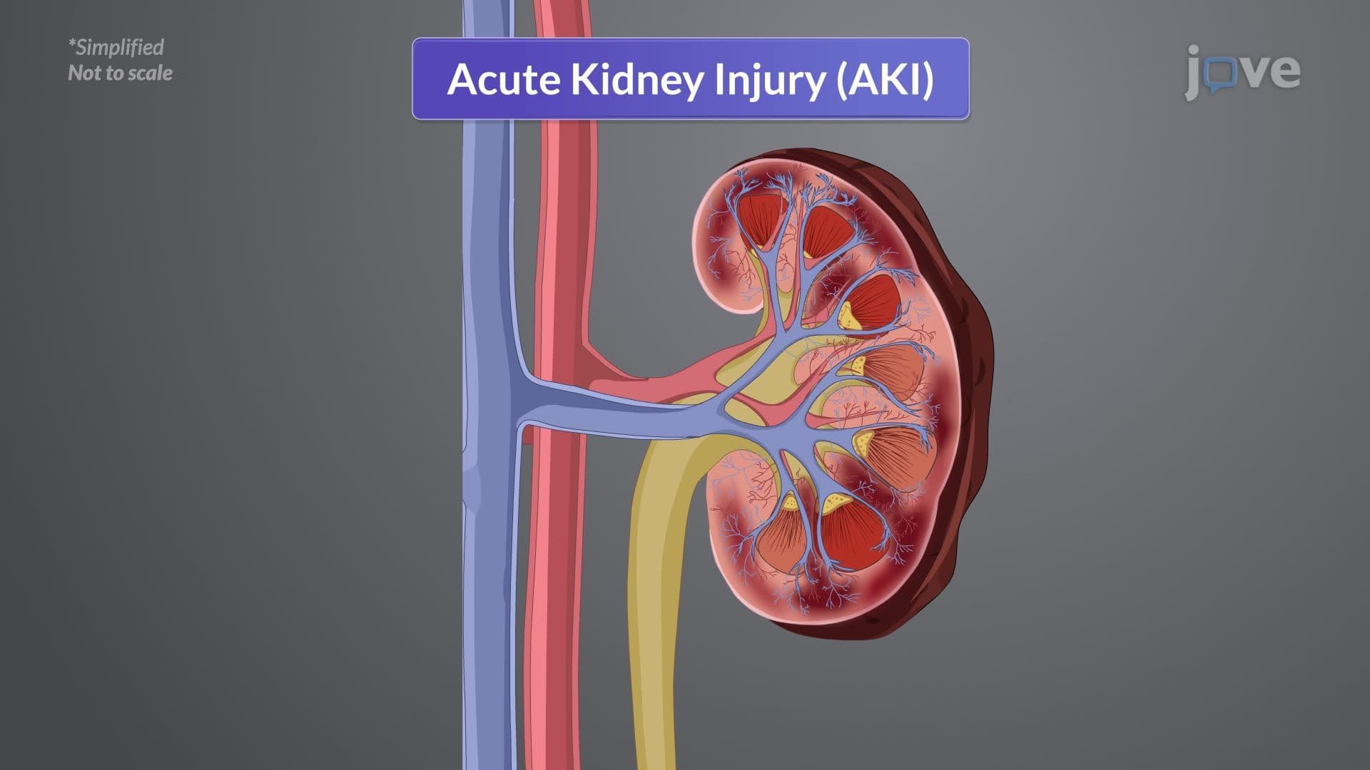
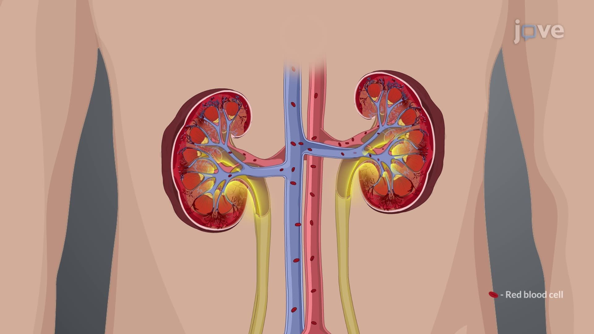
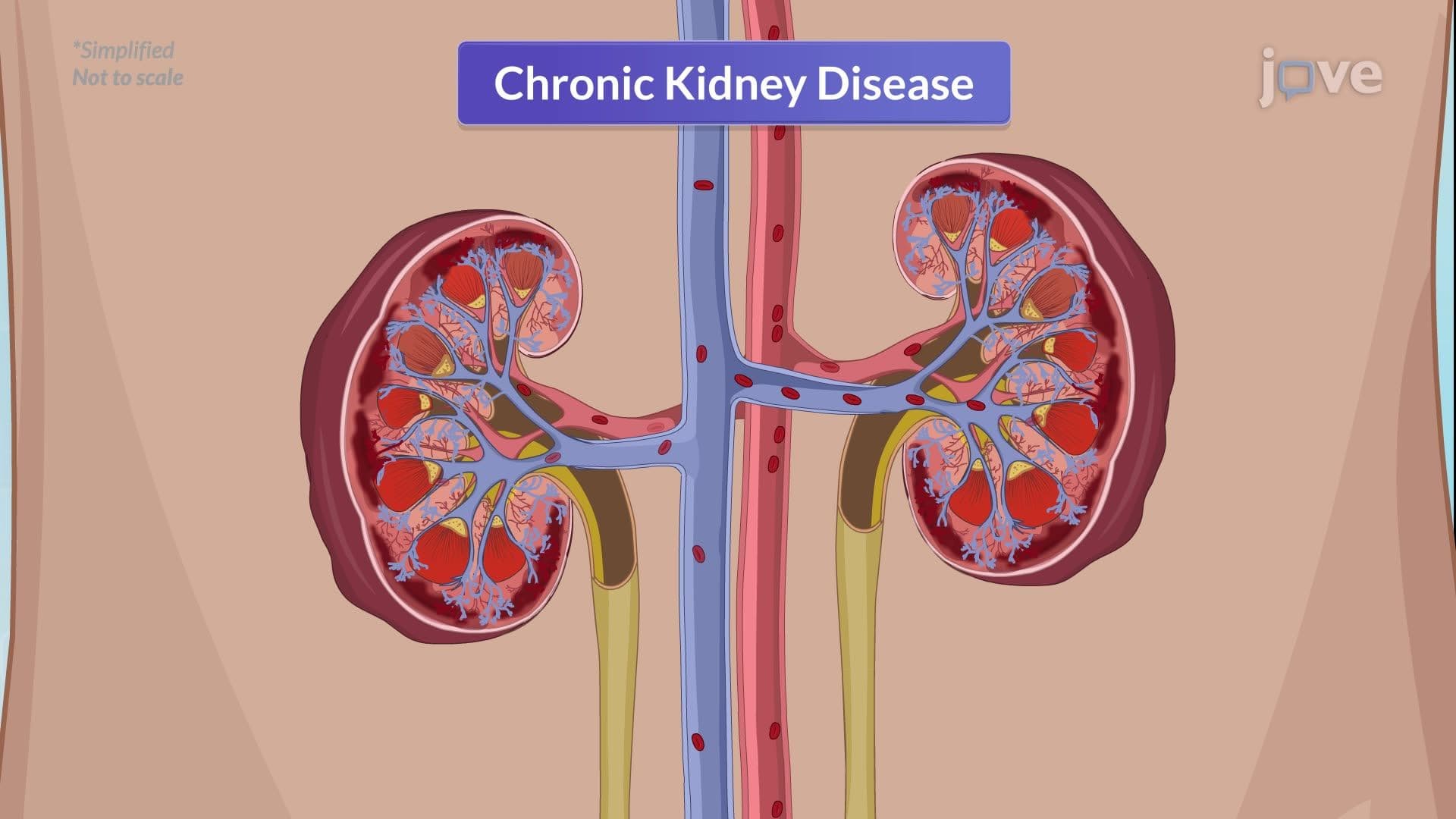
The Acute Kidney Injury IV Diagnostic Explained approach represents a systematic methodology that healthcare providers use to rapidly identify and categorize kidney dysfunction. This diagnostic framework has become essential in US hospitals, where AKI contributes to approximately 300,000 deaths annually. The systematic approach divides potential causes into three distinct categories: prerenal (blood flow issues), intrarenal (kidney tissue damage), and postrenal (urinary obstruction).
Serum creatinine elevation serves as the primary diagnostic marker, though it may lag behind actual kidney damage by 24-48 hours. Normal creatinine levels range from 0.7-1.3 mg/dL, with increases of 0.3 mg/dL or 50% above baseline indicating AKI. Urine output monitoring provides earlier detection, with oliguria (less than 400 mL/day) often preceding creatinine changes. At Cleveland Clinic, nephrologists emphasize that combining both markers increases diagnostic accuracy by 40% compared to using either alone.
The acute kidney injury iv diagnostic tutorial emphasizes urinalysis as a critical differentiation tool. Prerenal AKI typically shows concentrated urine with normal sediment, reflecting the kidney's attempt to conserve fluid. Conversely, intrarenal AKI reveals abnormal findings: muddy brown casts indicate acute tubular necrosis, red blood cell casts suggest glomerulonephritis, and white blood cell casts point to interstitial nephritis. These patterns help clinicians at institutions like Johns Hopkins rapidly determine treatment strategies.
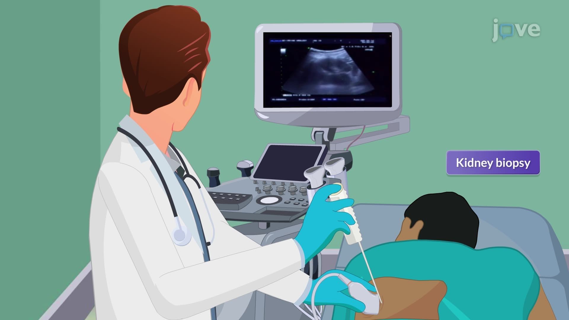
Renal ultrasound serves as the first-line imaging study, identifying hydronephrosis or structural abnormalities without contrast exposure. CT scans may be necessary for detailed evaluation but require careful consideration of contrast-induced nephropathy risk. Prevention strategies have proven highly effective in US healthcare settings, with IV normal saline hydration reducing contrast-induced AKI by up to 60%. The acute kidney injury iv diagnostic study guide emphasizes monitoring high-risk populations, particularly patients with baseline kidney disease, diabetes mellitus, and congestive heart failure who undergo procedures at major medical centers nationwide.
Related Micro-courses