Loading video…


 11 Concepts
 16.4K views


Video Summary: Physical Assessment Respiratory Tract I Explained
Did you know that a single respiratory assessment can reveal everything from hidden asthma triggers to early pneumonia signs? Physical assessment respiratory tract I combines systematic patient interviews with hands-on examination techniques to identify breathing disorders before they become critical. Emergency room nurses at Johns Hopkins use these exact methods to quickly assess patients with chest pain or shortness of breath. Watch the full video on JoVE Coach to master this concept with expert-led visuals and step-by-step explanations.
Physical assessment respiratory tract I represents the cornerstone of respiratory care, combining systematic data gathering with clinical examination skills. This assessment framework divides information collection into two essential categories: subjective data from patient interviews and objective data from physical examination and diagnostic testing.
Healthcare providers across the United States, from Mayo Clinic to community hospitals, rely on this structured approach to identify respiratory conditions ranging from seasonal allergies to life-threatening pneumonia. The assessment process begins before any hands-on examination, emphasizing the critical role of thorough patient communication.
The subjective component focuses on gathering comprehensive patient history through targeted questioning. Healthcare providers explore three main areas: past respiratory issues, medical history, and lifestyle factors. Past respiratory problems include recurrent sore throats, seasonal allergies, asthma episodes, and previous pneumonia diagnoses. Understanding frequency patterns and specific triggers helps clinicians predict future episodes and recommend preventive measures.
Medical history review encompasses current medications, previous respiratory treatments, and any oxygen therapy requirements. This information proves crucial for students preparing for nursing entrance exams like the HESI A2 or TEAS, where understanding medication interactions and treatment protocols frequently appears in test questions.
The objective assessment phase involves systematic physical examination and diagnostic interpretation. Vital sign assessment provides immediate insights into respiratory function, with abnormal respiratory rates or oxygen saturation levels indicating potential problems. Students studying for the MCAT or preparing for pre-health college courses should note how these measurements correlate with overall cardiovascular health.
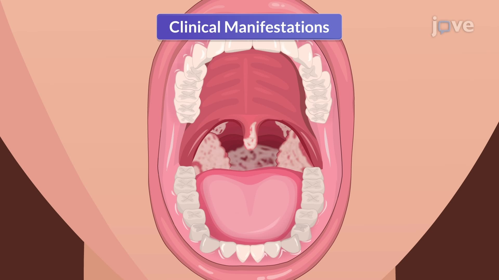
Physical examination proceeds systematically from the nasal cavity to the thoracic region. Nasal and throat inspection reveals inflammation, obstruction, or structural abnormalities that might compromise breathing. Tracheal palpation assesses positioning and mobility, detecting deviations that suggest underlying pathology.
Thoracic inspection evaluates chest symmetry, identifies skin lesions, and detects structural deformities like barrel chest associated with chronic lung disease. Sinus palpation over frontal and maxillary areas reveals tenderness indicating sinusitis, a common condition affecting millions of Americans annually.
The assessment concludes with hypoxia evaluation through cyanosis observation. This critical skill appears frequently on NCLEX examinations and college nursing program assessments. Recognition of central versus peripheral cyanosis helps differentiate cardiac from respiratory causes of oxygen deficiency.
For students preparing for AP Biology or college anatomy courses, understanding how these assessment techniques connect to respiratory physiology concepts enhances both exam performance and clinical application knowledge.
Related Micro-courses