Loading subject page…


78,050 views

Video Summary: The Structure of Intermediate Filaments Explained
Did you know that the protein structure keeping your skin cells intact is the same type found in your neurons? The structure of intermediate filaments reveals how these remarkable cytoskeletal proteins maintain cellular integrity through their unique rope-like architecture. In diseases like epidermolysis bullosa, mutations in intermediate filament proteins cause skin to blister at the slightest touch, demonstrating their critical structural role. These filaments get their name from being intermediate in size-about 10 nanometers wide-positioned between thin microfilaments and thick microtubules. Watch the full video on JoVE Coach to master this concept with expert-led visuals and step-by-step explanations.
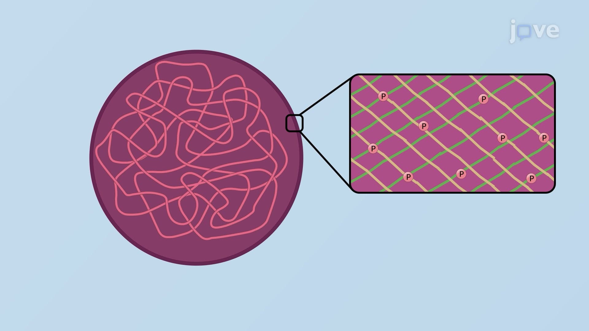
The structure of intermediate filaments represents one of biology's most elegant examples of how protein architecture determines cellular function. These cytoskeletal components derive their name from their intermediate diameter of approximately 10 nanometers, positioning them between the 7-nanometer microfilaments (actin) and 25-nanometer microtubules in the cellular structural hierarchy.
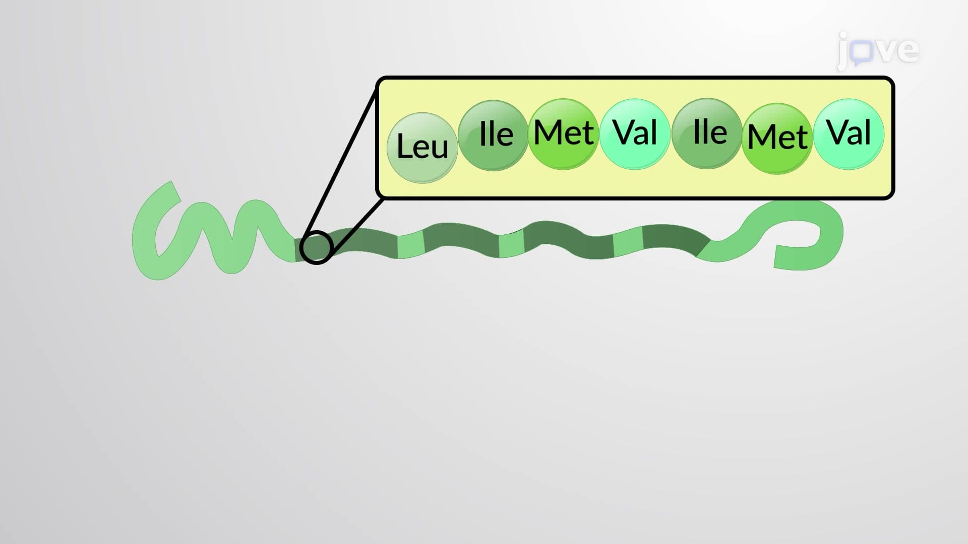
The fundamental building block of intermediate filaments is a protein monomer with three distinct structural regions. The central alpha-helical rod domain spans approximately 310 amino acids and forms the structural backbone of the filament. This rod contains characteristic heptad repeats-sequences of seven amino acids where positions 1 and 4 are typically occupied by hydrophobic residues like leucine, isoleucine, methionine, or valine. This hydrophobic core drives the coiled-coil formation essential for filament stability.
The N-terminal head domain and C-terminal tail domain flank the rod region and vary significantly between different intermediate filament types. In keratin proteins found in human skin and hair, these domains can extend for hundreds of amino acids and contain specific binding sites for cellular regulatory proteins. Students preparing for the AP Biology exam should note that this structure-function relationship exemplifies how protein domains enable specific molecular interactions.
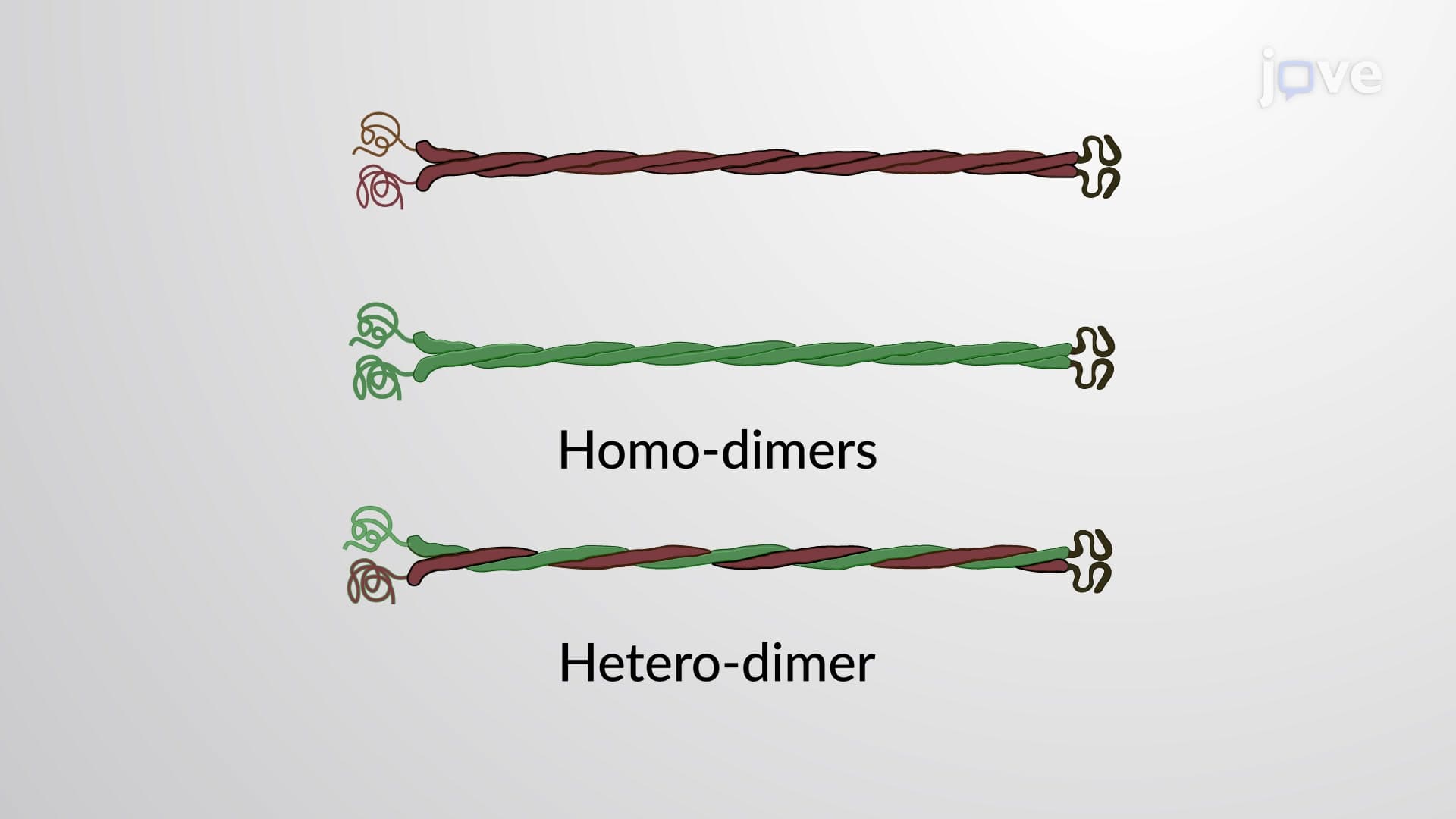
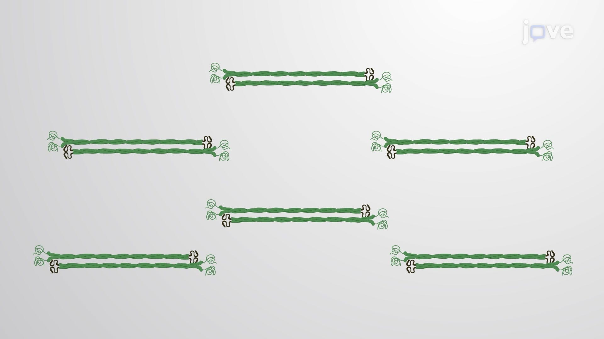
Intermediate filament assembly follows a precise hierarchical process crucial for understanding cellular organization. Two monomers first associate through their rod domains to form a parallel dimer, with their alpha-helices wrapping around each other in a coiled-coil configuration. Two dimers then align in an antiparallel arrangement to create a tetramer, the basic soluble unit of intermediate filament assembly.
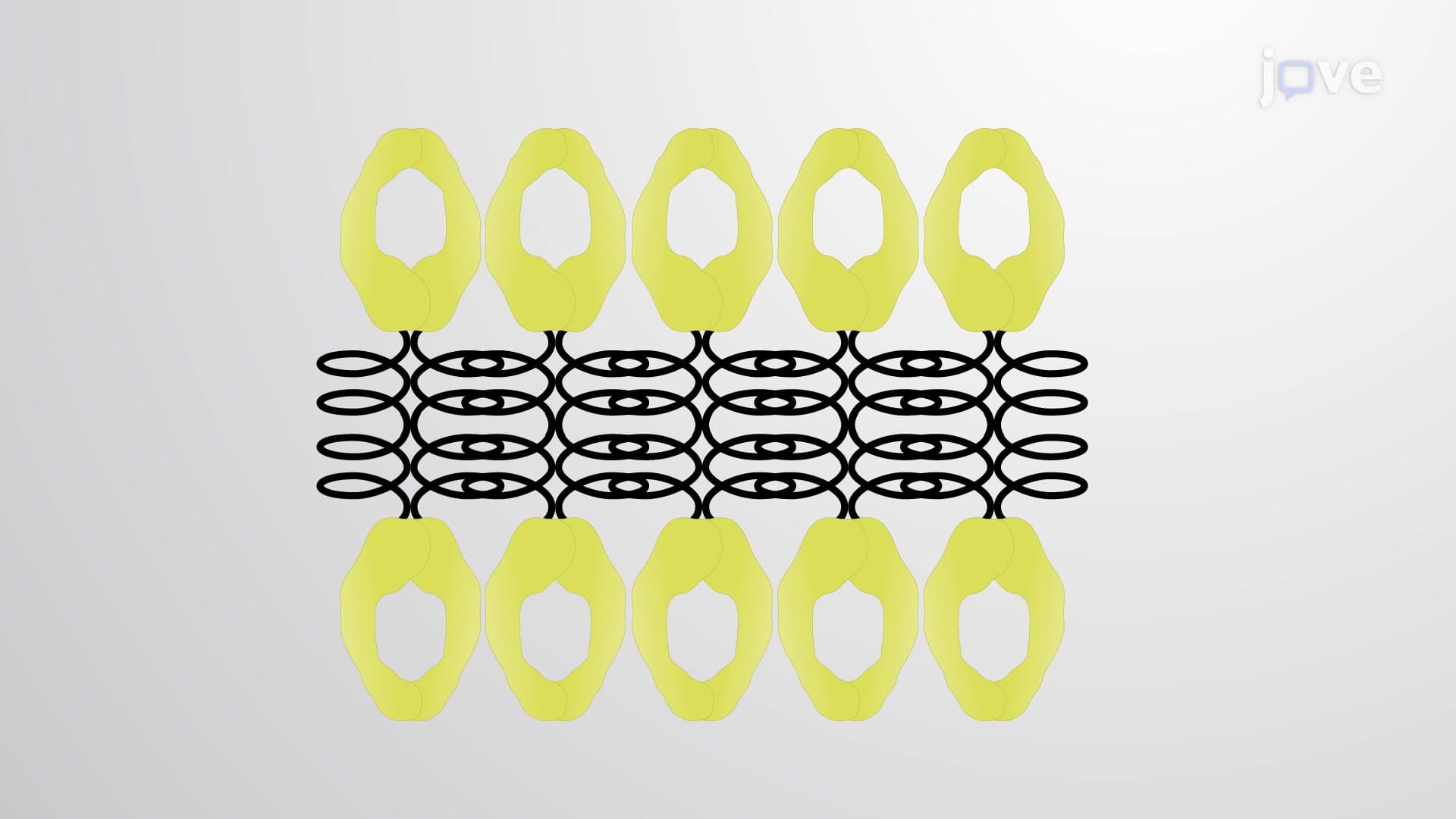
Eight tetramers associate laterally to form a unit-length filament approximately 60 nanometers long. These unit-length filaments then undergo end-to-end annealing and additional lateral associations to generate the mature intermediate filaments that can span the entire cell diameter. This assembly process differs significantly from actin and tubulin polymerization, making it a frequent topic on MCAT biochemistry sections.
Understanding intermediate filament structure has profound medical implications. Mutations in keratin genes cause epidermolysis bullosa simplex, a condition affecting approximately 1 in 50,000 Americans where skin blisters form from minor trauma. Research at institutions like Johns Hopkins University has shown that specific mutations in the rod domain's hydrophobic core disrupt filament assembly, compromising cellular mechanical strength.
Similarly, mutations in neurofilament proteins contribute to neurodegenerative diseases like amyotrophic lateral sclerosis (ALS), affecting roughly 16,000 Americans annually. The Mayo Clinic's research demonstrates how disrupted intermediate filament organization in motor neurons leads to cellular dysfunction and eventual cell death.
Related Micro-courses