Loading video…


 20 Concepts
 158.1K views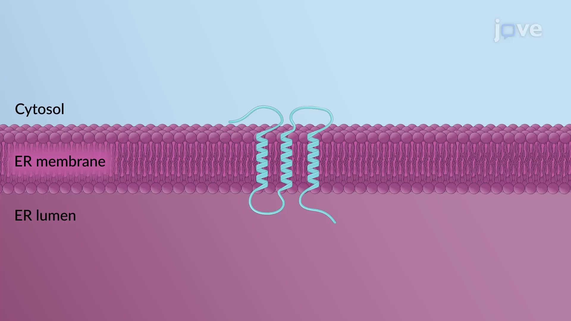

Video Summary: Insertion of Multi Pass Transmembrane Explained
Ever wondered how your cell phone GPS proteins navigate through cellular membranes? The insertion of multi pass transmembrane proteins is like threading a complex rope through multiple holes in a sheet, creating essential communication channels in cells. For instance, the CFTR protein defective in cystic fibrosis patients undergoes this precise insertion process at Johns Hopkins Hospital research labs. This intricate molecular assembly involves start-transfer signals, translocon machinery, and sequential membrane integration steps. Watch the full video on JoVE Coach to master this concept with expert-led visuals and step-by-step explanations.
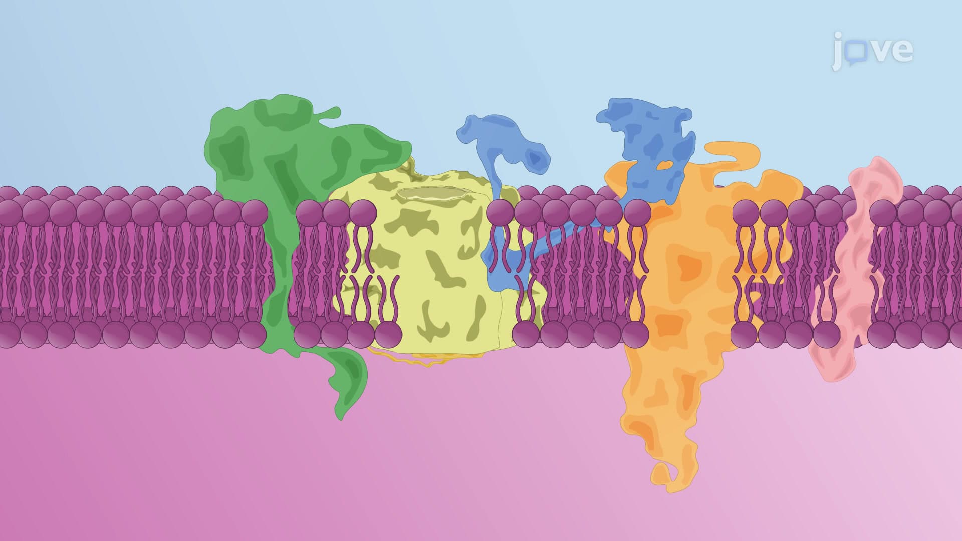
The insertion of multi pass transmembrane proteins represents one of cellular biology's most sophisticated assembly processes. Unlike single-pass proteins that cross membranes once, multipass proteins contain multiple hydrophobic domains that must be precisely positioned within lipid bilayers. This process is crucial for creating functional membrane proteins like ion channels, transporters, and receptors that maintain cellular homeostasis.
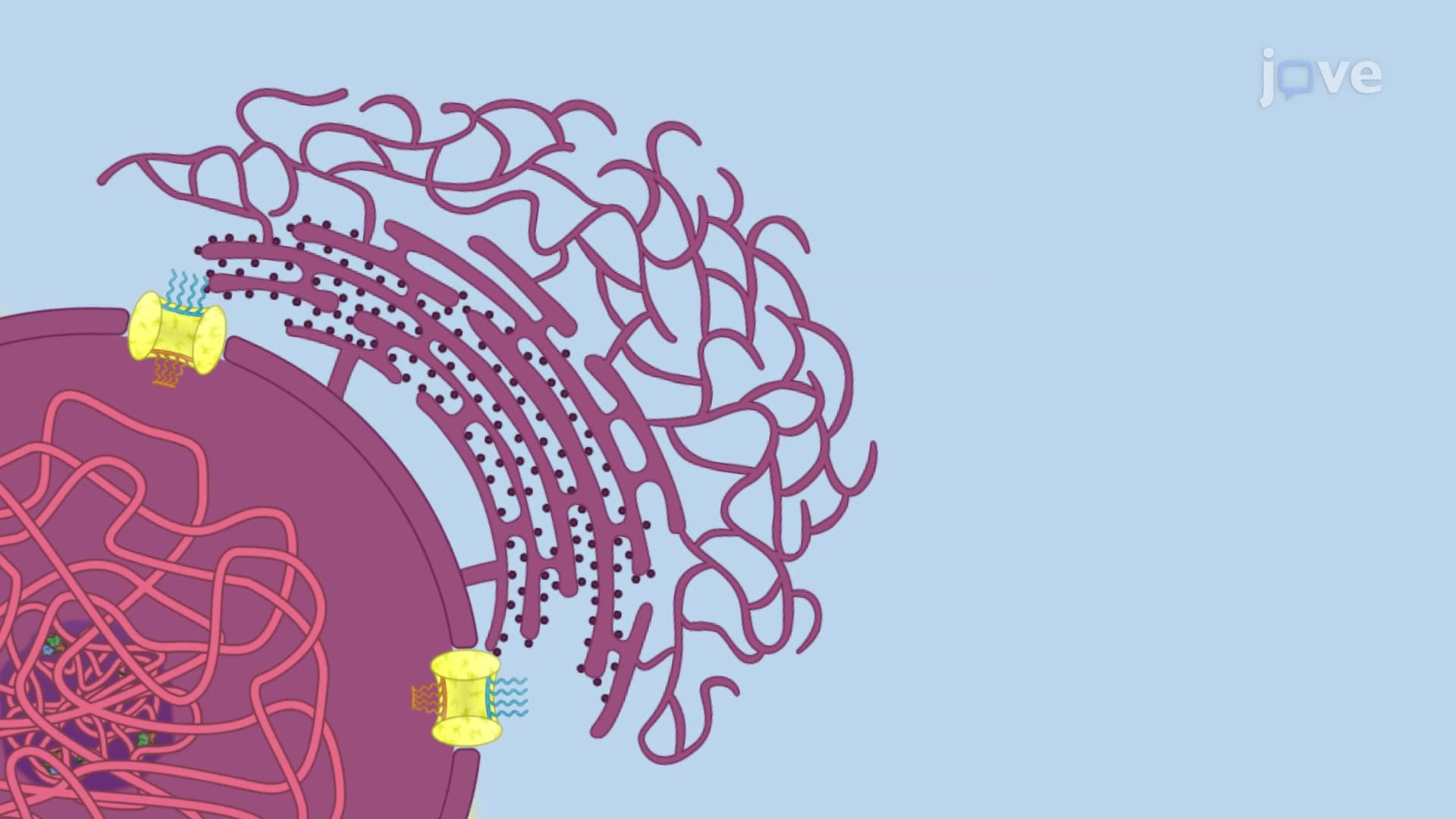
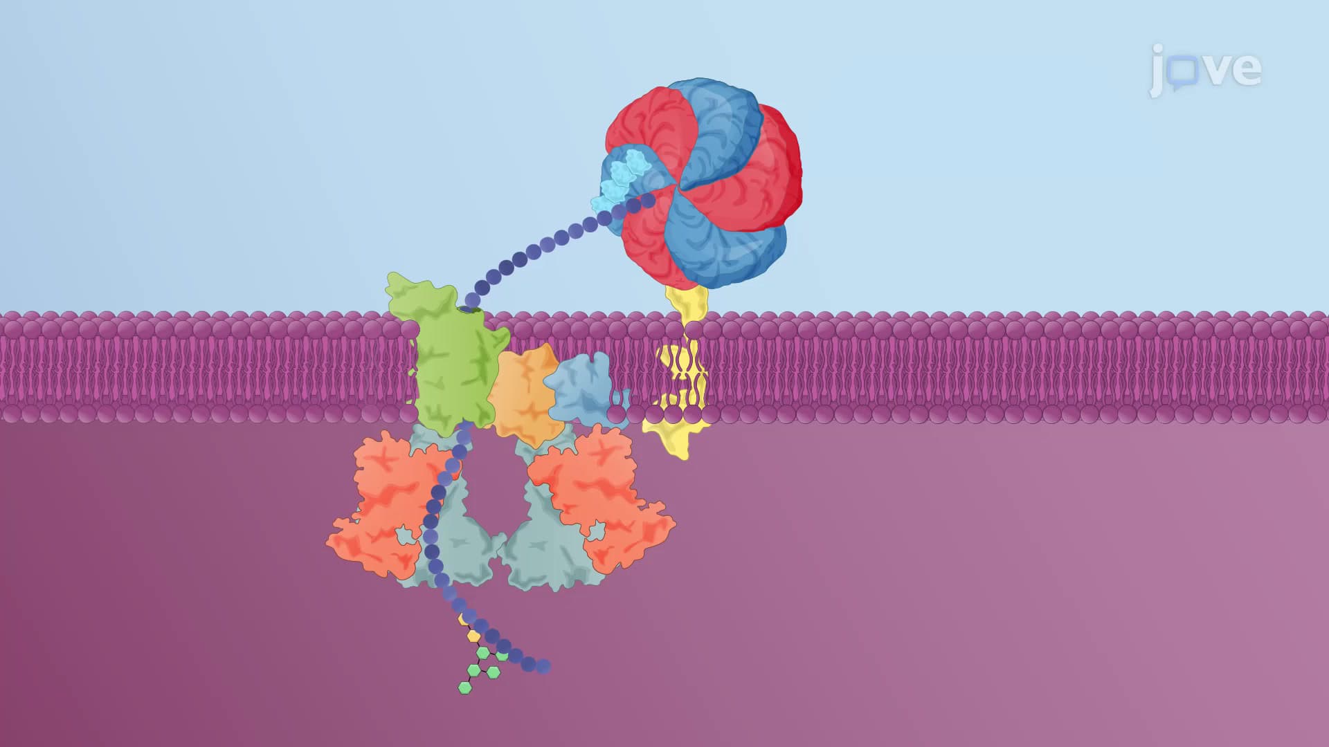
The endoplasmic reticulum (ER) serves as the primary site for transmembrane protein insertion. The process begins when ribosomes synthesizing multipass proteins recognize N-terminal signal sequences. These sequences act as molecular ZIP codes, directing the ribosome-nascent chain complex to ER-bound translocons. The Sec61 translocon complex functions as a dynamic protein-conducting channel, capable of both translocation and lateral membrane insertion.
Signal recognition particles (SRPs) first identify the signal sequence, halting translation until the ribosome docks with the ER membrane. This co-translational process ensures proper protein folding and membrane integration. Students preparing for the MCAT or AP Biology exams should note that this mechanism prevents misfolded proteins from accumulating in the cytoplasm.
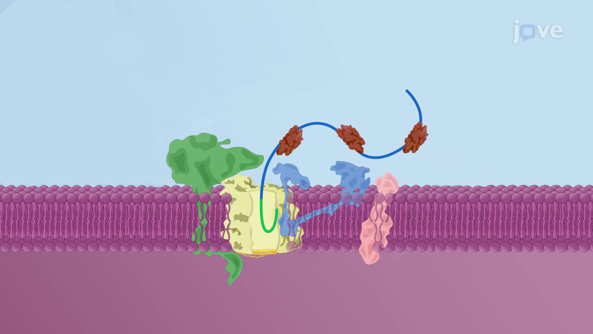
The insertion process follows a predictable pattern for proteins with multiple transmembrane domains. After the initial signal sequence guides the polypeptide into the translocon, each hydrophobic region triggers specific responses. When the translocon encounters a transmembrane domain, it temporarily halts translocation and opens its lateral gate, allowing the hydrophobic segment to partition into the lipid bilayer.
This stop-and-go mechanism creates alternating membrane-embedded and soluble domains. For example, G-protein coupled receptors (GPCRs) like those targeted by FDA-approved medications undergo this precise insertion process. The resulting protein topology directly influences pharmaceutical drug interactions studied in medical schools across institutions like Harvard Medical School and Mayo Clinic.
The final orientation of multipass transmembrane proteins follows predictable rules that appear frequently on standardized exams. Proteins with odd numbers of transmembrane domains position their N- and C-termini on opposite membrane sides, while even-numbered domain proteins align both termini on the same side. This topology principle helps predict protein function and drug binding sites.
Understanding these concepts proves essential for students pursuing healthcare careers. The CFTR chloride channel, defective in cystic fibrosis patients treated at centers like Cincinnati Children's Hospital, exemplifies how insertion defects cause disease. Similarly, insulin receptor insertion errors contribute to diabetes complications studied in endocrinology programs nationwide.
Related Micro-courses