Loading subject page…


52,500 views


Video Summary: What Is Computed Tomography
Ever wondered how doctors can see a blood clot deep inside your brain without surgery? Computed tomography explained reveals this remarkable imaging technology that creates detailed 3D pictures from X-ray slices. At Massachusetts General Hospital, CT scanners help diagnose everything from stroke patients in the ER to athletes with sports injuries. What is Computed Tomography transforms traditional X-rays into powerful diagnostic tools by rotating around your body and capturing cross-sectional images just millimeters thick. Watch the full video on JoVE Coach to master this concept with expert-led visuals and step-by-step explanations.
What is computed tomography in medical imaging represents one of the most significant advances in diagnostic technology since the discovery of X-rays. Unlike traditional radiography that produces flat, two-dimensional images where structures overlap and obscure each other, CT technology creates detailed cross-sectional views that reveal internal anatomy with remarkable clarity. This computed tomography principle has transformed medical diagnosis across virtually every specialty.
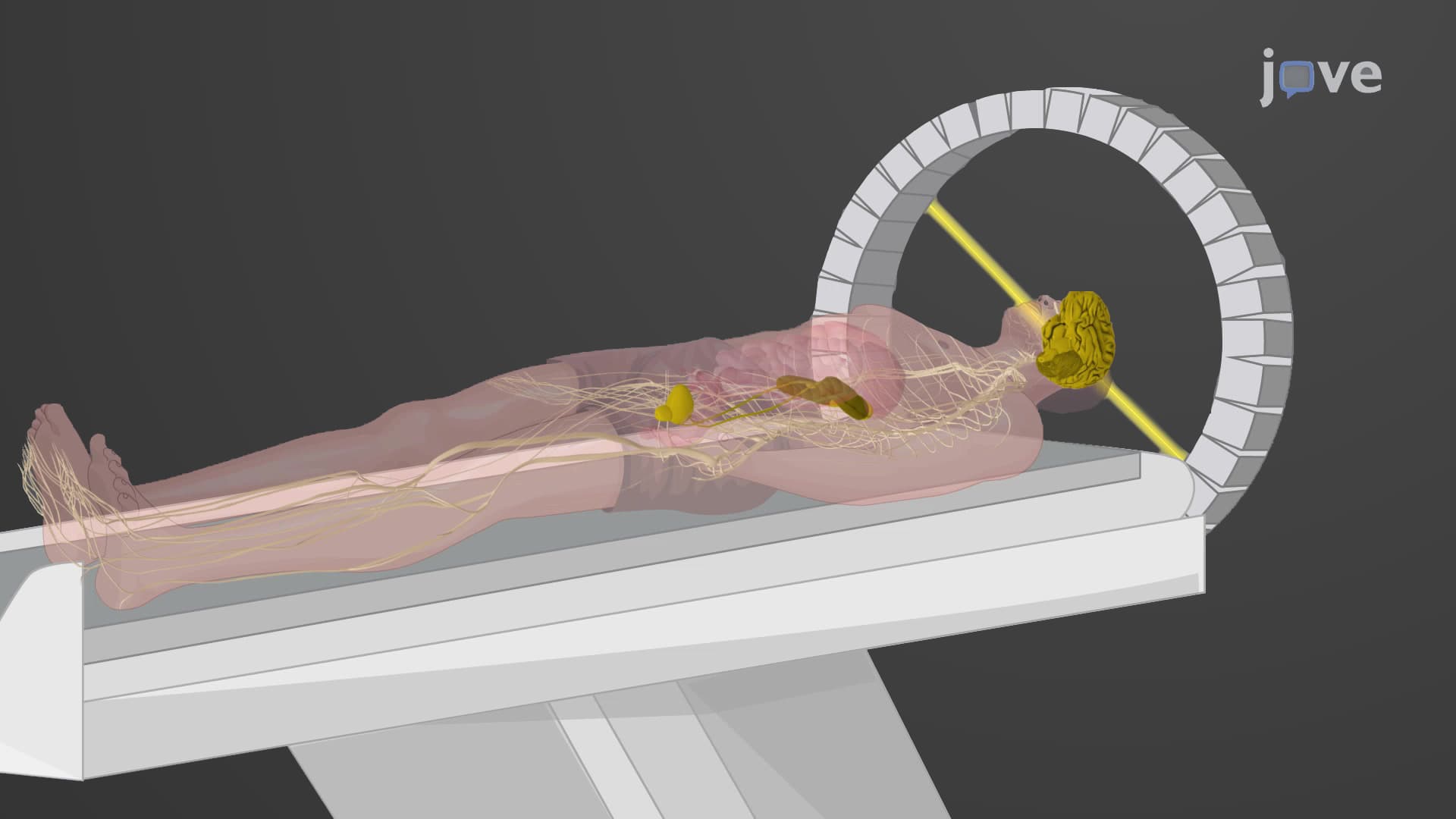
The CT imaging technique operates on an elegant principle: an X-ray tube rotates 360 degrees around the patient while detectors on the opposite side measure how much radiation passes through different tissues. As the tube spins, it captures thousands of measurements from multiple angles. Think of it like photographing a loaf of bread from every possible angle, then using those images to reconstruct what each individual slice looks like inside.
CT scanner components work in perfect coordination to achieve this feat. The gantry houses the rotating X-ray tube and detector array, while sophisticated computers process the massive amounts of data collected. Modern multi-detector CT scanners at hospitals like Cleveland Clinic can capture up to 320 slices simultaneously, allowing complete heart scans in just a few heartbeats.
CT Hounsfield units provide the quantitative foundation for image interpretation. Named after Sir Godfrey Hounsfield, who won the Nobel Prize for inventing CT scanning, this scale assigns numerical values to different tissue densities. Water measures exactly 0 HU, air reads -1000 HU, while dense bone can exceed +1000 HU. This standardization allows radiologists worldwide to communicate precise findings.
For students preparing for the MCAT or pursuing pre-med coursework, understanding Hounsfield units proves essential. Emergency room physicians at Johns Hopkins rely on these measurements to quickly distinguish between blood (50-90 HU) and brain tissue (20-40 HU) when evaluating stroke patients, where every minute counts.
CT scan imaging basics extend far beyond simple anatomy visualization. In trauma centers across the United States, whole-body CT scans can be completed in under 30 seconds, providing life-saving information about internal bleeding, fractures, and organ damage. Oncologists use contrast-enhanced CT to track tumor response to treatment, while cardiologists employ specialized cardiac CT to detect coronary artery disease non-invasively.
Students studying for AP Biology or college anatomy courses should appreciate how CT technology reveals anatomical relationships impossible to observe otherwise. The ability to "slice" through the human body digitally provides insights into spatial relationships between organs, blood vessels, and pathological processes that textbook diagrams cannot adequately convey.
Related Micro-courses