Loading video…


 6 Concepts
 52.5K views

Video Summary: What Is Magnetic Resonance Imaging
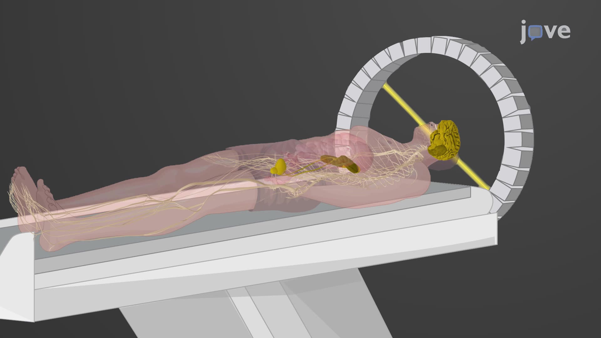
Did you know that magnetic resonance imaging basics rely on the hydrogen atoms in your body acting like tiny magnets? Every time you get an MRI at a hospital like Mayo Clinic, powerful magnetic fields align billions of these atoms before radio waves flip them to create detailed images of your internal structures. What is Magnetic Resonance Imaging reveals how this non-invasive technology revolutionized medical diagnosis by providing crystal-clear views of soft tissues that X-rays simply cannot detect. Watch the full video on JoVE Coach to master this concept with expert-led visuals and step-by-step explanations.
Magnetic resonance imaging basics center on a fascinating physics principle: your body is approximately 60% water, and each water molecule contains hydrogen atoms that behave like microscopic magnets. When placed in an MRI scanner's powerful magnetic field-typically 1.5 to 3 Tesla (30,000 times stronger than Earth's magnetic field)-these hydrogen nuclei align like compass needles pointing north.
The MRI scan explained simply involves three key steps. First, the magnetic field aligns hydrogen protons in your tissues. Second, radiofrequency pulses knock these protons out of alignment, causing them to absorb energy. Third, when the radio waves stop, protons "relax" back to their original position, releasing energy that MRI detectors capture to construct images.
This process, rooted in nuclear magnetic resonance (NMR) physics, allows radiologists at institutions like Johns Hopkins to distinguish between different tissue types based on their unique relaxation patterns. What is magnetic resonance imaging in medicine becomes clear when you realize that diseased tissues often have different water content and molecular environments than healthy tissues, making abnormalities visible on MRI scans.
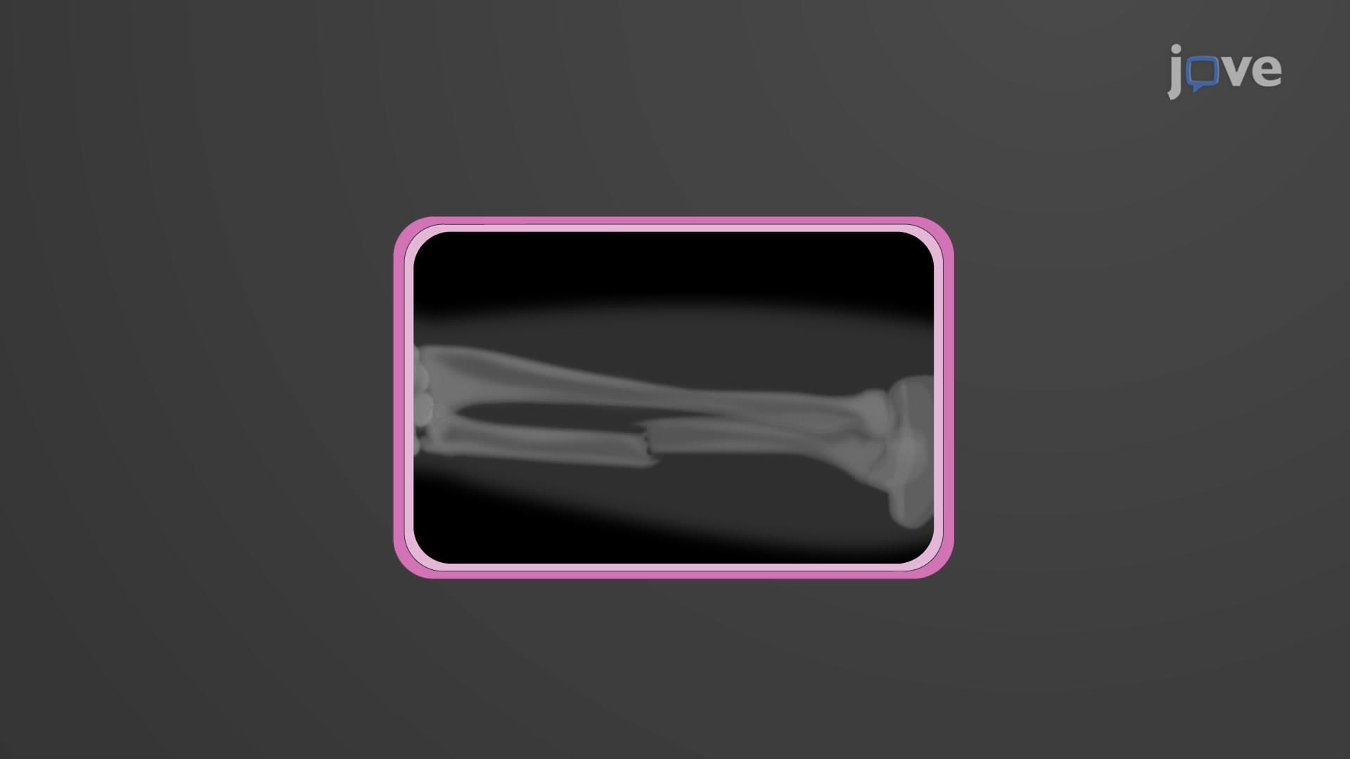
MRI excels in MRI soft tissue imaging, making it invaluable for diagnosing conditions invisible to X-rays. Neurologists at Cleveland Clinic rely on MRI to detect brain tumors, multiple sclerosis lesions, and stroke damage. Orthopedic surgeons use MRI to visualize torn ACL ligaments in athletes-something impossible with traditional radiography.
The T1 T2 MRI contrast mechanisms provide different types of tissue information. T1-weighted images excel at showing anatomy and fat, while T2-weighted images highlight fluid and inflammation. Gadolinium contrast agents, approved by the FDA, enhance blood vessels and help identify tumors by highlighting areas where the blood-brain barrier is compromised.
Students preparing for the MCAT will encounter MRI physics in their physical sciences section, particularly regarding electromagnetic radiation and nuclear physics concepts. AP Physics students should understand how MRI magnetic field interactions demonstrate real-world applications of electromagnetism. Pre-med students taking anatomy courses benefit from understanding how MRI's multiplanar imaging capabilities (axial, sagittal, coronal views) help visualize three-dimensional anatomical relationships that textbook diagrams cannot fully convey.
Related Micro-courses