Loading subject page…


52,500 views

Video Summary: What Is Positron Emission Tomography
Did you know that doctors can actually watch your body's cells consume sugar in real-time? Positron emission tomography explained reveals how this revolutionary medical imaging technique uses radioactive tracers to detect diseases like cancer before they're visible on traditional X-rays. At Mayo Clinic, PET scans help oncologists identify tumors as small as 4mm by tracking abnormal cellular metabolism. What is positron emission tomography becomes clear when you understand it captures the body's biochemical processes, not just anatomical structures. Watch the full video on JoVE Coach to master this concept with expert-led visuals and step-by-step explanations.
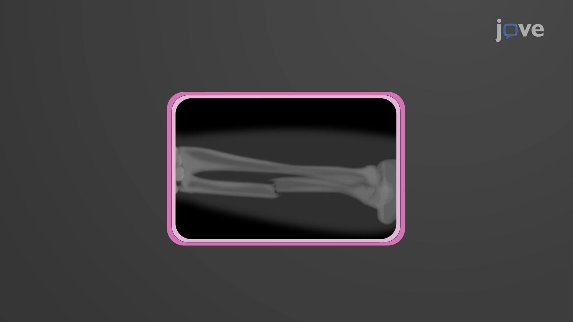
Positron emission tomography explained begins with understanding its unique approach to medical imaging. Unlike X-rays or CT scans that show anatomical structures, PET reveals metabolic activity at the cellular level. This functional imaging capability makes it invaluable for detecting diseases in their earliest stages, often before structural changes occur.
The technology relies on radioactive tracers-biological molecules tagged with positron-emitting isotopes. These tracers are designed to mimic natural substances your body uses, allowing physicians to track specific biological processes. PET scan imaging basics center on the principle that diseased tissues often have altered metabolism compared to healthy tissues.
PET scan radiotracer molecules contain unstable isotopes like fluorine-18, carbon-11, or oxygen-15. The most commonly used tracer is fluorodeoxyglucose (FDG), which contains fluorine-18. FDG mimics glucose, the primary fuel for cellular energy production.
When these unstable isotopes decay, they emit positrons-the antimatter counterparts of electrons. Within millimeters of emission, each positron encounters an electron, resulting in mutual annihilation. This reaction converts both particles into pure energy, producing two high-energy photons (511 keV each) that travel in exactly opposite directions at the speed of light.
FDG PET imaging exploits a fundamental characteristic of cancer cells: they consume glucose at rates 10-20 times higher than normal cells. This hypermetabolism makes tumors appear as "hot spots" on PET images. At institutions like MD Anderson Cancer Center, oncologists use PET scans to stage cancers, monitor treatment response, and detect recurrence.
Beyond oncology, PET functional imaging serves crucial roles in cardiology and neurology. Cardiologists use it to assess heart muscle viability after heart attacks, while neurologists employ specialized tracers to study Alzheimer's disease progression and epilepsy.
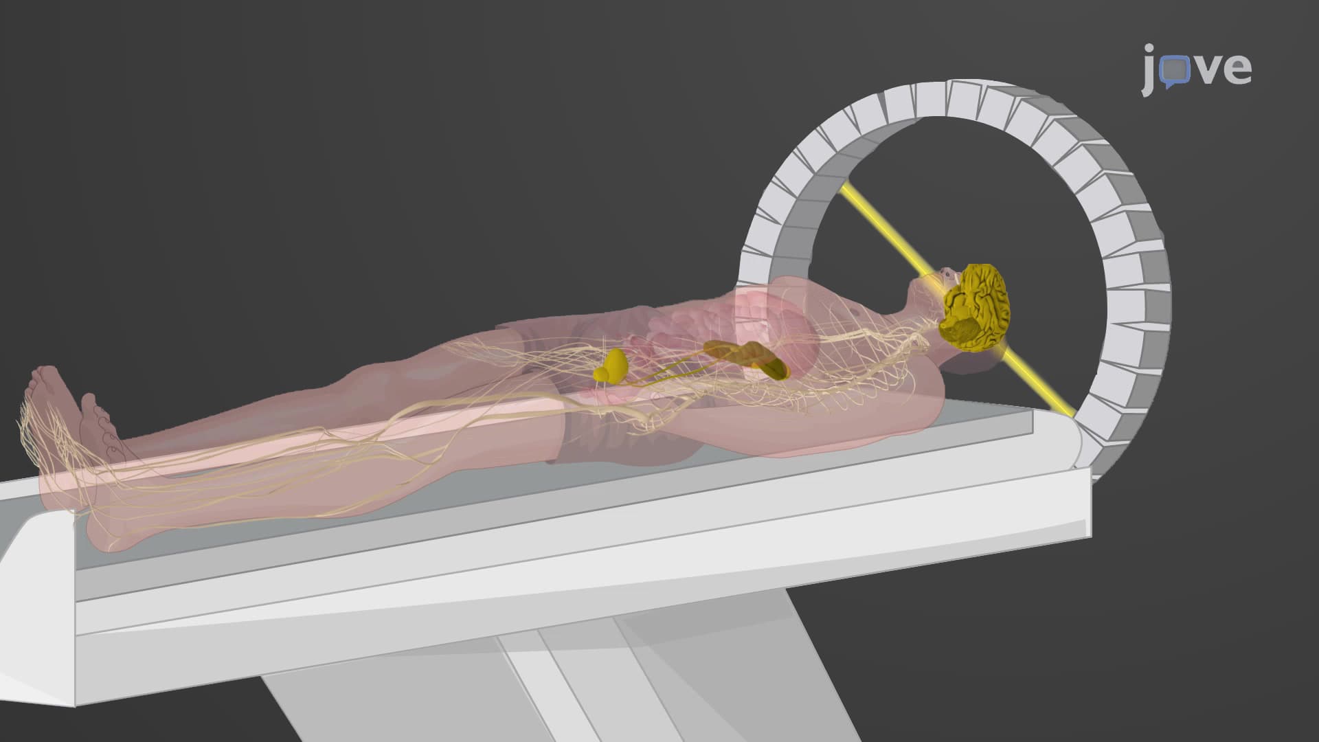
The PET scanner contains thousands of detector crystals arranged in rings around the patient. When the annihilation photons strike these detectors simultaneously, sophisticated algorithms calculate the exact location where the annihilation occurred. Millions of these "coincidence events" are processed to create detailed three-dimensional images.
For students preparing for the MCAT or advanced placement biology exams, understanding these physics principles is crucial. The 511 keV photon energy and 180-degree emission pattern are frequently tested concepts that demonstrate practical applications of Einstein's mass-energy equivalence (E=mc²).
Related Micro-courses