Loading subject page…


52,500 views

Video Summary: What Is X Ray Imaging
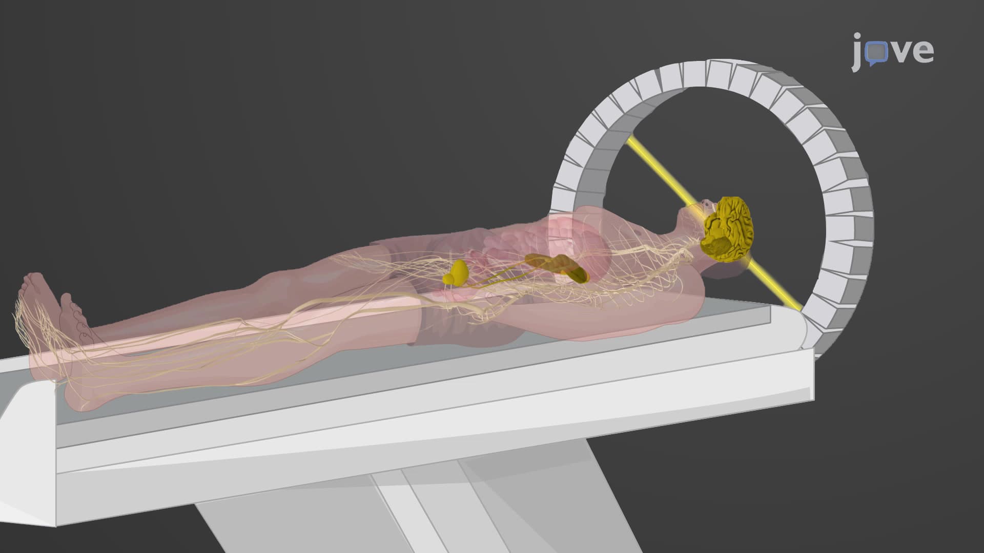
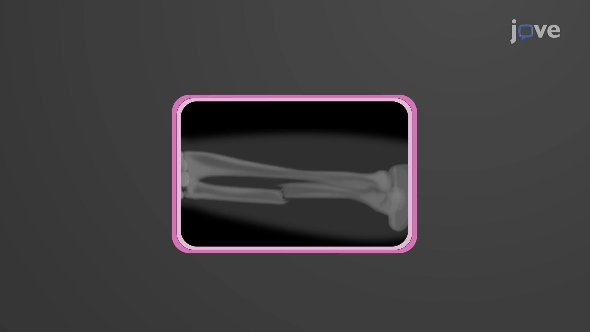
Every time you break a bone playing sports or need a dental checkup, chances are you'll encounter X ray imaging basics in action. This fundamental diagnostic technique uses high-energy electromagnetic radiation to create detailed images of your internal structures, helping doctors at hospitals like Mayo Clinic and Johns Hopkins diagnose everything from fractures to infections. What is X Ray Imaging reveals how dense tissues like bones appear white while soft tissues show up in gray shades on the resulting radiograph. Watch the full video on JoVE Coach to master this concept with expert-led visuals and step-by-step explanations.
X ray imaging basics form the cornerstone of modern medical diagnostics, representing one of the most widely used imaging techniques in healthcare facilities across the United States. This technology harnesses the power of ionizing radiation to peer inside the human body without invasive procedures, making it invaluable for emergency departments, dental offices, and routine medical examinations.
The principle behind X ray imaging explained medicine lies in the differential absorption of electromagnetic radiation by various body tissues. When X-rays pass through the human body, dense structures like bones and teeth absorb more radiation, creating the characteristic white appearance on radiographs. Meanwhile, soft tissues such as muscles, organs, and blood vessels absorb less radiation, appearing in various shades of gray. Air-filled spaces like the lungs appear black since they offer minimal resistance to X-ray penetration.
Diagnostic X ray technique operates on the fundamental principle that different tissues have varying radiopacities. This concept is crucial for students preparing for advanced coursework in biology, chemistry, or pre-medical studies. The radiograph X ray principle demonstrates how electromagnetic radiation interacts with matter at the atomic level, providing practical applications of physics concepts taught in AP Physics and college-level courses.
Healthcare professionals at institutions like Cleveland Clinic and Massachusetts General Hospital rely on precise X ray image interpretation skills to diagnose conditions ranging from pneumonia to bone fractures. The technology's ability to provide immediate results makes it essential for emergency medicine, where rapid diagnosis can be life-saving.
X ray bone imaging represents the most common application of this technology, particularly in orthopedic medicine and sports medicine. When athletes suffer injuries during high school or college sports, X-rays quickly reveal fractures, dislocations, or other bone abnormalities. This immediate diagnostic capability has revolutionized emergency care in American hospitals.
The controlled use of ionizing radiation X ray technology ensures patient safety while maximizing diagnostic benefits. Modern digital X-ray systems used in facilities like Johns Hopkins Hospital and UCLA Medical Center have significantly reduced radiation exposure compared to traditional film-based methods, addressing safety concerns while maintaining image quality.
Understanding what is X ray imaging in medical diagnostics prepares students for advanced studies in radiology, medical technology, or healthcare administration. This knowledge proves essential for MCAT preparation and provides foundational understanding for various healthcare career paths available in the robust American medical system.
Related Micro-courses