Loading subject page…


654,700 views


Video Summary: What Is Anatomy Circulatory System
Your heart beats 100,000 times daily, pumping 2,000 gallons of blood through a 60,000-mile network of vessels-equivalent to circling Earth twice! Understanding what anatomy circulatory system encompasses reveals how this incredible biological highway delivers oxygen and nutrients to every cell while removing metabolic waste. Consider how athletes at the Boston Marathon rely on their circulatory efficiency to maintain peak performance throughout the 26.2-mile race. Watch the full video on JoVE Coach to master this concept with expert-led visuals and step-by-step explanations.
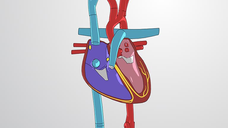
The anatomy circulatory system represents one of biology's most sophisticated transport networks, comprising the heart as a central pump, an extensive vessel system, and blood as the transport medium. This closed-loop system ensures every cell in your body-from brain neurons to muscle fibers-receives essential nutrients and oxygen while removing carbon dioxide and metabolic waste products.
The circulatory system operates through three interconnected circuits, each serving distinct physiological needs. The coronary circuit supplies the heart muscle itself, crucial for maintaining cardiac function. When coronary arteries become blocked, heart attacks occur-a leading cause of death in the United States, affecting over 800,000 Americans annually.
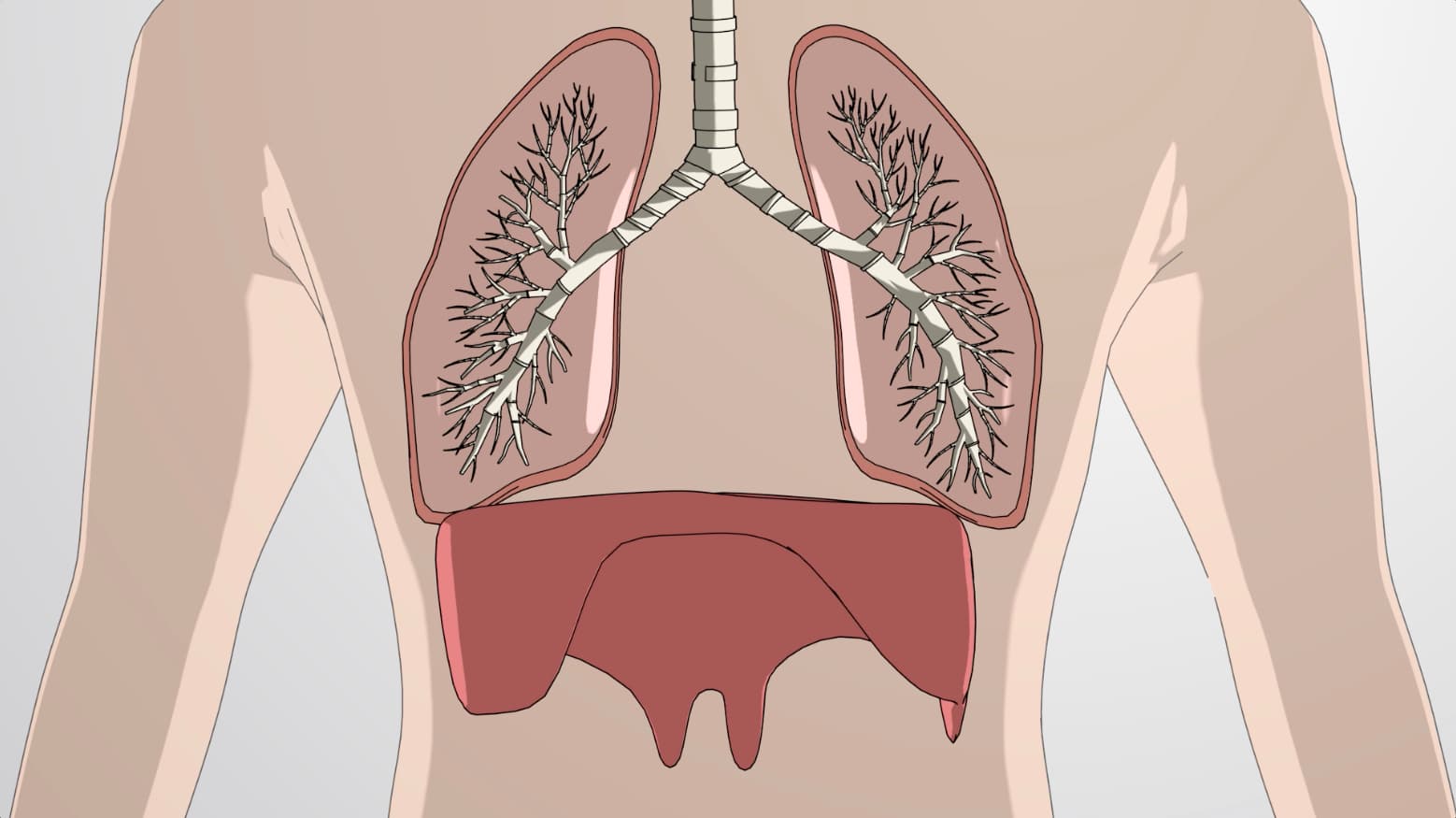
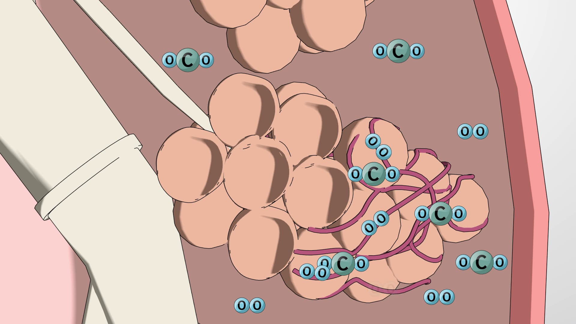
The pulmonary circuit creates a short loop between the heart and lungs, where deoxygenated blood picks up oxygen and releases carbon dioxide. This circuit operates under lower pressure than systemic circulation, explaining why pulmonary vessels are thinner-walled than their systemic counterparts.
The systemic circuit represents the body's major highway system, distributing oxygenated blood from the left ventricle through the aorta to all body tissues. Major arteries branch strategically: carotid arteries supply the brain, brachial arteries serve the arms, and iliac arteries feed the legs. This organization ensures that if one pathway becomes compromised, collateral circulation can often maintain blood flow.
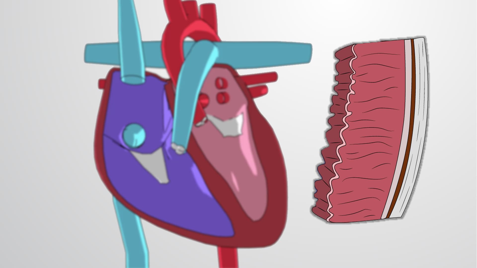
Blood vessels demonstrate remarkable structural diversity reflecting their functions. Arteries feature thick, muscular walls capable of withstanding high pressure from ventricular contractions. The aorta, your body's largest artery, experiences pressures reaching 120 mmHg during systole.
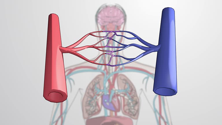
As arteries branch into arterioles, smooth muscle allows precise flow regulation-critical for directing blood to active tissues. During exercise, arterioles in working muscles dilate while those in digestive organs constrict, redistributing blood where needed most.
Capillaries, despite being microscopic, represent the circulatory system's most important component. Their thin walls facilitate rapid nutrient and gas exchange, while their vast network ensures no cell lies more than a few micrometers from a blood supply. In conditions like diabetes, capillary damage leads to serious complications affecting vision, kidney function, and wound healing.
This anatomical understanding proves essential for students preparing for AP Biology exams, MCAT sections on cardiovascular physiology, and nursing school prerequisites. Healthcare professionals regularly apply this knowledge when interpreting blood pressure readings, understanding medication distribution, and recognizing symptoms of circulatory disorders affecting millions of Americans.
Related Micro-courses