Loading concepts…


8 Concepts
21 Concepts
9 Concepts
16 Concepts
16 Concepts
14 Concepts
15 Concepts
14 Concepts
7 Concepts
9 Concepts
5 Concepts
19 Concepts
13 Concepts
12 Concepts
15 Concepts
7 Concepts
8 Concepts
11 Concepts
12 Concepts
11 Concepts
6 Concepts
8 Concepts
8 Concepts
9 Concepts
8 Concepts
10 Concepts
12 Concepts
12 Concepts
11 Concepts
5 Concepts
4 Concepts
5 Concepts
7 Concepts
21 Concepts
7 Concepts
8 Concepts
 8 Concepts
 654.7K Views
 Popular topics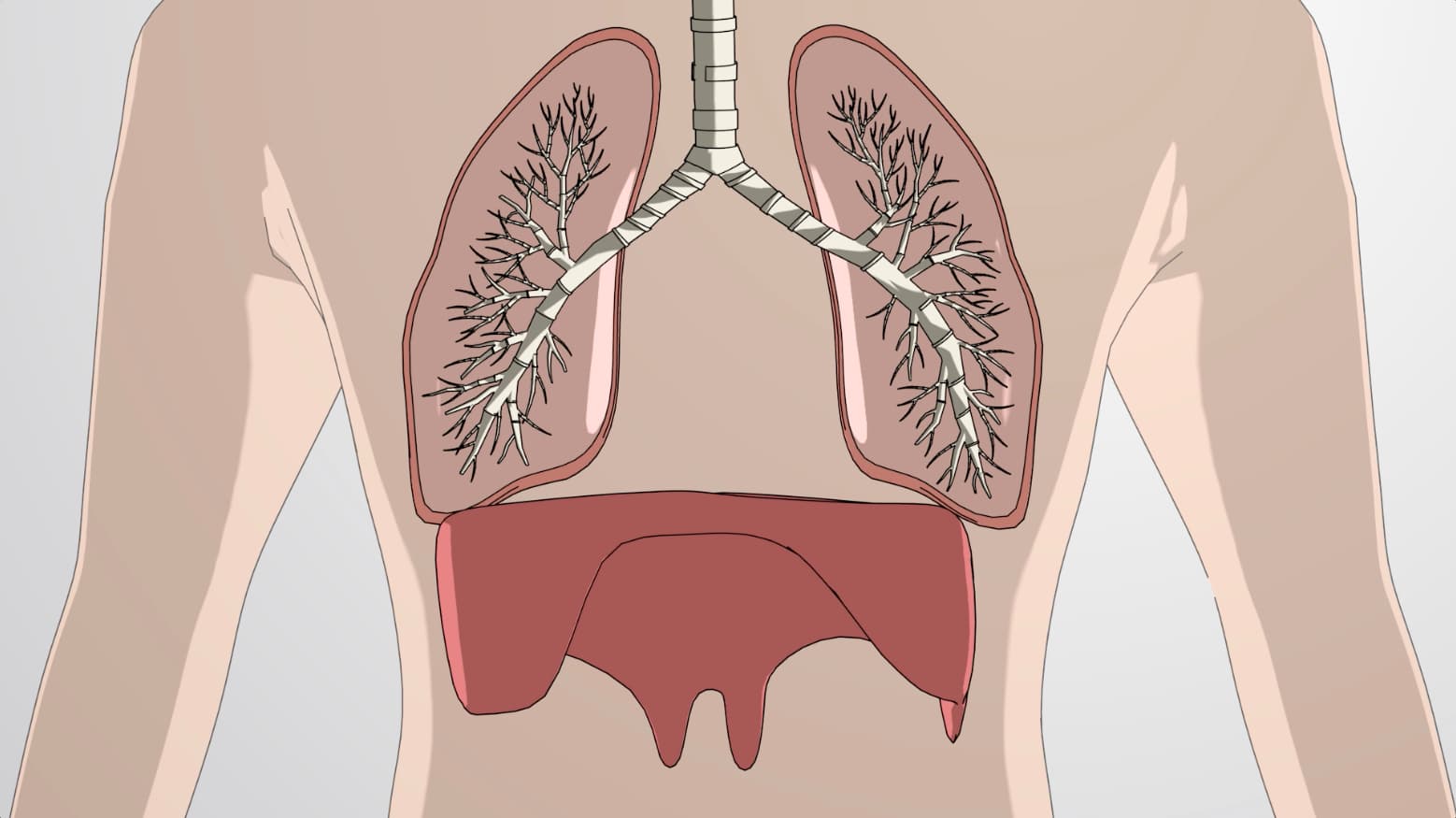



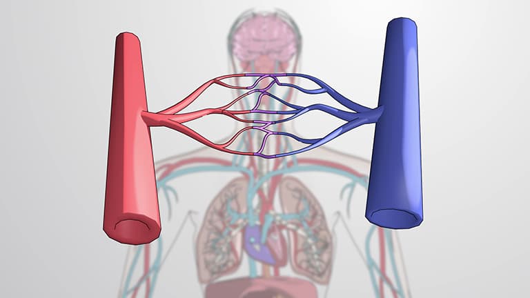
Circulatory systems and respiratory mechanisms work together to deliver oxygen and remove carbon dioxide throughout the human body. This comprehensive course explores heart and lung function, examining how the circulatory and respiratory systems interact through blood circulation, gas exchange, and cardiac cycles. Master these essential physiological processes with JoVE Coach's expert guidance.
1. Respiratory System Architecture and Air Flow Pathways The respiratory system creates an intricate network of airways that condition and transport air to gas exchange sites. Air enters through nasal cavities where mucus and hair filter particles, then travels through the pharynx and larynx into the trachea. The trachea bifurcates into bronchi that branch into progressively smaller bronchioles, ending in alveolar ducts and sacs. This branching pattern, similar to an inverted tree, maximizes surface area for efficient gas exchange. The pleural membranes surrounding each lung create a sealed environment, while the diaphragm forms the thoracic cavity floor. Understanding this anatomy helps explain respiratory diseases like asthma, where bronchiole constriction impairs airflow, commonly seen in American students during allergy seasons.
2. Breathing Mechanics and Pressure Dynamics Breathing relies on coordinated muscle contractions that create pressure gradients driving air movement. During inspiration, the diaphragm and intercostal muscles contract, expanding the thoracic cavity and decreasing internal pressure below atmospheric levels. This pressure differential draws air into the lungs following Boyle's Law principles. Expiration occurs when these muscles relax, reducing cavity volume and increasing pressure to push air out. This mechanical bellows system operates continuously, with typical resting rates of 12-20 breaths per minute in healthy adults. Athletes often demonstrate enhanced breathing efficiency through trained respiratory muscles, particularly relevant for American sports medicine and exercise physiology applications.
3. Lung Volumes, Capacities, and Clinical Applications Pulmonary function testing measures specific air volumes that indicate respiratory health and disease states. Tidal volume represents normal breathing (approximately 500mL), while inspiratory and expiratory reserve volumes show additional air movement capacity. Residual volume prevents lung collapse by maintaining approximately 1,200mL of air. Total lung capacity combines all volumes, averaging 6,000mL in healthy adults. Vital capacity, the maximum air movement in one breath, serves as a key diagnostic measure. These measurements help diagnose conditions like chronic obstructive pulmonary disease (COPD), affecting millions of Americans, particularly those with smoking histories. Spirometry testing in American hospitals and clinics routinely uses these parameters for patient assessment.
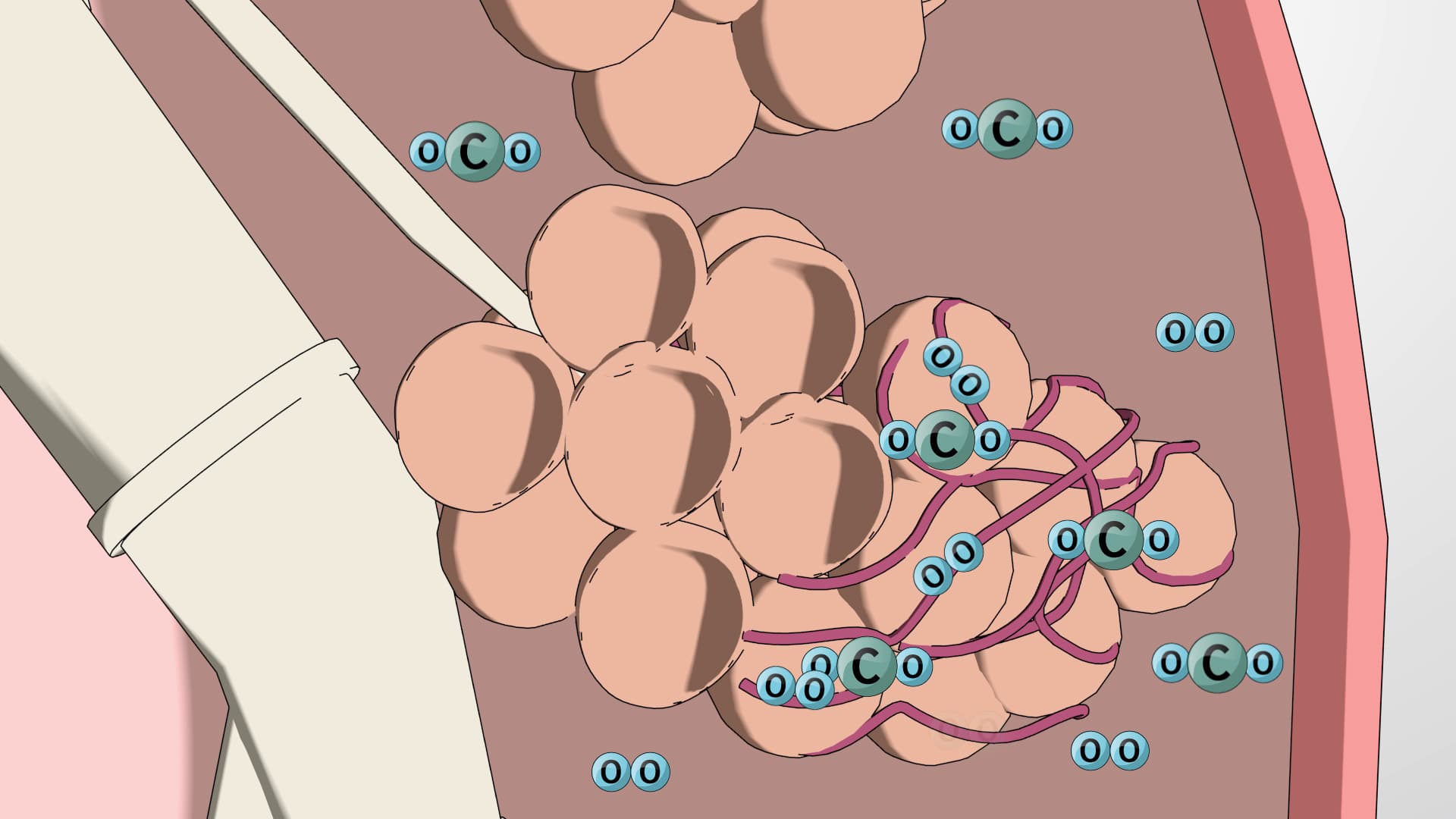
4. Gas Exchange Mechanisms and Hemoglobin Transport Efficient oxygen delivery and carbon dioxide removal depend on partial pressure gradients driving diffusion across respiratory and circulatory interfaces. Oxygen diffuses from alveolar air into pulmonary capillaries, then binds to hemoglobin in red blood cells for transport. At tissue sites, lower oxygen partial pressure causes hemoglobin to release oxygen for cellular use. Carbon dioxide follows the reverse pathway, moving from tissues to blood, then to alveoli for elimination. This process maintains cellular respiration essential for ATP production. Altitude changes, common when Americans travel to locations like Denver (5,280 feet elevation), demonstrate these principles as the body adapts to reduced atmospheric oxygen pressure.
5. Circulatory System Organization and Blood Flow Circuits The human circulatory system operates through three distinct circuits serving different body regions. The pulmonary circuit moves deoxygenated blood from the right heart to lungs for gas exchange, then returns oxygenated blood to the left heart. The systemic circuit distributes oxygenated blood from the left heart throughout the body via the aorta and its branches, including carotid arteries to the brain, brachial arteries to arms, and iliac arteries to legs. The coronary circuit supplies the heart muscle itself through specialized arteries arising from the aortic root. This organization ensures efficient oxygen delivery to all tissues while maintaining separate oxygenated and deoxygenated blood streams, crucial for supporting high metabolic demands in active Americans.
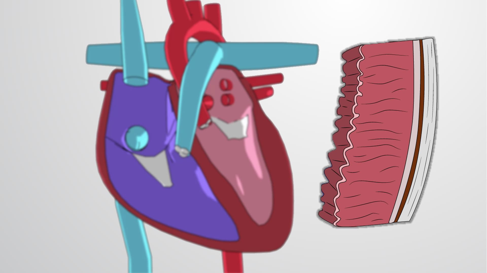
6. Heart Anatomy and Valve Function The heart's four-chamber design with specialized valves ensures unidirectional blood flow through both pulmonary and systemic circuits. The right atrium receives deoxygenated blood from the superior and inferior vena cavae, while the left atrium accepts oxygenated blood from pulmonary veins. Ventricles provide pumping force, with the left ventricle's thicker muscular wall generating higher pressures for systemic circulation. Atrioventricular valves (tricuspid and mitral) prevent backflow between chambers, while semilunar valves (pulmonary and aortic) control outflow from ventricles. Heart murmurs, detected in routine American medical examinations, often result from valve dysfunction, highlighting the clinical importance of understanding normal valve operation and timing.
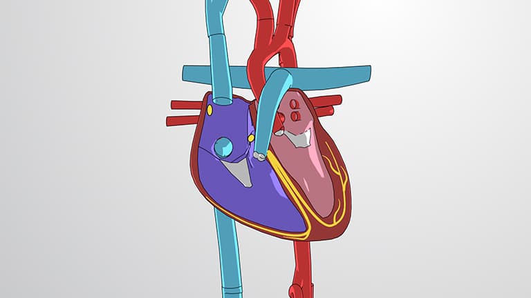
7. Cardiac Cycle Electrical Conduction and Timing The cardiac cycle coordinates atrial and ventricular contractions through a specialized electrical conduction system ensuring optimal blood flow. The sinoatrial (SA) node initiates each heartbeat, causing simultaneous atrial contraction that fills the ventricles. The signal then reaches the atrioventricular (AV) node, which delays transmission by 0.1 seconds, allowing complete atrial emptying. Electrical impulses then travel through the Bundle of His and Purkinje fibers, triggering powerful ventricular contraction that ejects blood into the aorta and pulmonary artery. This electrical activity, recorded as electrocardiograms (ECGs) in American hospitals, reveals heart rhythm abnormalities and guides cardiac treatments, making understanding of normal conduction patterns essential for healthcare professionals.