Loading concepts…


8 Concepts
21 Concepts
9 Concepts
16 Concepts
16 Concepts
14 Concepts
15 Concepts
14 Concepts
7 Concepts
9 Concepts
5 Concepts
19 Concepts
13 Concepts
12 Concepts
15 Concepts
7 Concepts
8 Concepts
11 Concepts
12 Concepts
11 Concepts
6 Concepts
8 Concepts
8 Concepts
9 Concepts
8 Concepts
10 Concepts
12 Concepts
12 Concepts
11 Concepts
5 Concepts
4 Concepts
5 Concepts
7 Concepts
21 Concepts
7 Concepts
8 Concepts
 11 Concepts
 665.9K Views
 Popular topics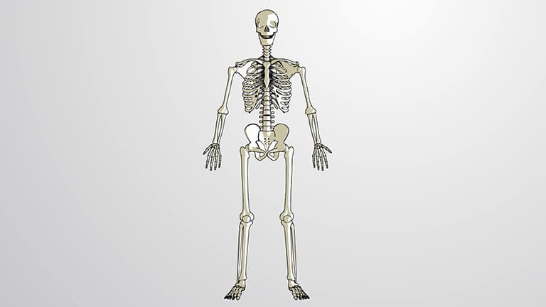
The musculoskeletal system forms the structural foundation of human movement and protection, combining bones, muscles, joints, and connective tissues in coordinated function. This comprehensive course examines muscle and bone biology, exploring how muscles and bones work together through skeletal muscle contraction, bone remodeling, neural control, and pain perception-essential concepts for understanding human anatomy and physiology in clinical and research applications.
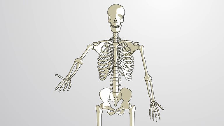
1. Skeletal System Organization and Function The human skeleton divides into axial and appendicular components, each serving distinct protective and locomotive functions. The axial skeleton, including the skull, vertebral column, and rib cage, protects vital organs like the brain, spinal cord, heart, and lungs. Meanwhile, the appendicular skeleton supports limb movement through arm and leg bones plus their connecting girdles. This organizational structure enables both stability and mobility, allowing complex movements like throwing a baseball while protecting delicate internal structures from injury during physical activity.
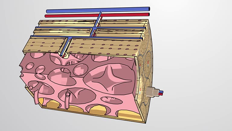
2. Bone Structure and Cellular Organization Long bones like the femur demonstrate the sophisticated architecture of osseous tissue, featuring dense cortical bone surrounding a medullary cavity filled with yellow bone marrow. The periosteum provides blood supply and contains bone-forming cells, while the endosteum lines internal surfaces. Microscopic osteons arrange in concentric lamellae around Haversian canals, housing osteocytes in lacunae. Cancellous bone at bone ends contains red bone marrow for blood cell production, with trabecular patterns optimizing strength-to-weight ratios essential for activities like basketball jumping.
3. Joint Classification and Movement Mechanics Joints enable movement through three main categories with varying mobility levels. Fibrous joints like skull sutures provide stability without movement, while cartilaginous joints between vertebrae allow controlled bending during spinal flexion. Synovial joints, exemplified by the shoulder's ball-and-socket design, permit extensive movement through articular cartilage and synovial fluid lubrication. This joint variety enables activities from precise handwriting to powerful swimming strokes, with ligaments and muscles providing stability and control throughout movement ranges.
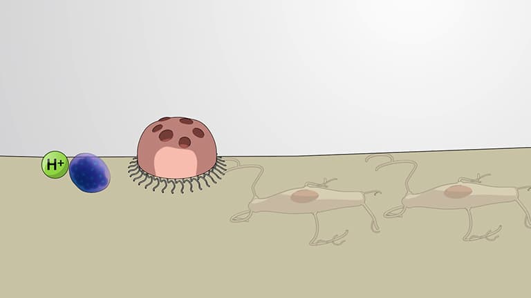
4. Bone Remodeling and Adaptation Living bone tissue continuously remodels through coordinated osteoclast resorption and osteoblast formation phases, replacing skeletal regions like femur ends every six months. Mechanical stress triggers osteocyte signaling, initiating bone removal where osteoclasts create erosion cavities through enzyme secretion. Subsequently, osteoblasts fill cavities with new osteoid matrix containing collagen fibers. This process maintains bone strength during activities like marathon running while releasing calcium for metabolic functions, demonstrating bone's dual role as structural support and mineral reservoir.
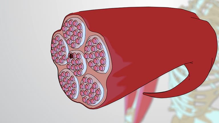
5. Skeletal Muscle Architecture and Fiber Types Skeletal muscles contain hierarchical organization from whole muscle through fascicles to individual sarcomeres, the contractile units containing actin and myosin filaments. Three distinct fiber types serve different functional demands: slow-twitch oxidative fibers excel in endurance activities like distance cycling through abundant myoglobin and mitochondria; fast-twitch oxidative fibers provide power for sprinting through rapid aerobic metabolism; fast-twitch glycolytic fibers generate maximum force for powerlifting through anaerobic glycogen breakdown. Genetic factors and training adaptations determine individual muscle fiber composition.
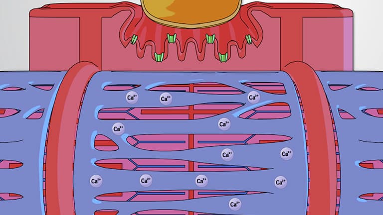
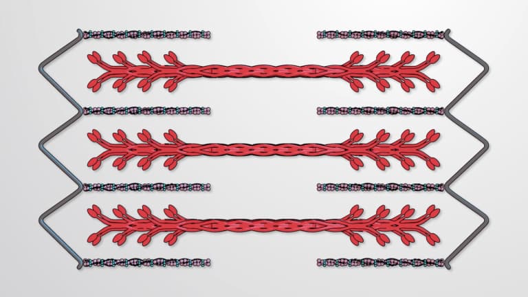
6. Muscle Contraction and Cross-Bridge Cycling Voluntary muscle contraction begins with motor cortex signals activating spinal motor neurons, which release acetylcholine at neuromuscular junctions. Calcium ion release from sarcoplasmic reticulum exposes myosin-binding sites on actin filaments by moving tropomyosin. ATP hydrolysis energizes myosin heads for actin binding, creating cross-bridges that pull actin filaments toward sarcomere centers during the power stroke. New ATP molecules dissociate cross-bridges, enabling repeated cycling until calcium removal and ATP depletion end contraction, as occurs when stopping a bicep curl.
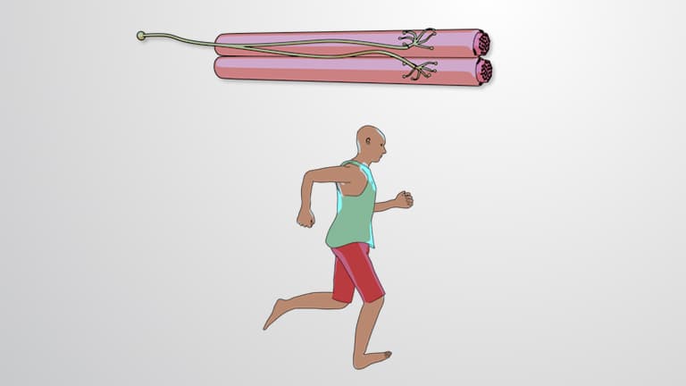
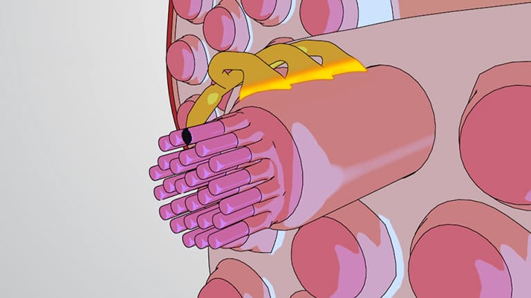
7. Motor Unit Organization and Neural Control Motor units consist of single spinal motor neurons innervating multiple muscle fibers, with unit sizes varying by precision requirements. Eye movement muscles contain small motor units with few fibers for precise control, while postural muscles like those supporting standing contain hundreds of fibers per unit. Motor neuron activation causes simultaneous contraction of all innervated fibers, producing graded muscle force through motor unit recruitment. This organization enables both delicate tasks like suturing wounds and powerful activities like weightlifting through selective motor unit activation patterns.
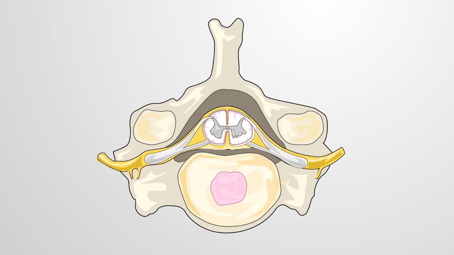
8. Spinal Cord Structure and Function The spinal cord forms a critical nervous system component, featuring butterfly-shaped gray matter containing motor neurons surrounded by white matter tracts carrying ascending sensory and descending motor information. Extending from the brainstem through vertebral foramina to the L1-2 level, paired spinal nerves emerge at each vertebral level. Ventral nerve roots carry motor signals to muscles, while dorsal roots transmit sensory information from skin dermatomes. This organization enables reflexes, voluntary movement, and sensation, with dermatome maps helping clinicians diagnose neurological conditions.
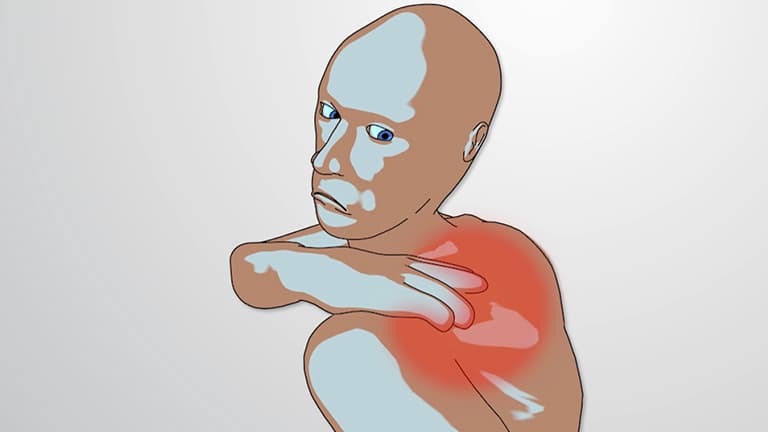
9. Pain Processing and Nociception Pathways Nociception begins when tissue damage activates free nerve endings, triggering inflammatory responses involving mast cell histamine release and macrophage cytokine secretion. Two fiber types transmit pain signals: myelinated A-delta fibers conduct sharp, localized pain rapidly for immediate withdrawal responses, while unmyelinated C fibers carry slower, burning pain sensations. Signals cross in the spinal cord before ascending to brainstem, thalamus, and somatosensory cortex for location identification. Additional processing in amygdala and prefrontal cortex creates emotional and cognitive pain components, explaining individual pain perception variations.