Loading video…


 8 Concepts
 654.7K views

Video Summary: What Is Anatomy of the Heart
Every minute, your heart pumps approximately 5 liters of blood through your body-that's enough to fill a large soda bottle! Understanding the anatomy heart structure reveals how this fist-sized muscle accomplishes this remarkable feat through its three tissue layers, four chambers, and specialized valves. For instance, during cardiac catheterization procedures at Johns Hopkins Hospital, cardiologists navigate through these precise anatomical structures to diagnose and treat heart conditions. Watch the full video on JoVE Coach to master this concept with expert-led visuals and step-by-step explanations.
The anatomy of the heart represents one of medicine's most crucial concepts, forming the foundation for understanding cardiovascular physiology, disease processes, and therapeutic interventions. Medical students preparing for the MCAT and nursing students studying for the NCLEX must master these anatomical relationships to succeed in their healthcare careers.
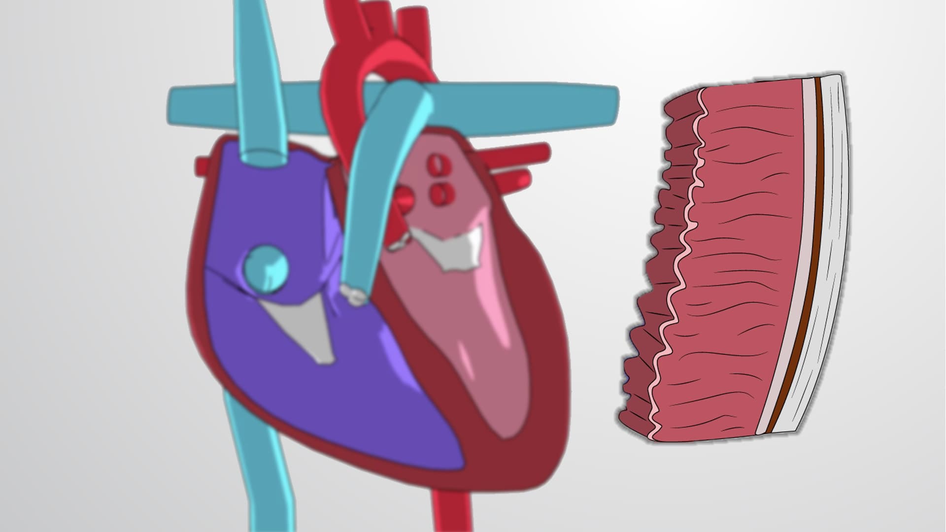
The heart wall's tri-layered architecture provides both protection and functionality. The outermost epicardium contains coronary blood vessels and nerves, while the protective pericardium creates a fluid-filled sac that prevents friction during heartbeats-a critical concept tested on AP Biology exams. The middle myocardium contains specialized cardiac muscle cells with unique properties: they contract rhythmically without nervous stimulation and communicate through intercalated discs. The innermost endocardium provides a smooth surface for blood flow and lines the heart valves, preventing clot formation that could lead to stroke or heart attack.
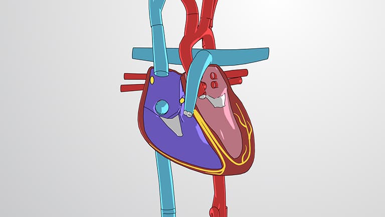
The anatomy heart overview reveals four distinct chambers working in coordinated sequence. The right atrium receives deoxygenated blood from three sources: the superior vena cava (from upper body), inferior vena cava (from lower body), and coronary sinus (from heart muscle itself). Meanwhile, the left atrium receives oxygenated blood exclusively from pulmonary veins returning from the lungs. This separation prevents oxygen-rich and oxygen-poor blood from mixing-a concept frequently tested on college anatomy midterms and the HESI A2 entrance exam.
Four strategically positioned valves ensure unidirectional blood flow, preventing the backflow that would compromise cardiac efficiency. The tricuspid valve (right side) and mitral valve (left side) separate atria from ventricles, while the pulmonary valve and aortic valve control blood exit from the heart. Understanding valve dysfunction helps explain common conditions like mitral valve prolapse, affecting approximately 2-3% of Americans, or aortic stenosis, frequently encountered in elderly patients at Cleveland Clinic and Mayo Clinic cardiology departments.
Related Micro-courses