Loading subject page…


235,304 views

Video Summary: What Is Ligand Binding and Linkage
Ever wonder how insulin knows exactly where to bind in your body to regulate blood sugar? Ligand binding linkage governs these precise molecular interactions that keep us alive. When studying What is Ligand Binding And Linkage, you're exploring how molecules like drugs, hormones, and neurotransmitters find their target proteins and influence cellular behavior. Consider how caffeine blocks adenosine receptors in your brain to keep you alert-this exemplifies ligand binding in action. Watch the full video on JoVE Coach to master this concept with expert-led visuals and step-by-step explanations.
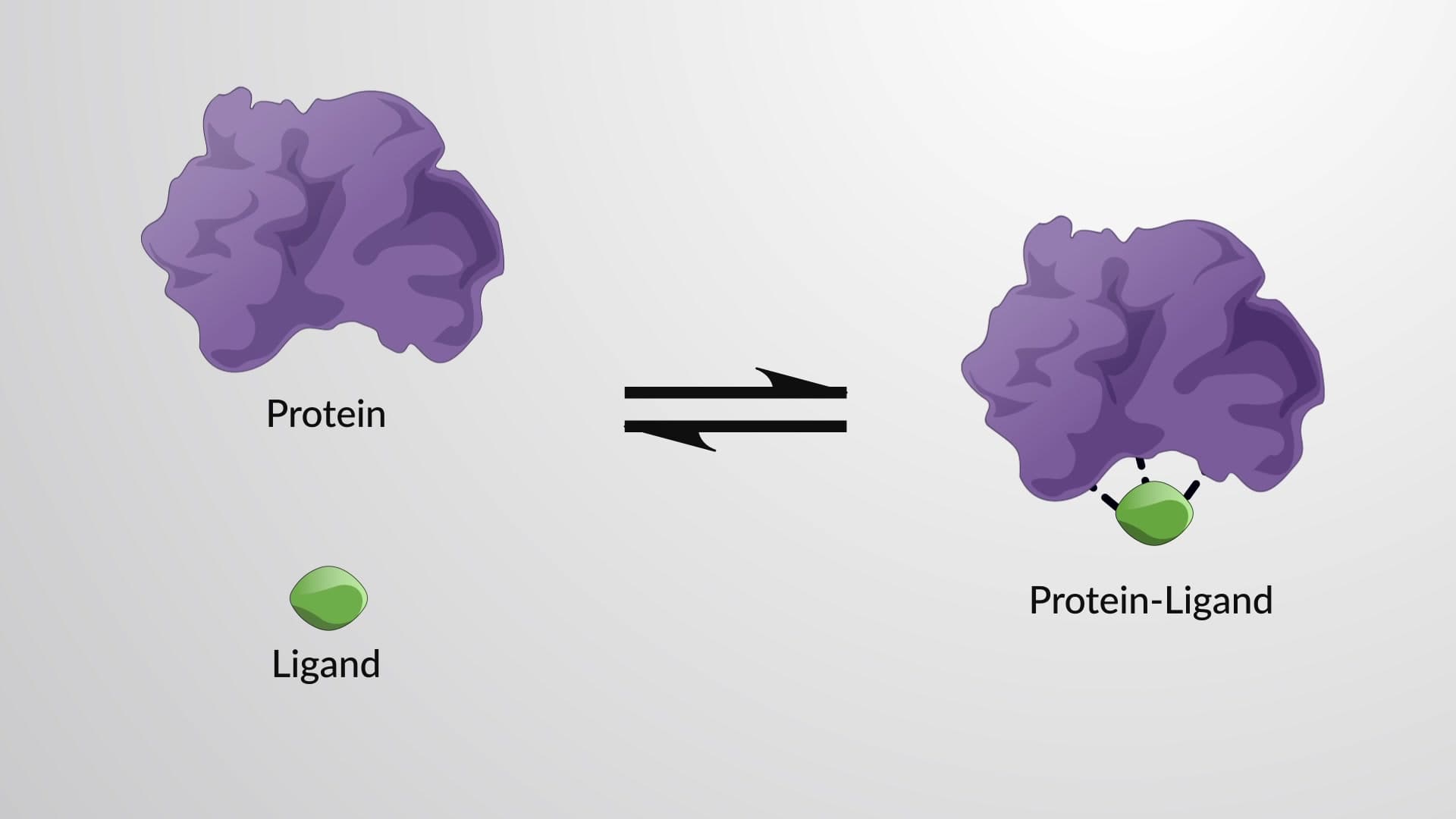
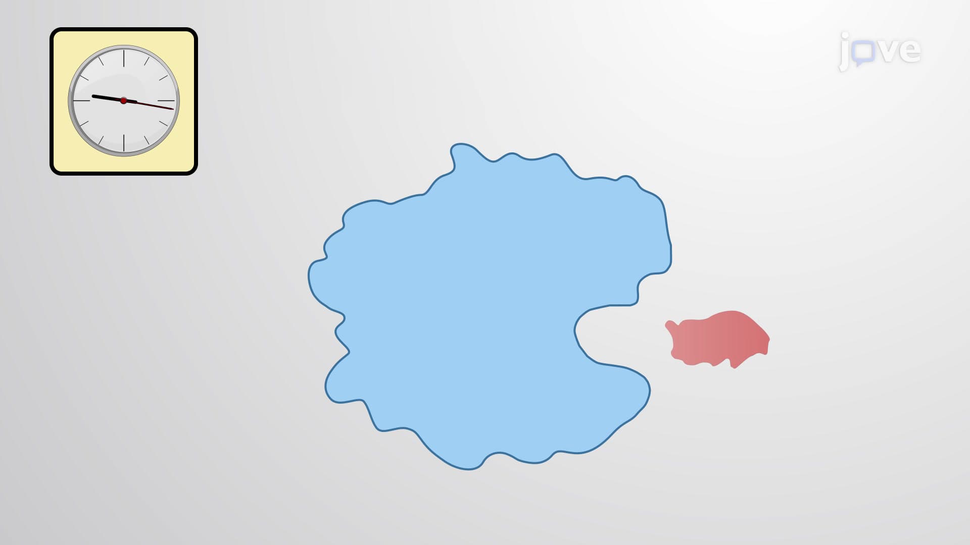
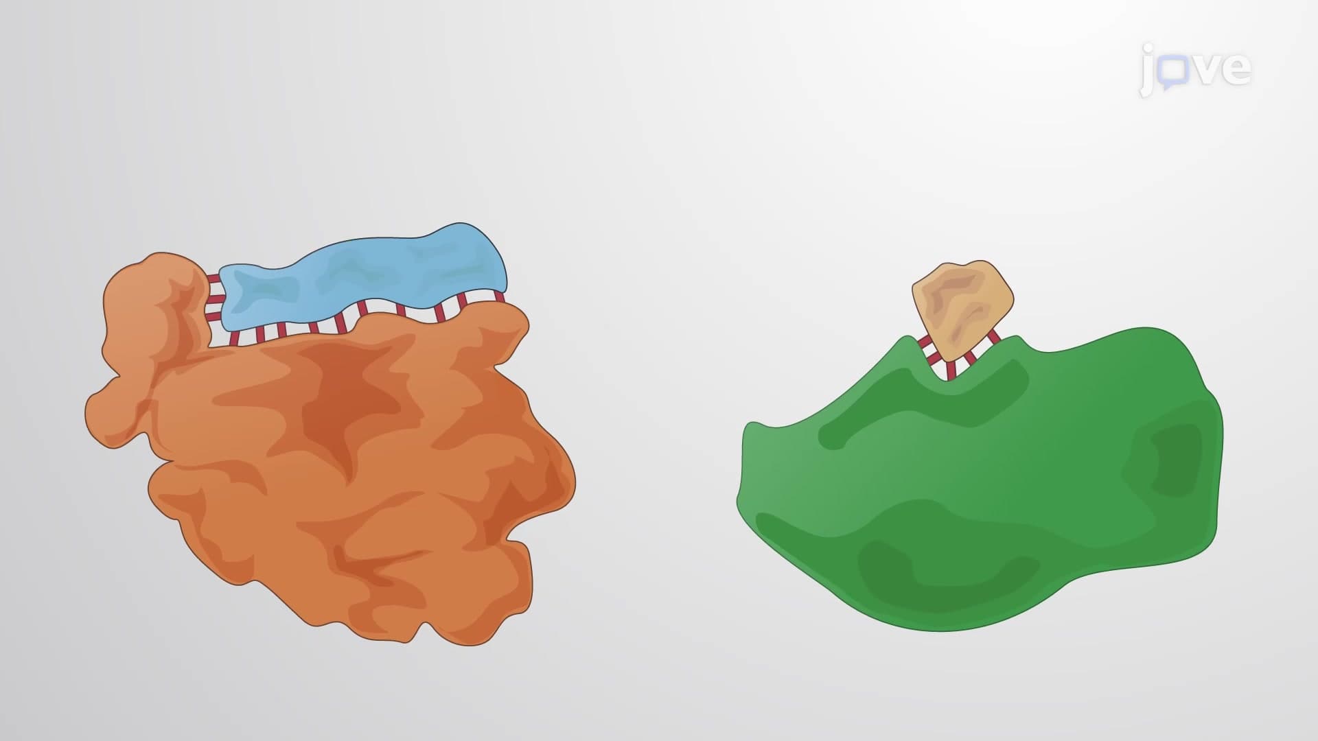
Ligand binding linkage represents one of biochemistry's most elegant regulatory mechanisms, where molecular recognition drives cellular function. A ligand-any molecule that binds to a protein-can be as simple as oxygen binding to hemoglobin or as complex as a multi-domain growth factor activating a receptor tyrosine kinase. The "linkage" aspect refers to how these binding events connect to influence other binding sites or cellular processes, creating sophisticated regulatory networks.
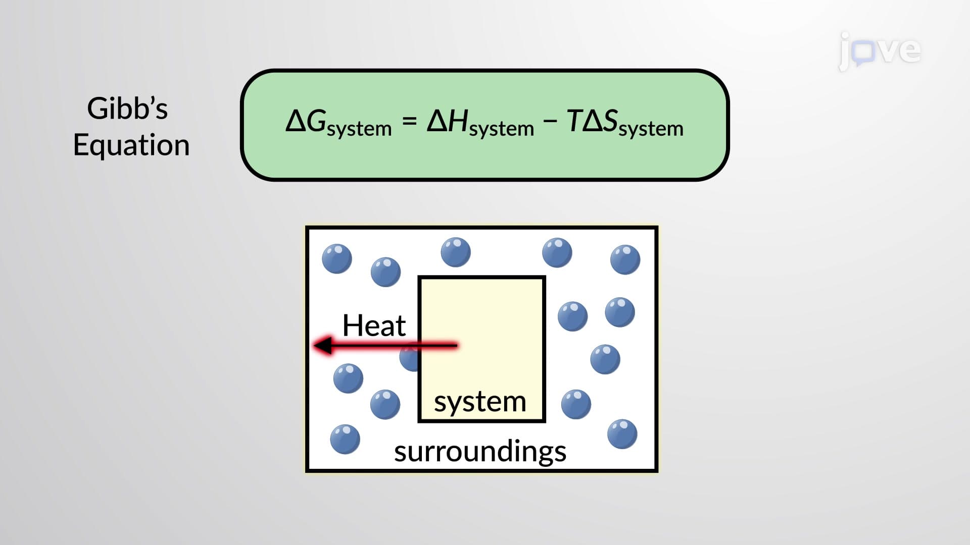
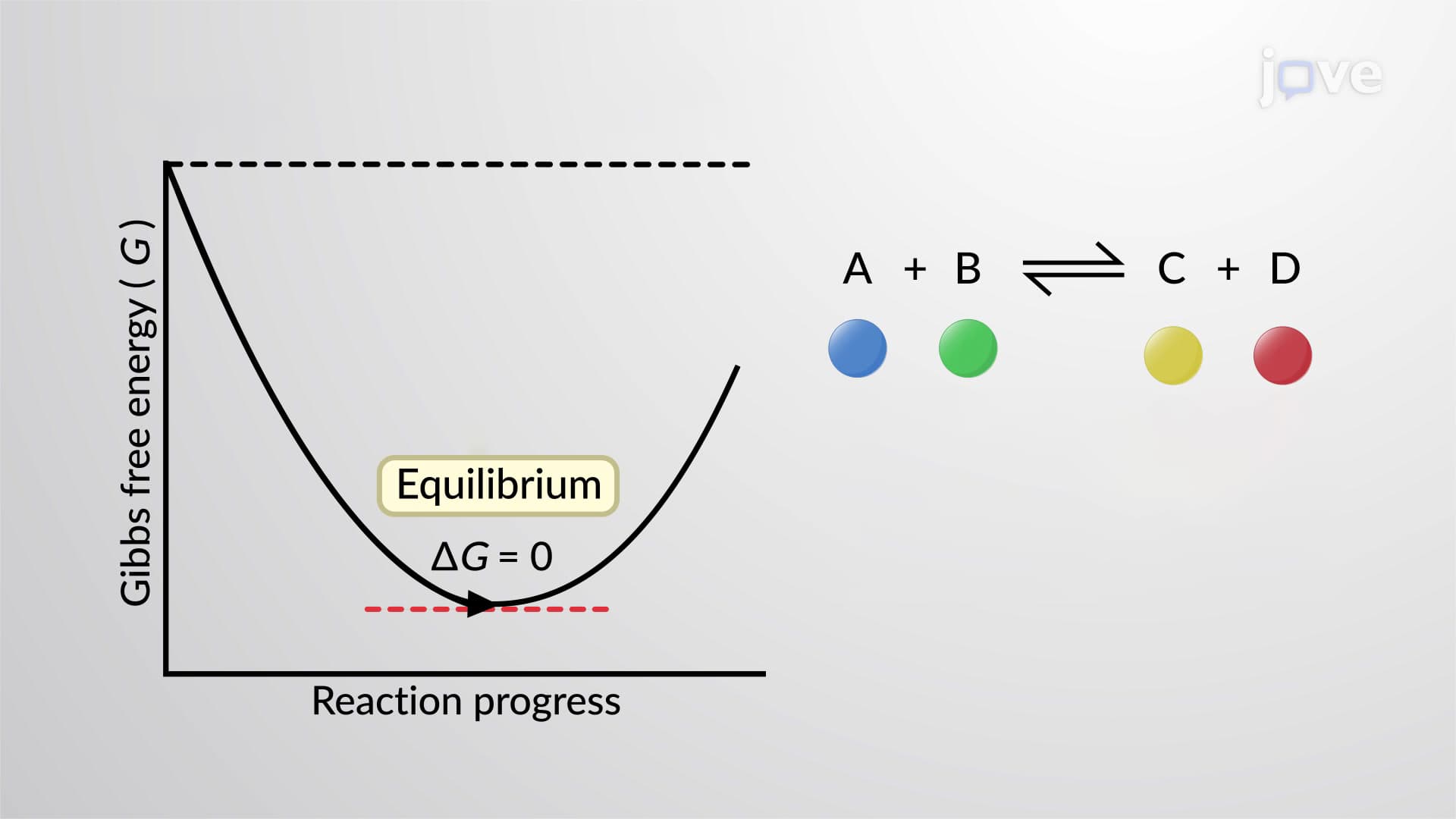
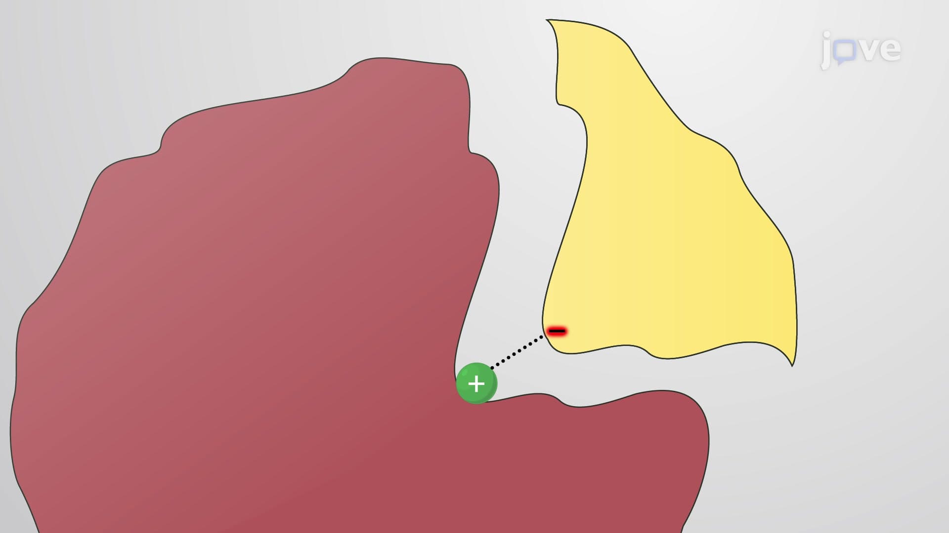
The strength of ligand-protein interactions depends on binding affinity, quantified by the dissociation constant (Kd). A lower Kd indicates stronger binding-for example, insulin binds its receptor with a Kd of approximately 0.1 nM, reflecting the hormone's potent biological activity. Free energy changes (ΔG) determine binding favorability, with negative values indicating spontaneous complex formation. Students preparing for the MCAT should master the relationship: ΔG = -RT ln(Ka), where Ka = 1/Kd.
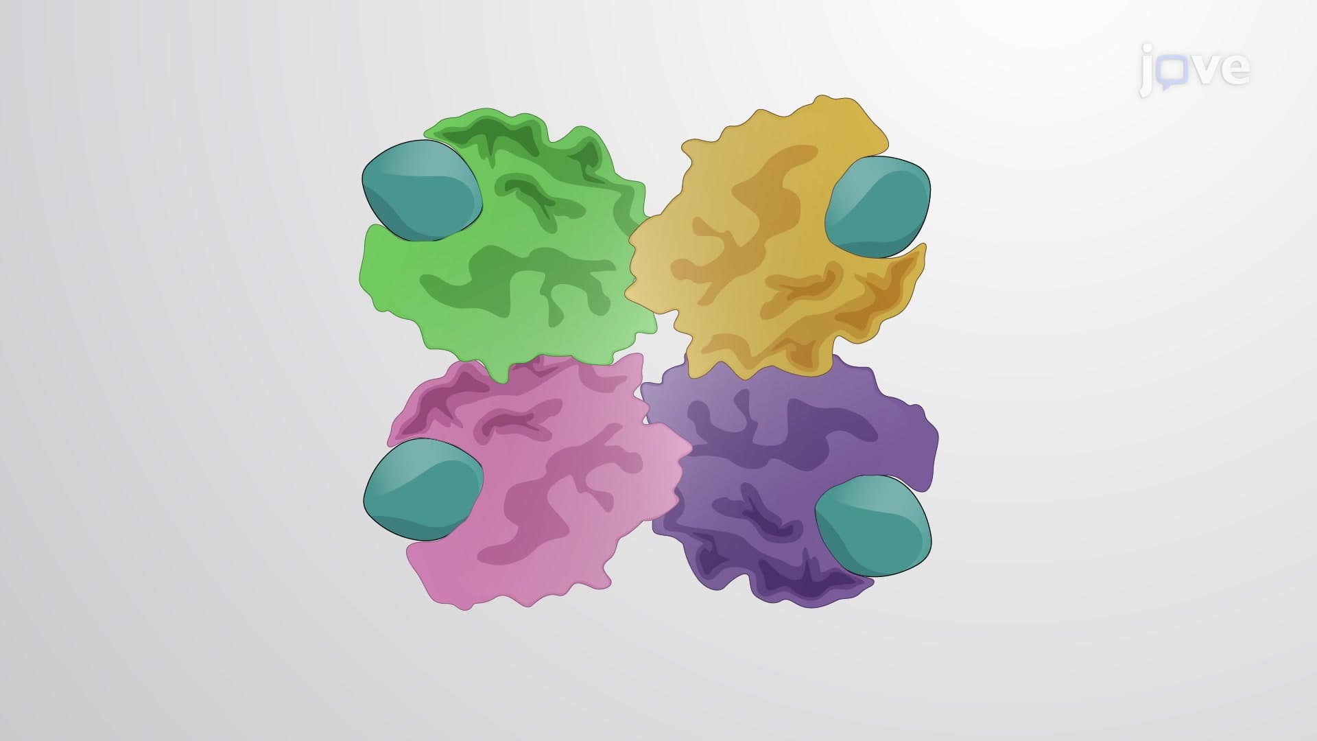
Ligand binding and linkage definition explained includes cooperative phenomena where initial ligand binding enhances subsequent binding events. Hemoglobin exemplifies positive cooperativity-oxygen binding to one subunit increases oxygen affinity at remaining subunits, creating the characteristic sigmoidal binding curve. The Hill coefficient (nH) quantifies cooperativity: nH > 1 indicates positive cooperativity, while nH < 1 suggests negative cooperativity.
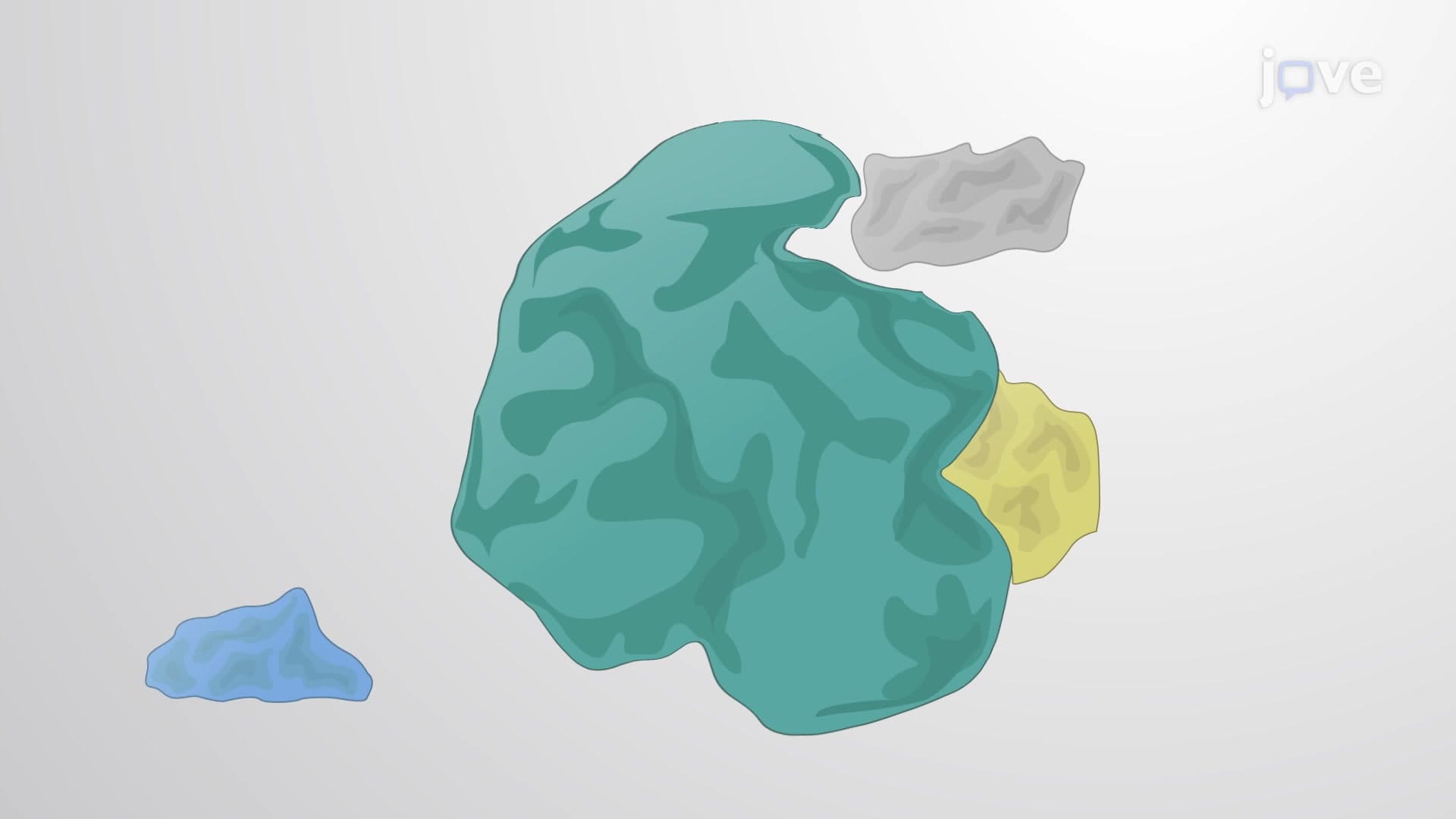
Two competing models explain allosteric transitions. The Monod-Wyman-Changeux (MWC) model proposes proteins exist in equilibrium between tense (T) and relaxed (R) states, with ligands stabilizing one conformation. Conversely, the Koshland-Némethy-Filmer (KNF) model suggests ligand binding induces sequential conformational changes. Both models appear in AP Biology curricula and college biochemistry courses, with real-world applications in understanding how aspirin inhibits cyclooxygenase enzymes or how caffeine blocks adenosine receptors.
Understanding ligand binding linkage proves essential for pharmaceutical development. Drug designers optimize binding affinity, specificity, and allosteric effects to create therapeutic agents. For instance, monoclonal antibodies used in cancer immunotherapy rely on precise antigen recognition, while allosteric inhibitors offer advantages over competitive inhibitors by avoiding direct competition with natural substrates. Students pursuing healthcare careers encounter these principles in pharmacology coursework and on exams like the USMLE Step 1.
Related Micro-courses