Loading subject page…


78,033 views

Video Summary: Assembly of Complex Microtubule Structures Explained
Did you know that a single neuron in your brain contains thousands of microscopic highways that must organize perfectly for you to think, move, or remember? The assembly of complex microtubule structures orchestrates this cellular architecture through specialized motor proteins and microtubule-associated proteins (MAPs). These intricate networks enable everything from the neural pathways that help Stanford University researchers study memory formation to the precise chromosome separation during cell division in cancer research at Johns Hopkins. Watch the full video on JoVE Coach to master this concept with expert-led visuals and step-by-step explanations.
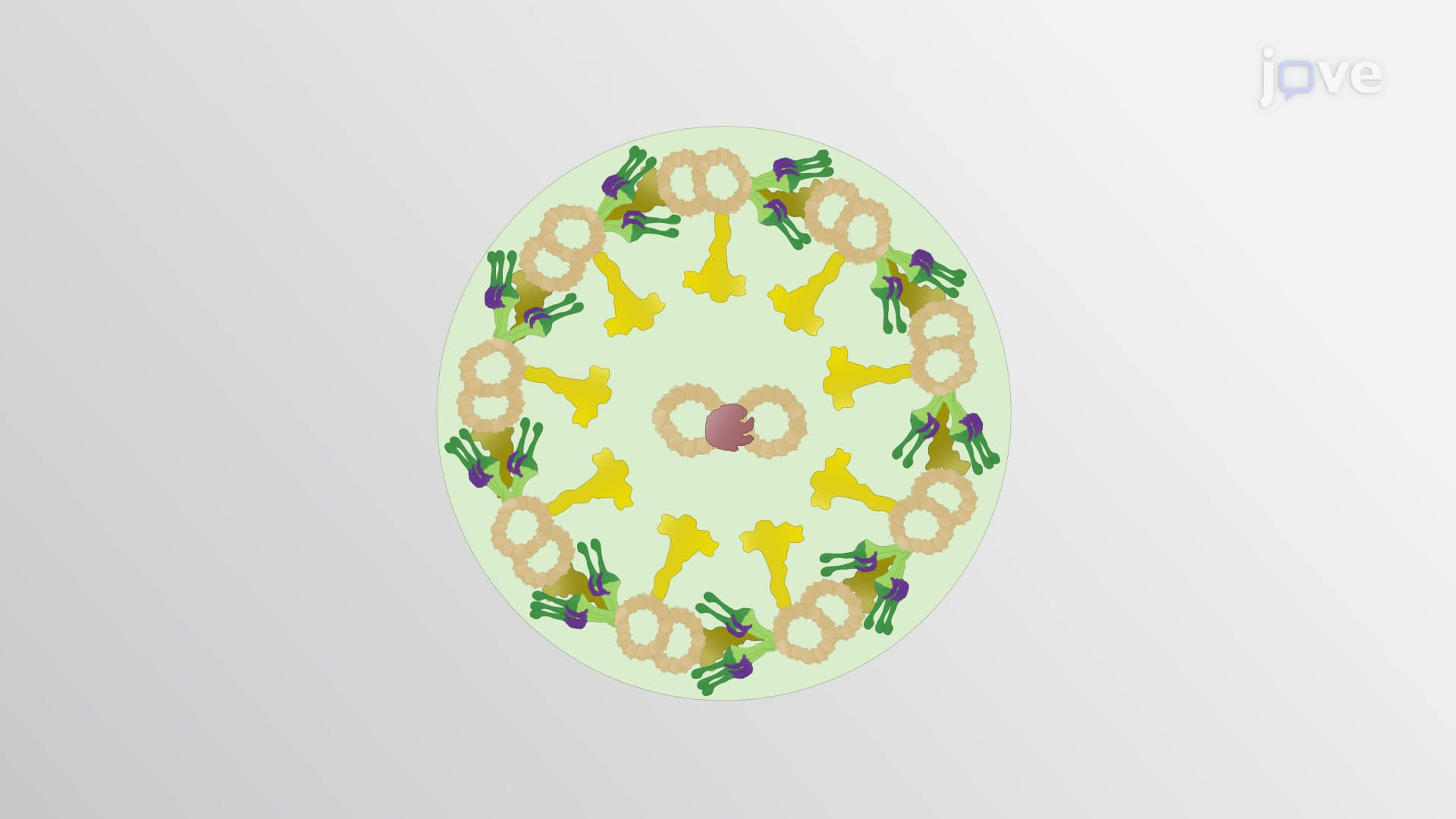
The assembly of complex microtubule structures represents one of cell biology's most sophisticated organizational systems. Unlike simple protein polymers, these structures require precise coordination between multiple protein families to create functional cellular machinery. Think of it as constructing a highway system where the roads (microtubules) must be built with specific traffic patterns, supported by appropriate infrastructure (MAPs), and maintained by specialized crews (motor proteins).
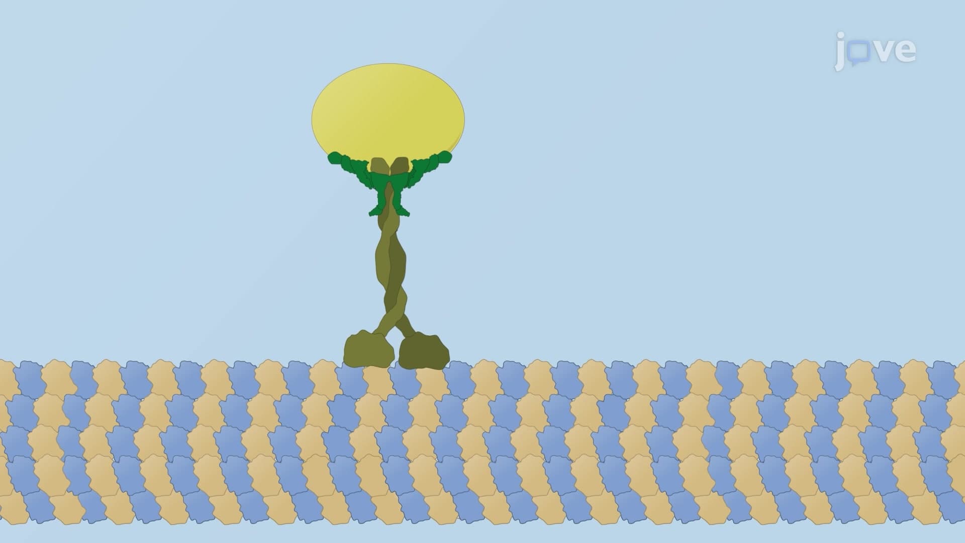
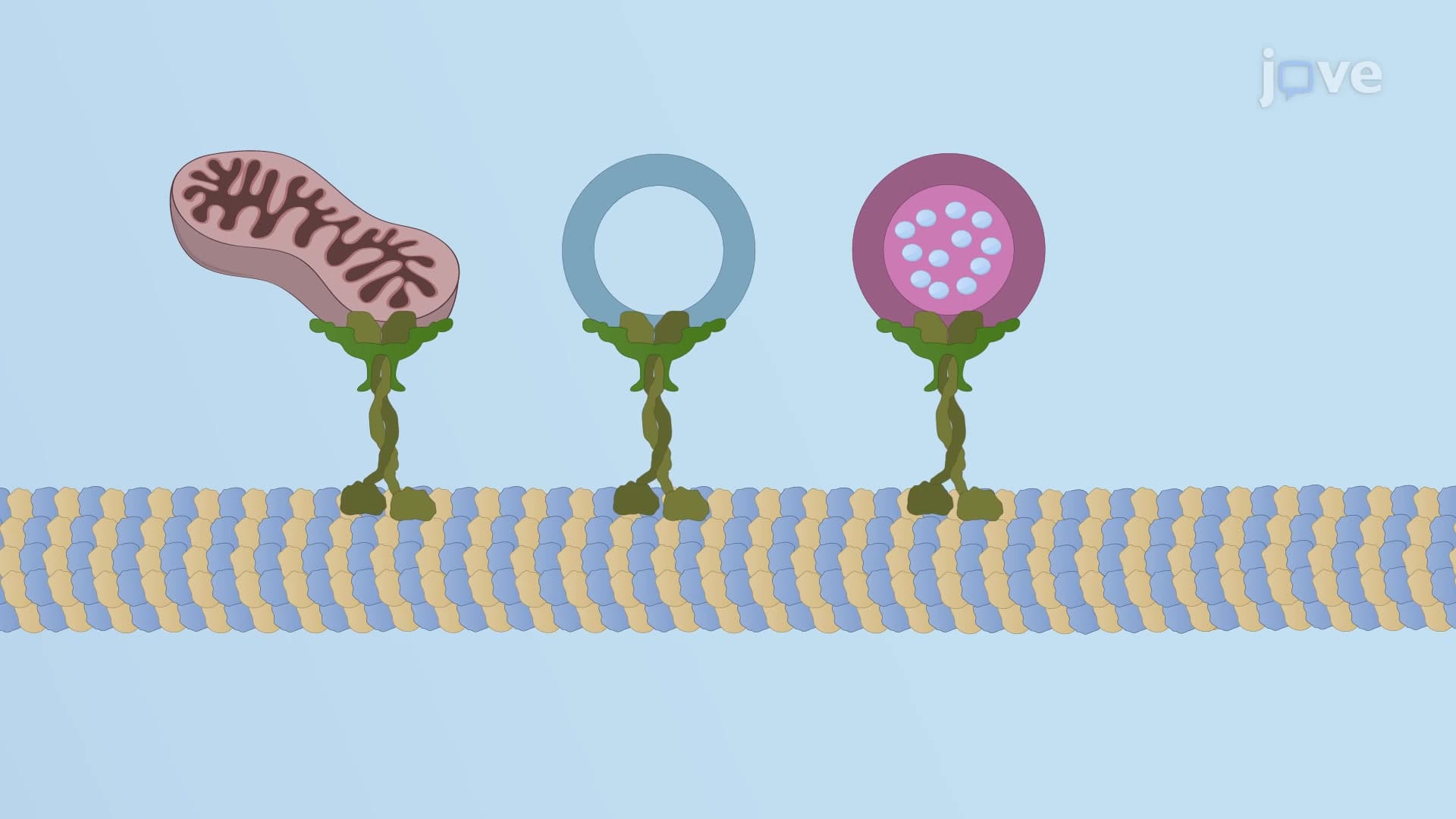
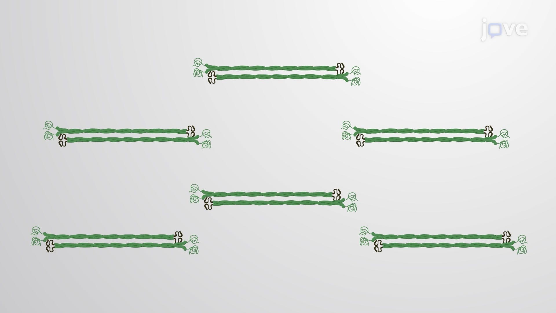
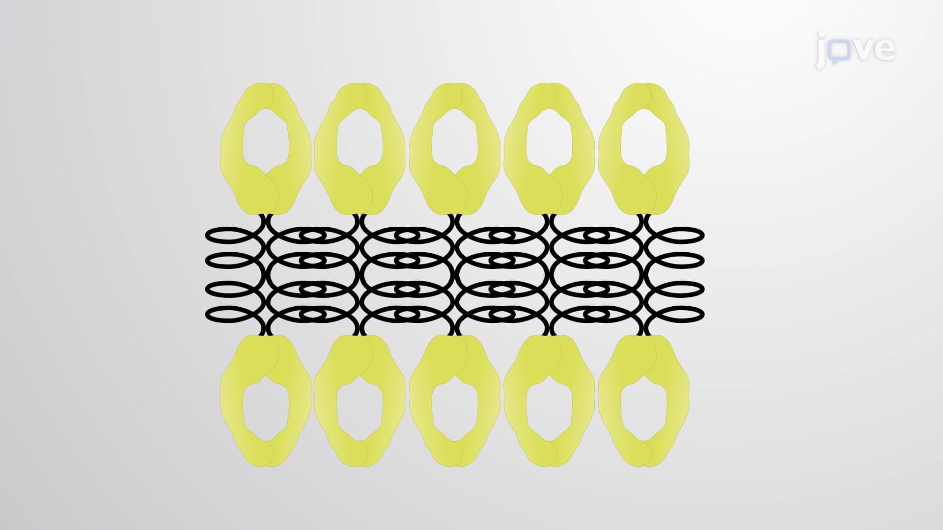
Motor proteins serve as the dynamic workforce, actively transporting cargo and organizing microtubule arrays. Kinesin motors typically move toward the plus-end, while dynein motors travel toward the minus-end, creating bidirectional transport systems essential for cellular function. Meanwhile, microtubule-associated proteins act as structural scaffolds and regulatory elements. For example, tau proteins stabilize axonal microtubules-a process that becomes critically important when studying neurodegenerative diseases like Alzheimer's, extensively researched at institutions like the Mayo Clinic and Harvard Medical School.
The most striking example of complex microtubule assembly appears in neurons, where axons and dendrites display dramatically different organizational patterns. Axonal microtubules maintain uniform polarity with plus-ends pointing toward the synaptic terminal, creating efficient highways for neurotransmitter vesicle transport. This organization proves crucial for understanding action potential propagation-a key concept tested on the MCAT and featured prominently in AP Biology curricula.
Conversely, dendritic microtubules exhibit mixed polarity, reflecting their role as signal receivers rather than transmitters. This arrangement supports the complex protein trafficking required for synaptic plasticity and learning-processes extensively studied in neuroscience programs at universities like MIT and Caltech.
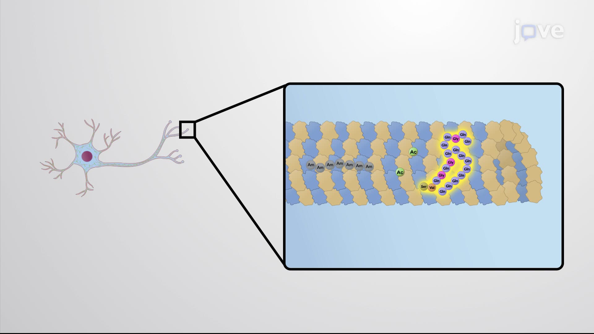
Post-translational modifications add another layer of complexity to microtubule assembly. Axonal microtubules undergo acetylation, polyamination, and long-chain glutamylation, modifications that increase stability and promote long-distance transport. These modifications become particularly relevant when studying peripheral neuropathies or preparing for USMLE examinations, where understanding cytoskeletal pathology proves essential.
Dendritic modifications, including tyrosination and short-chain glutamylation, support the dynamic reorganization required for synaptic plasticity. This differential modification pattern explains why certain neurological conditions affect axons and dendrites differently-knowledge crucial for medical school coursework and board examinations.
Related Micro-courses