Loading video…


 19 Concepts
 78.1K views

Video Summary: What are Destabilization of Microtubules
Did you know that every time a cancer cell divides, it relies on precisely controlling microtubule breakdown? The destabilization of microtubules occurs when cellular proteins accelerate the disassembly of these crucial structural components faster than they can rebuild. In cancer treatment centers across the US, drugs like taxanes exploit this process to halt tumor growth by disrupting normal microtubule dynamics. This cellular mechanism involves specialized proteins that either sequester building blocks or directly promote filament shortening. Watch the full video on JoVE Coach to master this concept with expert-led visuals and step-by-step explanations.
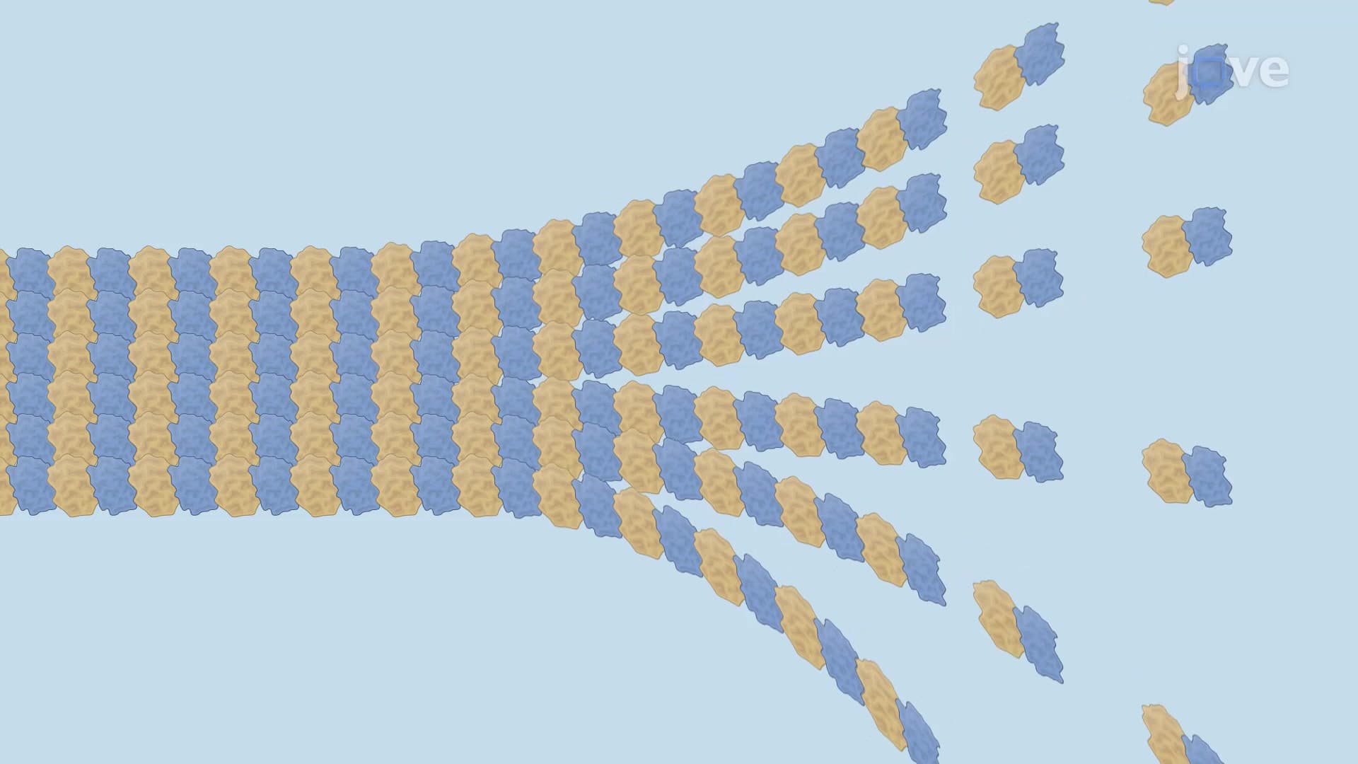
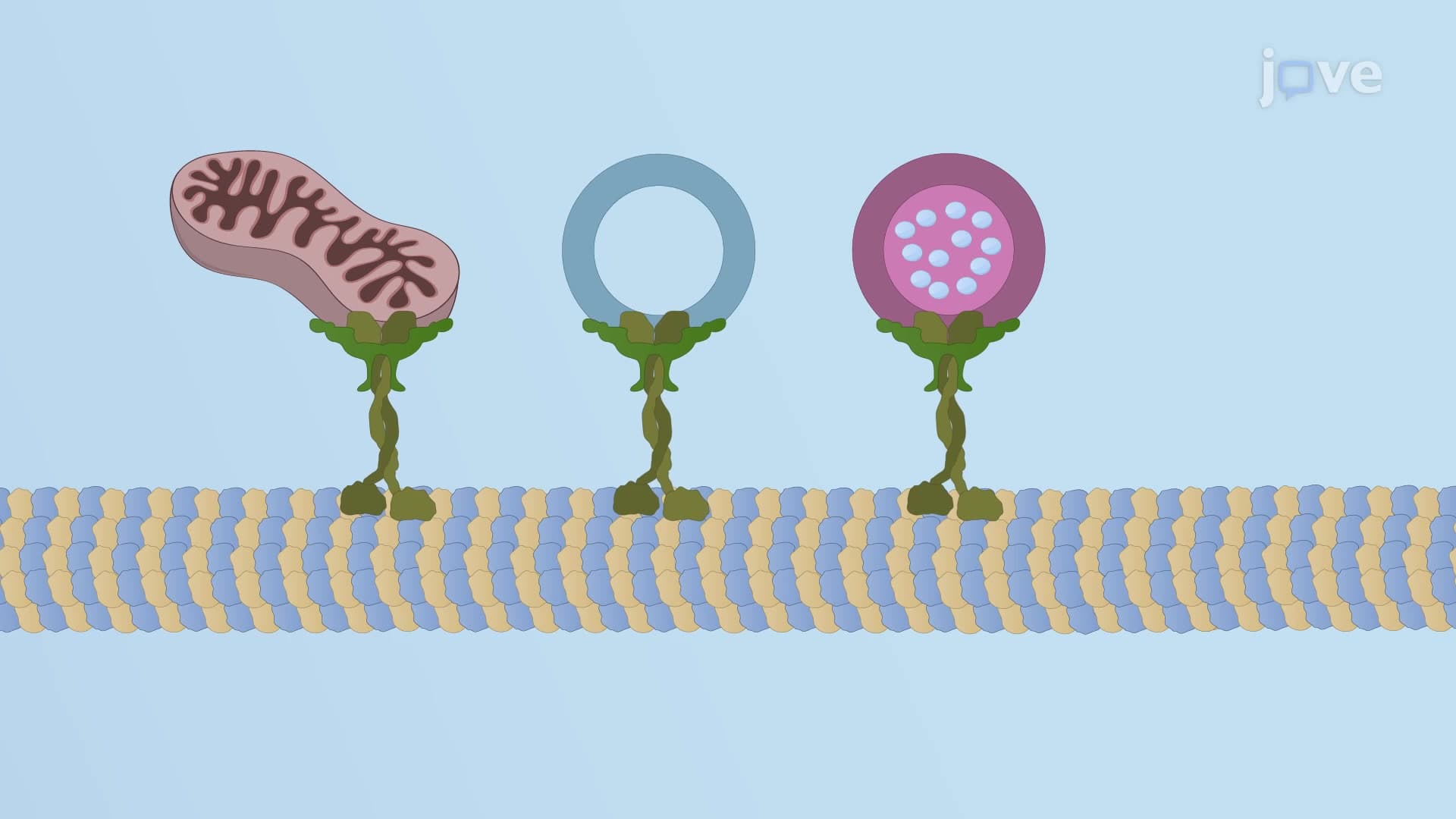
The destabilization of microtubules represents a carefully orchestrated cellular process where specialized proteins accelerate the breakdown of these essential cytoskeletal structures. Unlike passive degradation, this active process involves sophisticated molecular machinery that cells use to rapidly reorganize their internal architecture. This mechanism proves crucial during cell division, migration, and response to environmental changes.
In healthy cells, microtubules exist in dynamic equilibrium between growth (rescue) and shrinkage (catastrophe). Destabilization tips this balance decisively toward catastrophe, enabling rapid cytoskeletal remodeling when cells need to change shape, divide, or respond to signals.
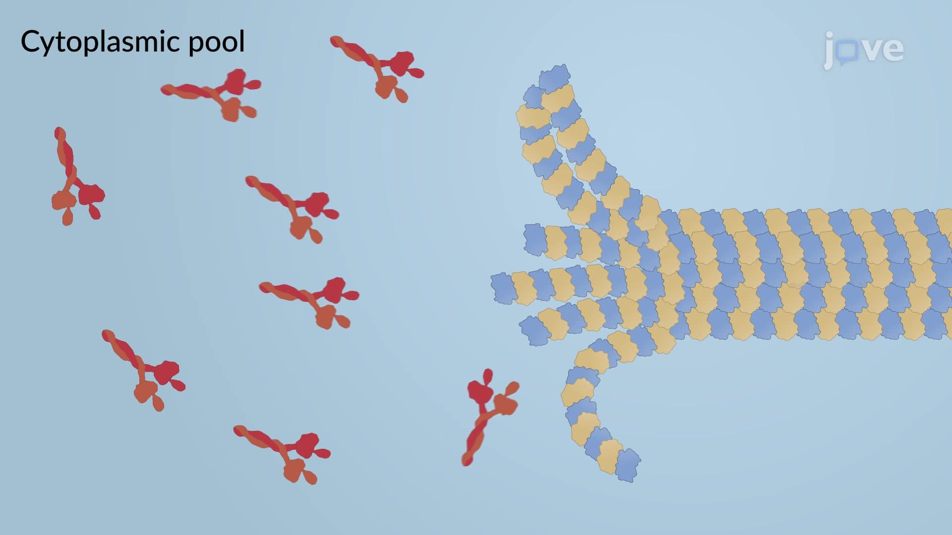
Stathmin operates as a molecular scavenger in the cell's cytoplasm, binding directly to tubulin heterodimers before they can incorporate into growing microtubules. This protein's dual action-sequestering free tubulin and binding to filament tips-makes it exceptionally effective at promoting destabilization. When stathmin levels increase, cells experience widespread microtubule shortening, which researchers at institutions like Johns Hopkins University have linked to enhanced cell motility in certain cancer types.
The conformational changes stathmin induces in tubulin dimers prevent proper assembly, effectively starving growing microtubules of their building materials. This mechanism becomes particularly relevant during mitosis when cells need to completely reorganize their cytoskeleton.
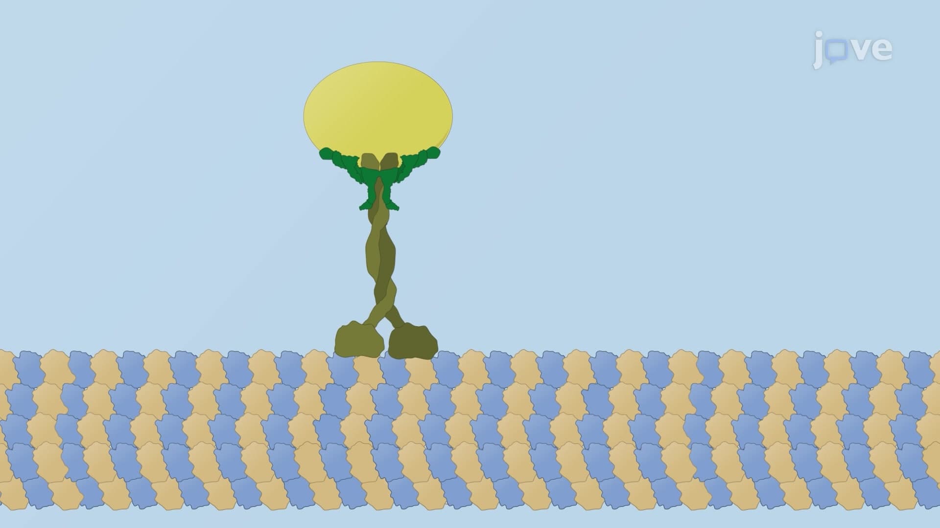
Unlike its motile kinesin relatives, kinesin-13 functions as a stationary destabilization machine. This protein family harnesses ATP energy to actively strip tubulin subunits from microtubule ends through a sophisticated biochemical process. The conversion of GTP-bound β-tubulin to GDP-bound β-tubulin weakens intermolecular bonds, allowing kinesin-13 to efficiently remove dimers.
Research conducted at Stanford University has shown that kinesin-13 proteins can completely depolymerize microtubules within minutes under physiological conditions. This rapid action proves essential during prometaphase of mitosis when cells must disassemble their interphase microtubule network.
Understanding microtubule destabilization directly impacts medical training and research in the United States. Medical students preparing for the MCAT encounter questions about cytoskeletal dynamics, while those taking the USMLE must understand how chemotherapy drugs exploit these pathways. In AP Biology courses, students analyze how cells control their shape and division through cytoskeletal regulation, making this concept fundamental to contemporary cell biology education.
Related Micro-courses