Loading subject page…


78,052 views


Video Summary: What are Microtubules
Did you know the same cellular highways that help your neurons transmit signals also power the tiny flagella of sperm cells racing toward an egg? Microtubules cell biology explained reveals how these dynamic protein tubes, made of tubulin alpha beta microtubule subunits, form the cellular equivalent of railroad tracks throughout your body. From Stanford University's groundbreaking research on Alzheimer's disease to understanding fertility treatments at Johns Hopkins, these cytoskeletal structures are fundamental to life itself. Watch the full video on JoVE Coach to master this concept with expert-led visuals and step-by-step explanations.
Microtubules represent one of biology's most elegant transportation systems, functioning as dynamic protein highways that maintain cellular organization and facilitate essential processes. These hollow cylindrical structures consist of globular protein subunits called tubulins-specifically alpha-tubulin and beta-tubulin-that combine to form heterodimers. With their 25-nanometer diameter, microtubules are the thickest components of the cytoskeleton, making them easily distinguishable under electron microscopy in research laboratories at institutions like Harvard Medical School and UCLA.
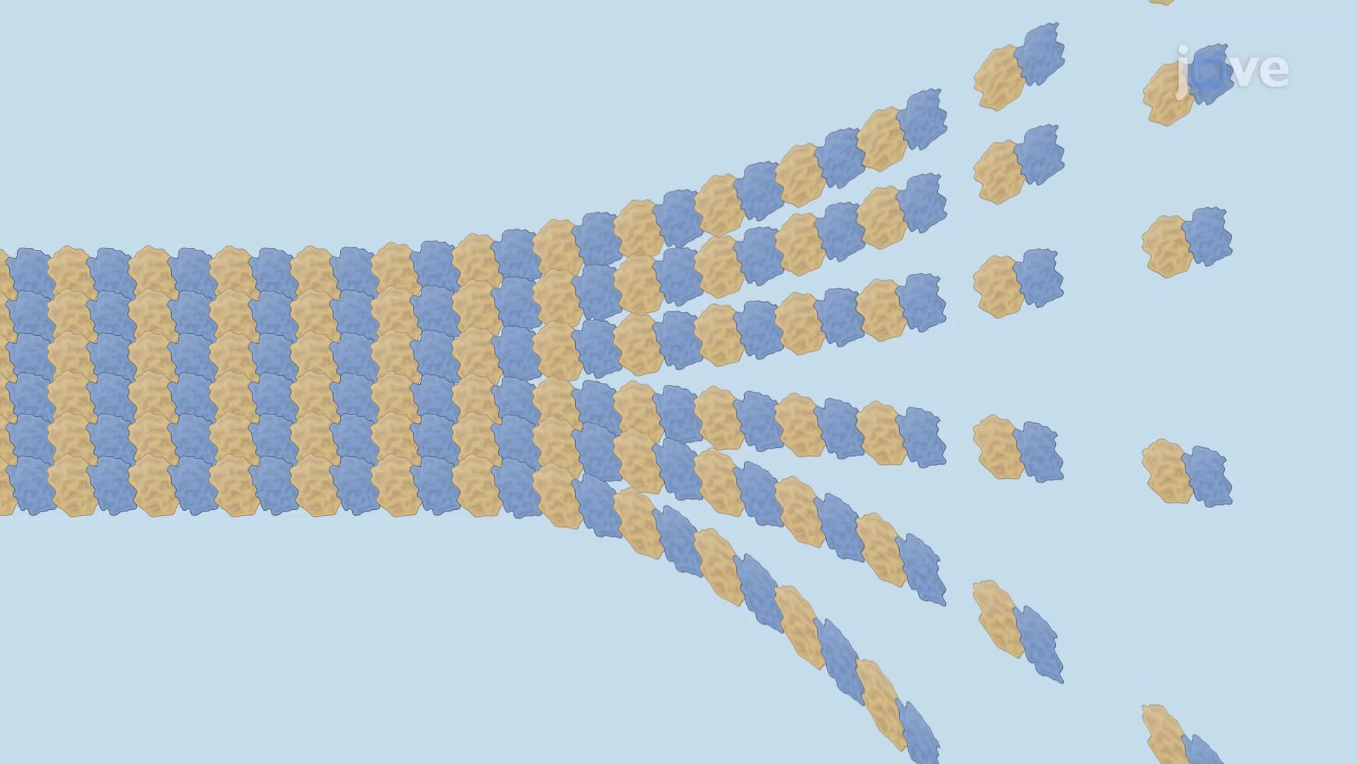
The microtubule structure function biology becomes fascinating when examining how thirteen protofilaments arrange in parallel to create the characteristic hollow tube. Each protofilament displays inherent polarity, with the minus-end (alpha-tubulin exposed) typically anchored at microtubule organizing centers (MTOCs) like the centrosome, while the plus-end (beta-tubulin exposed) extends toward the cell periphery. This polarity is crucial for understanding questions commonly found on AP Biology exams and MCAT Cell Biology sections, where students must identify directional transport mechanisms.
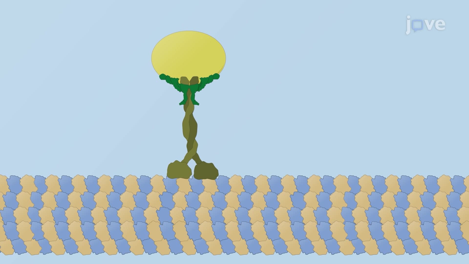
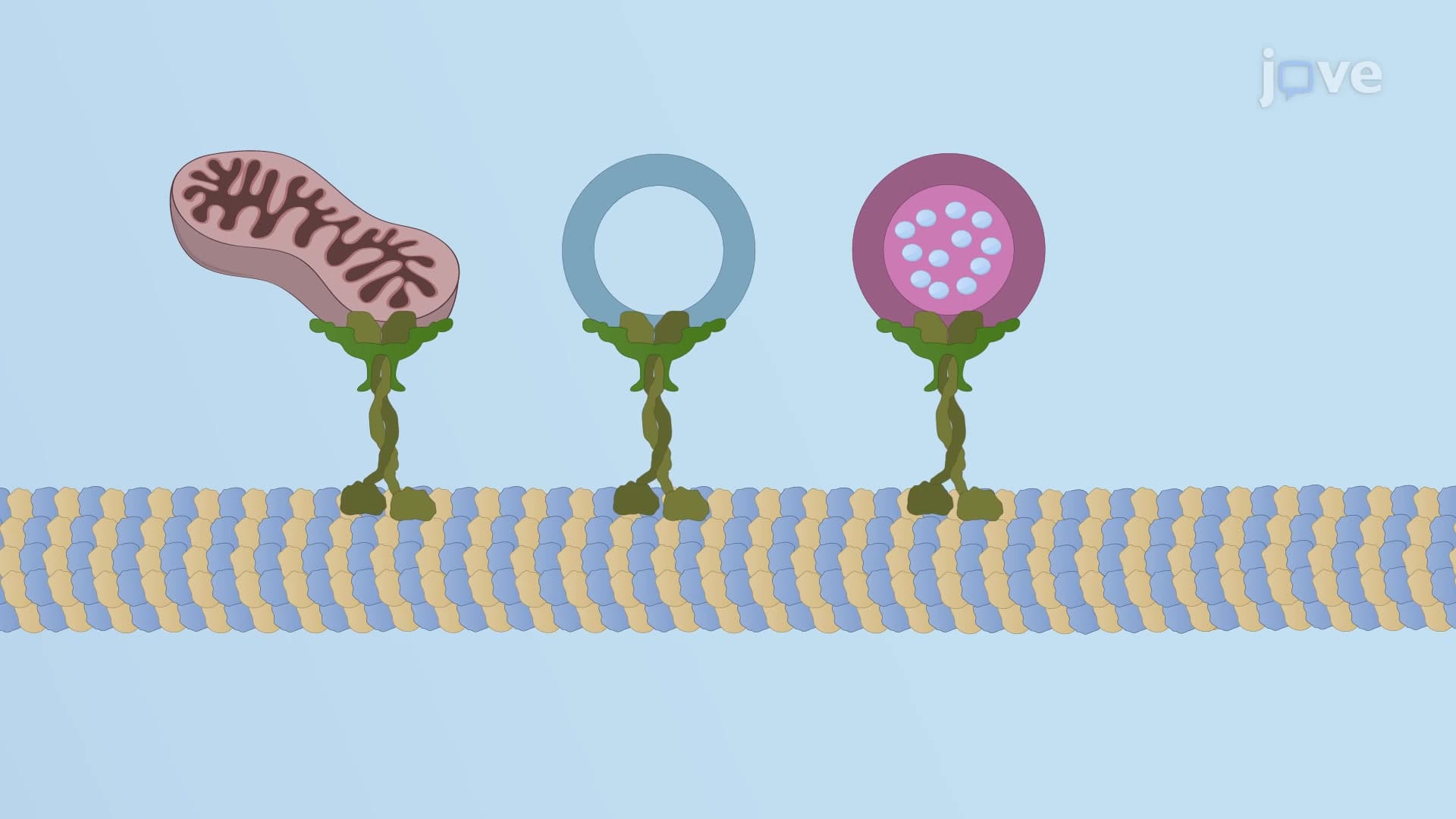
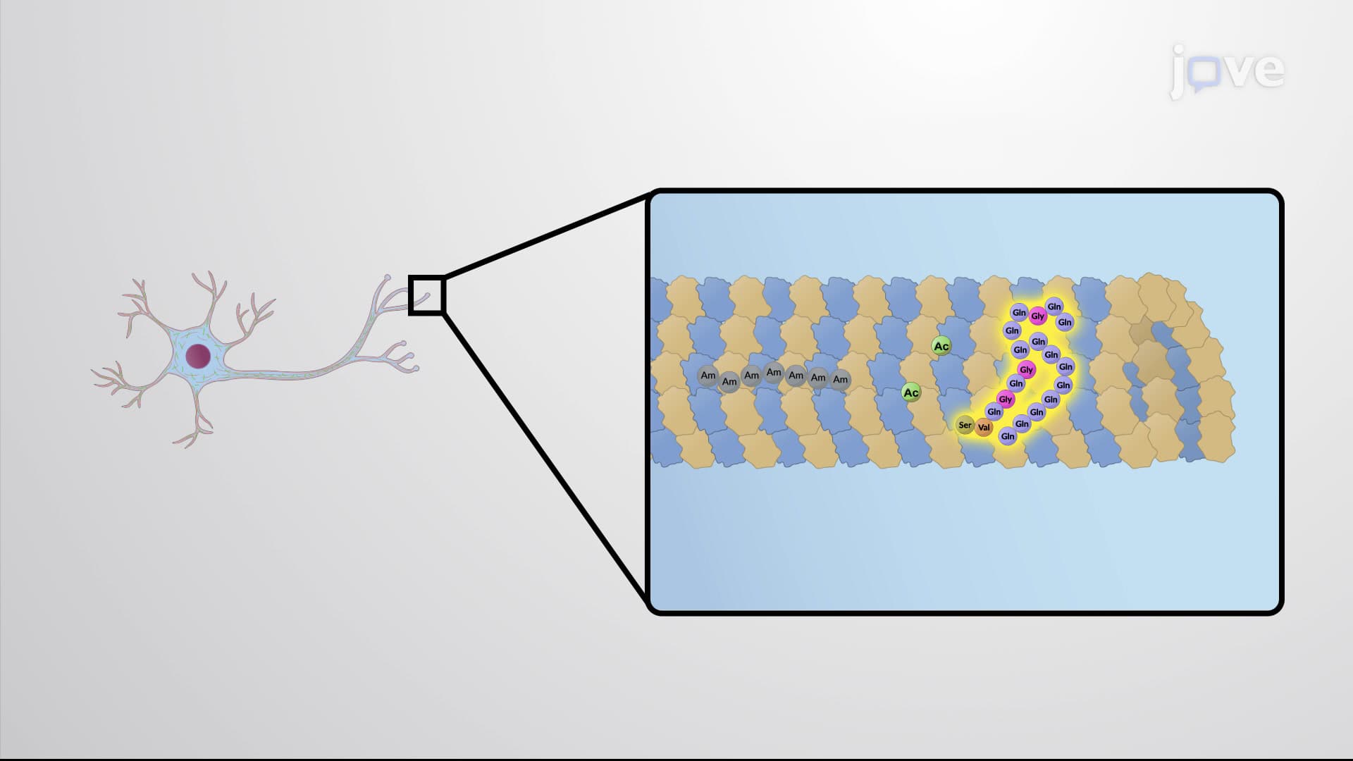
The microtubule cytoskeleton track system enables sophisticated cargo transport through two primary motor proteins. Kinesin motors move toward the plus-end, typically transporting materials from the cell center to the periphery, while dynein motors travel toward the minus-end, bringing materials back to the cell center. This bidirectional highway system is essential for neurons-a single motor neuron axon can extend over three feet in tall individuals, requiring efficient transport of neurotransmitters, mitochondria, and other cellular components. Research at institutions like the Mayo Clinic has shown that disrupted microtubule transport contributes to neurodegenerative diseases like ALS and Alzheimer's.
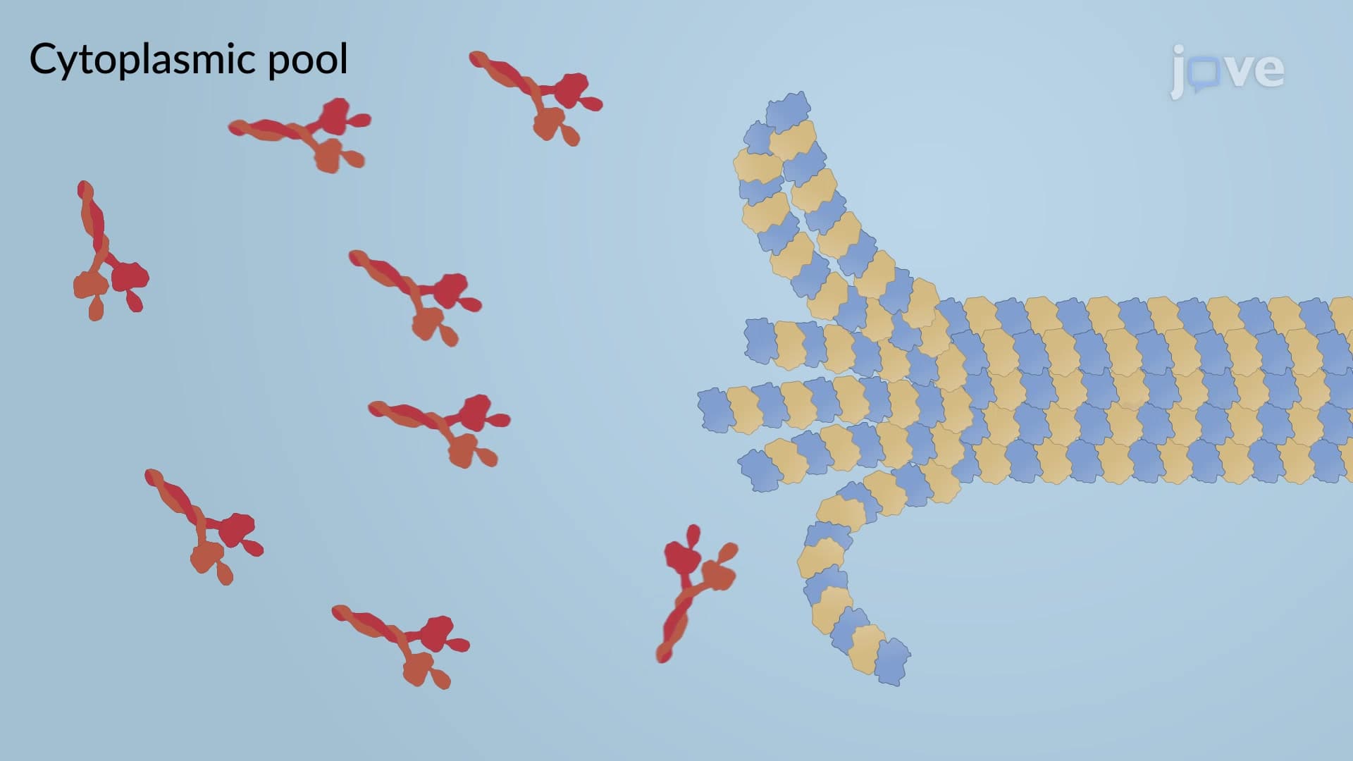
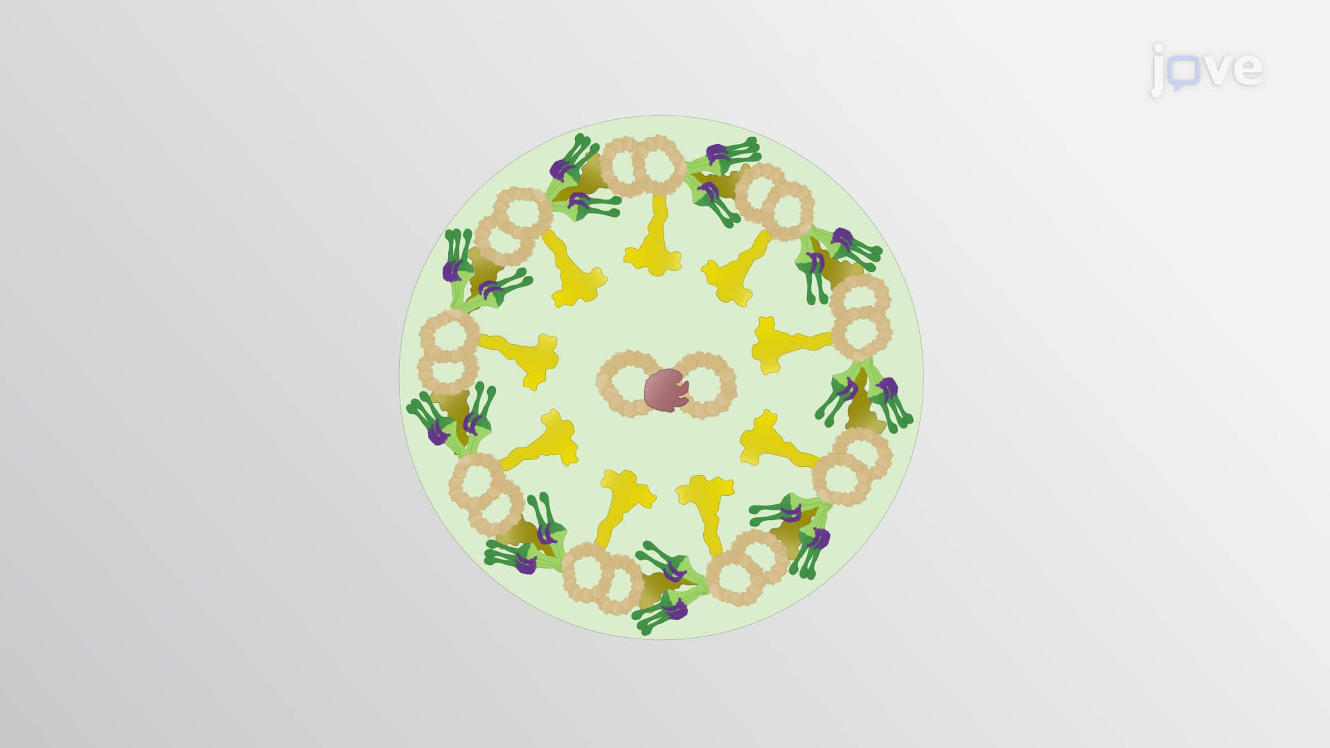
During mitosis, spindle fiber microtubule formation creates the mitotic spindle apparatus, one of the most critical structures in cell biology. This process involves dramatic microtubule reorganization, with centrosomes serving as spindle poles and microtubules extending to capture chromosomes at their kinetochores. The concept of microtubule dynamic instability-where microtubules rapidly switch between growth and shrinkage phases-allows precise chromosome positioning and segregation. Additionally, cilia flagella microtubule structures follow a characteristic "9+2" arrangement, where nine doublet microtubules surround two central singlets, enabling the coordinated beating motions essential for cellular motility and fluid movement in organs like the respiratory tract.
Related Micro-courses