Loading video…


 19 Concepts
 78.1K views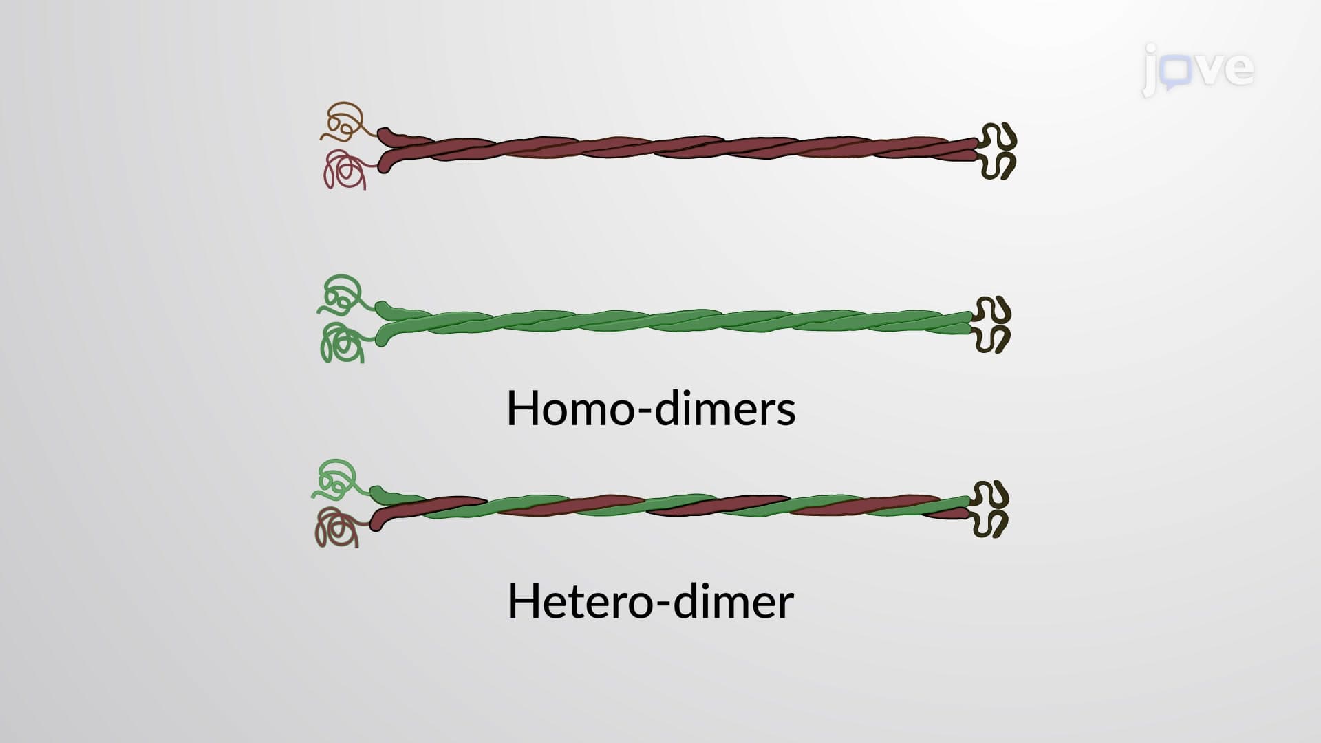

Video Summary: What are Types of Intermediate Filaments
Ever wondered why your hair and nails can withstand daily abuse while maintaining their structure? The secret lies in understanding the types of intermediate filaments that form the cellular scaffolding throughout your body. From the keratin proteins strengthening a marathon runner's skin in Boston to the neurofilaments supporting nerve signals in medical students studying at Johns Hopkins, these six distinct types of intermediate filaments create the backbone of cellular architecture. Watch the full video on JoVE Coach to master this concept with expert-led visuals and step-by-step explanations.
The types of intermediate filaments represent a sophisticated classification system that reveals how cells customize their structural support based on functional demands. Unlike actin filaments and microtubules, intermediate filaments provide mechanical resilience and tissue-specific architectural solutions. This classification becomes particularly relevant when studying cellular biology for AP courses or preparing for the MCAT, where understanding structure-function relationships is crucial.
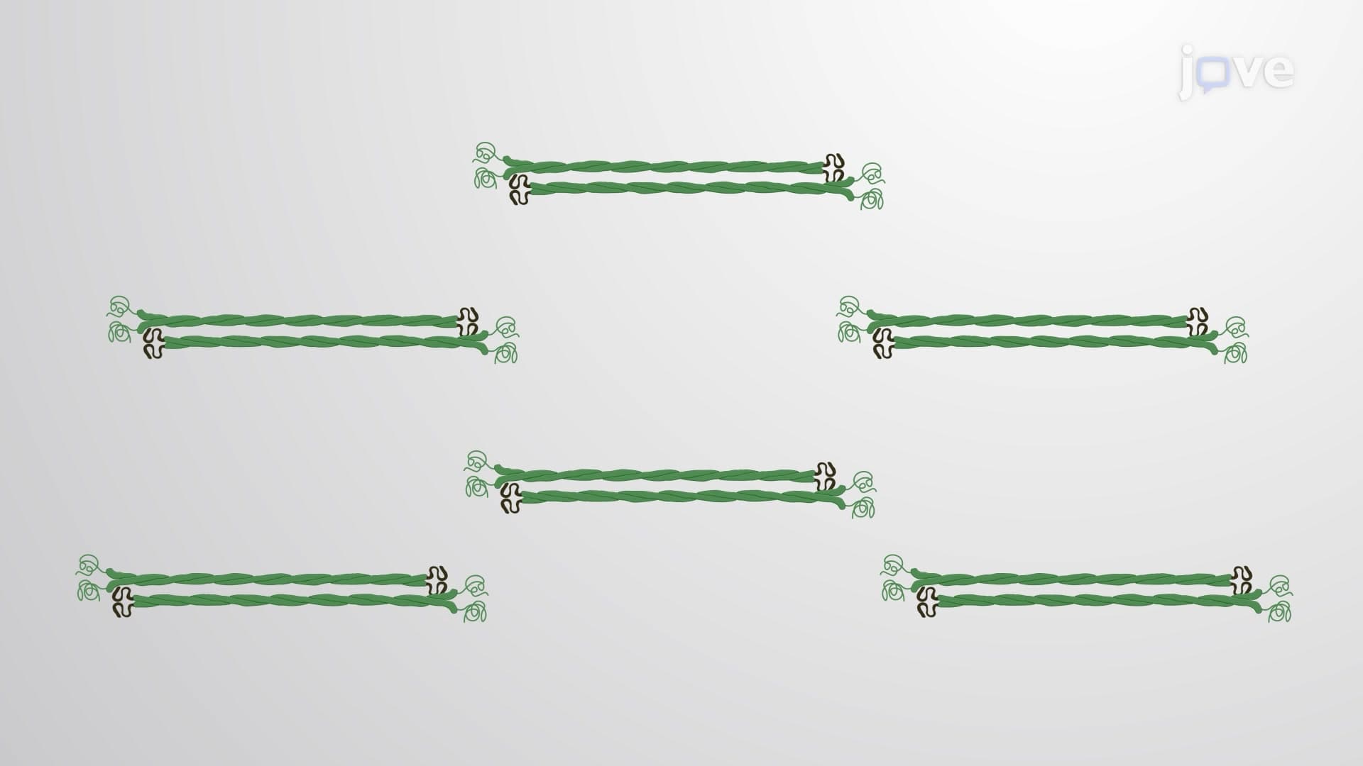
Types 1 and 2 intermediate filaments demonstrate elegant molecular cooperation through their keratin protein composition. These interdependent proteins form heterodimeric filaments exclusively found in epithelial cells. Consider the practical application: a dermatologist treating patients at the Mayo Clinic relies on understanding how these keratins provide the high tensile strength needed in constantly abraded tissues like palms and soles. The abundance of these filaments in skin and nails explains why these structures can withstand mechanical stress that would destroy other cellular components.
Type 3 intermediate filaments showcase remarkable versatility through their four distinct proteins: vimentin, desmin, glial fibrillary acidic protein (GFAP), and peripherin. Unlike Types 1 and 2, these can form both homodimers and heterodimers, providing cellular flexibility. Vimentin appears in mesenchymal cells, while desmin specifically supports muscle architecture. Medical students studying pathology learn that GFAP serves as a critical biomarker for brain tumors, as it's predominantly expressed in astrocytes.
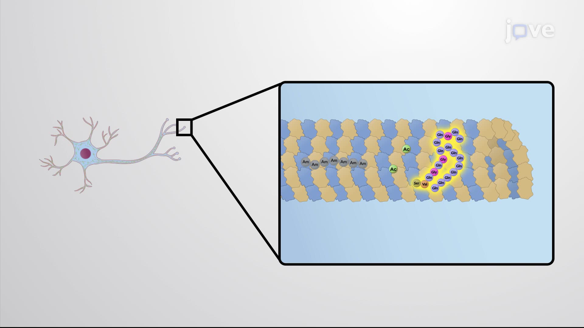
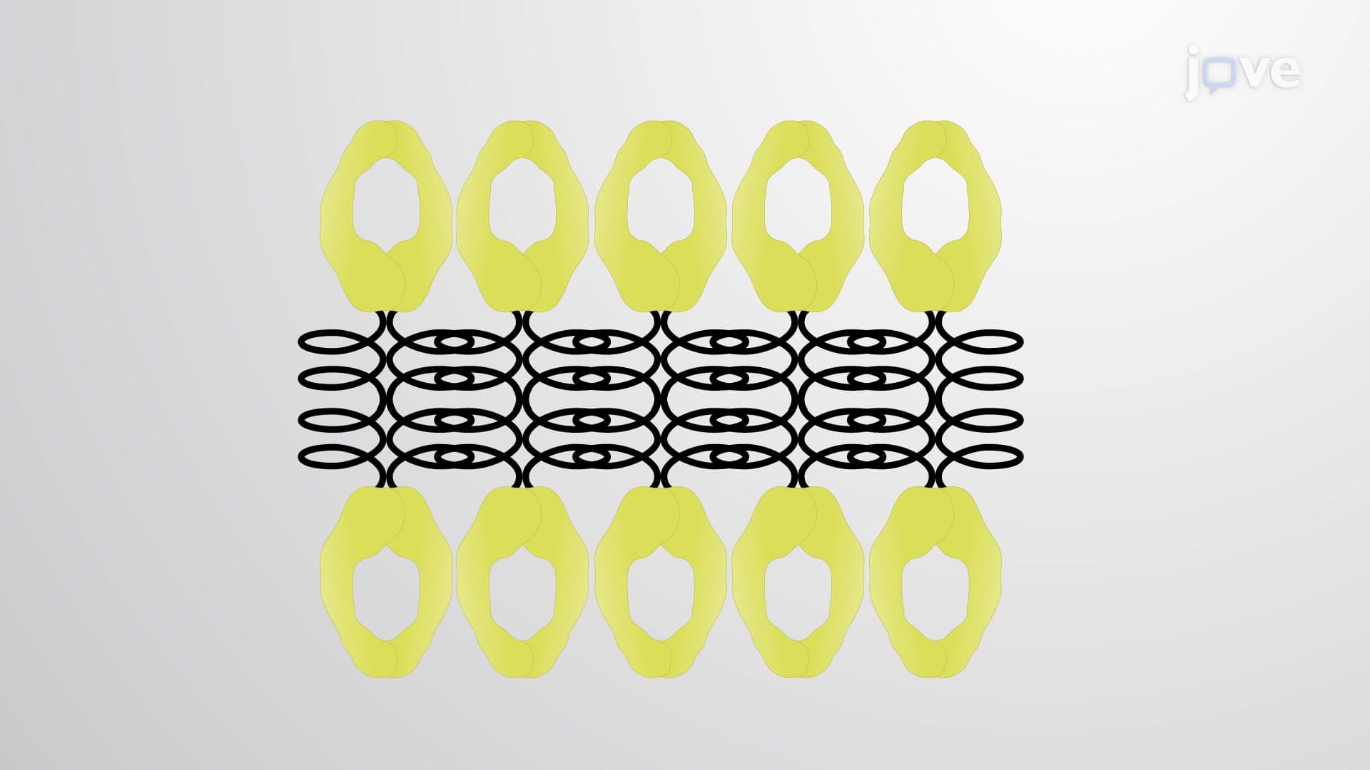
Type 4 neurofilaments represent highly specialized structures essential for nervous system function. Their glutamic acid-rich tails and characteristic short sidearms directly influence axon diameter, which determines nerve conduction velocity. Neurologists at institutions like Harvard Medical School study how neurofilament dysfunction contributes to neurodegenerative diseases, making this knowledge clinically relevant for future healthcare professionals.
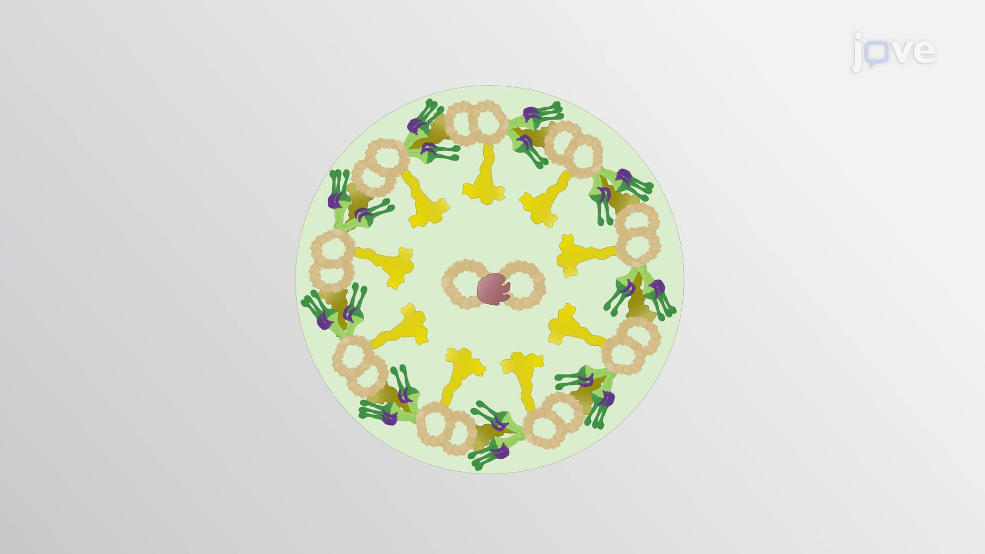
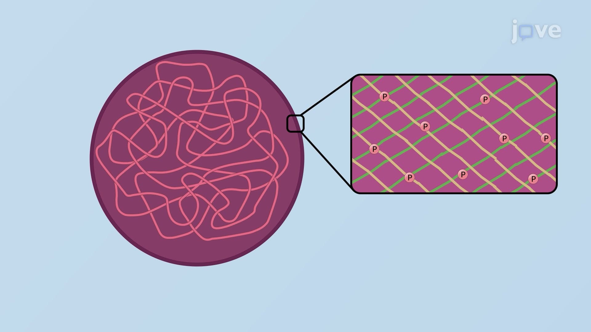
Type 5 nuclear lamins occupy a unique position within the nuclear envelope's inner membrane. Their immunoglobulin-fold structure and distinctive CAAX box (cysteine-aliphatic-aliphatic-X amino acid sequence) enable crucial nuclear functions. College biochemistry courses emphasize how lamin mutations cause laminopathies, including premature aging disorders studied at research centers like the National Institutes of Health.
The recently discovered Type 6 beaded intermediate filaments represent cutting-edge cell biology research, predominantly found in lens cells and neuronal stem cells. Their ability to form both homodimers and heterodimers suggests specialized roles in cellular differentiation and maintenance, areas actively investigated at US research universities.
Related Micro-courses