Loading video…


 19 Concepts
 78.1K views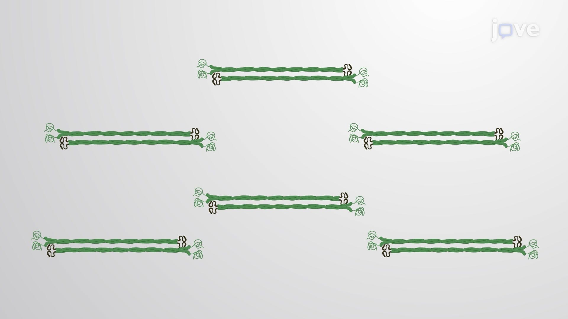

Video Summary: What Is Formation of Intermediate Filaments
Did you know that your skin cells contain protein cables stronger than steel wire that help them resist mechanical stress? The formation of intermediate filaments follows a precise assembly process where protein monomers build into coiled-coil dimers, then tetramers, and finally polymerize into sturdy 10-nanometer filaments. These structures are essential in conditions like epidermolysis bullosa, where faulty keratin intermediate filaments cause severe skin blistering in patients treated at hospitals like Johns Hopkins. Watch the full video on JoVE Coach to master this concept with expert-led visuals and step-by-step explanations.
The formation of intermediate filaments represents one of cell biology's most elegant examples of hierarchical protein assembly. Unlike actin filaments or microtubules, intermediate filaments follow a unique construction pathway that creates remarkably stable cellular structures. This process is particularly important for pre-med students preparing for the MCAT, as intermediate filament dysfunction underlies numerous human diseases.
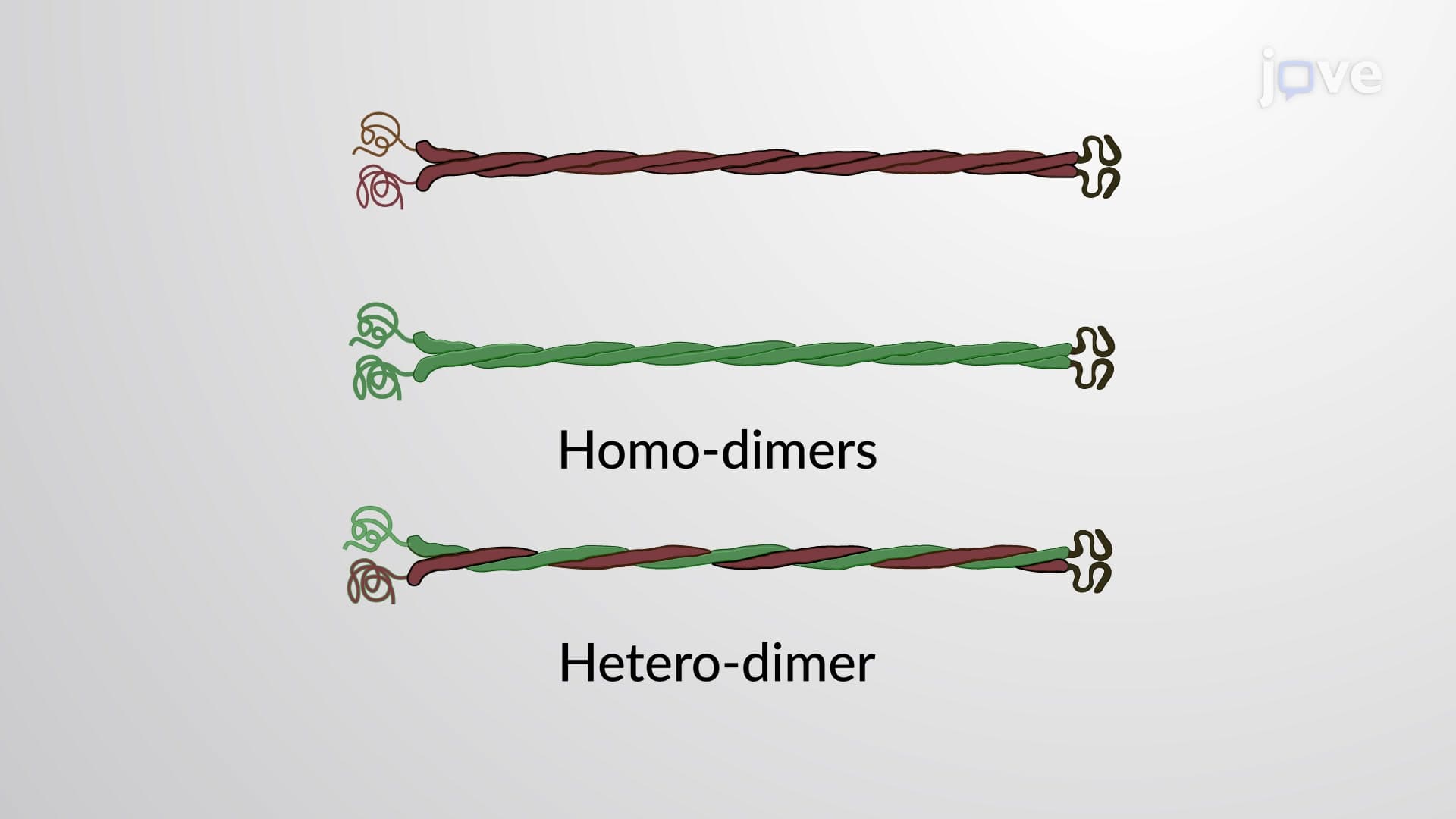
The assembly begins with two monomeric protein subunits that interact laterally to form a coiled-coil dimer. This coiled-coil structure, similar to the structure found in muscle myosin, provides the basic building block's stability. In human keratinocytes (skin cells), keratin monomers assemble this way to begin forming the protective intermediate filament network that helps skin resist mechanical damage.
Two oppositely oriented polar dimers associate to create a nonpolar tetramer-the fundamental unit of all intermediate filaments. This design eliminates the polarity found in actin filaments and microtubules, contributing to intermediate filaments' unique properties. Tetramers then bind end-to-end, forming protofilaments that serve as the scaffold for further assembly. Students studying for AP Biology exams should note how this differs from actin polymerization, which involves monomer addition rather than pre-formed tetramer units.
Protofilaments associate laterally to create protofibrils measuring 2-3 nanometers in width. When four protofibrils coil together laterally, they form a unit-length filament approximately 60 nanometers long and 10 nanometers wide. These unit-length filaments then polymerize head-to-tail to create the mature intermediate filament. This multi-step process explains why intermediate filaments are more stable than other cytoskeletal elements-they're essentially molecular ropes with multiple levels of structural redundancy.
Understanding this assembly process helps explain clinical conditions like Hutchinson-Gilford progeria syndrome, where defective lamin intermediate filaments in the nuclear envelope cause premature aging, affecting patients treated at specialized centers like the NIH Clinical Center.
Related Micro-courses