Loading video…


 19 Concepts
 78.1K views

Video Summary: What Is Microtubule Associated Proteins Maps
Microtubule associated proteins maps reveal how cellular "highways" control everything from brain function to cancer cell division. In Alzheimer's disease, the breakdown of Tau proteins-key microtubule stabilizers-leads to the characteristic neurofibrillary tangles found in patient brain tissue at Johns Hopkins Medical Center. Understanding what is microtubule associated proteins maps helps explain how cells maintain their structural integrity and coordinate complex movements. Watch the full video on JoVE Coach to master this concept with expert-led visuals and step-by-step explanations.
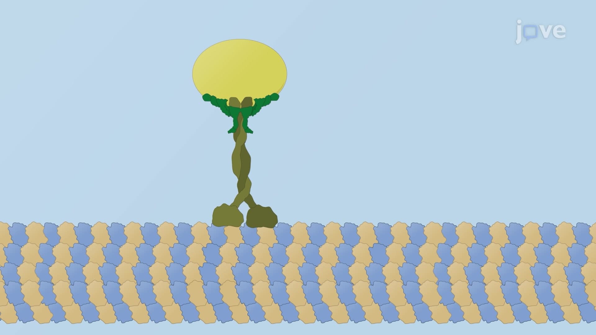
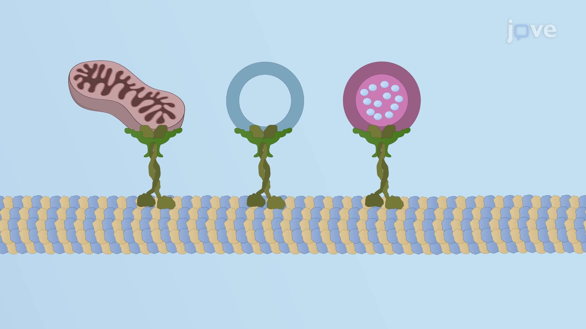
Microtubule associated proteins maps represent a sophisticated regulatory system that controls one of the cell's most dynamic structures. These specialized proteins act like molecular traffic controllers, determining when, where, and how microtubules assemble and disassemble. Unlike static structural components, MAPs respond to cellular signals, making them crucial for processes ranging from mitosis to neuronal signaling.
The diversity of MAP functions reflects the complexity of microtubule regulation. Each MAP type has evolved specific binding domains and regulatory mechanisms that allow precise control over microtubule behavior. This precision becomes critical in specialized cells like neurons, where microtubule organization directly impacts synaptic function and axonal transport.
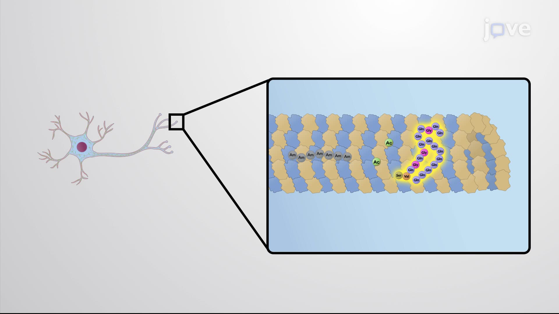
Stabilizing MAPs like Tau protein demonstrate how electrostatic interactions drive biological function. Tau's positively charged domains bind to microtubules' negatively charged surface, reducing catastrophic depolymerization events. In healthy neurons at institutions like Stanford Medical School, researchers observe how proper Tau function maintains axonal integrity over decades.
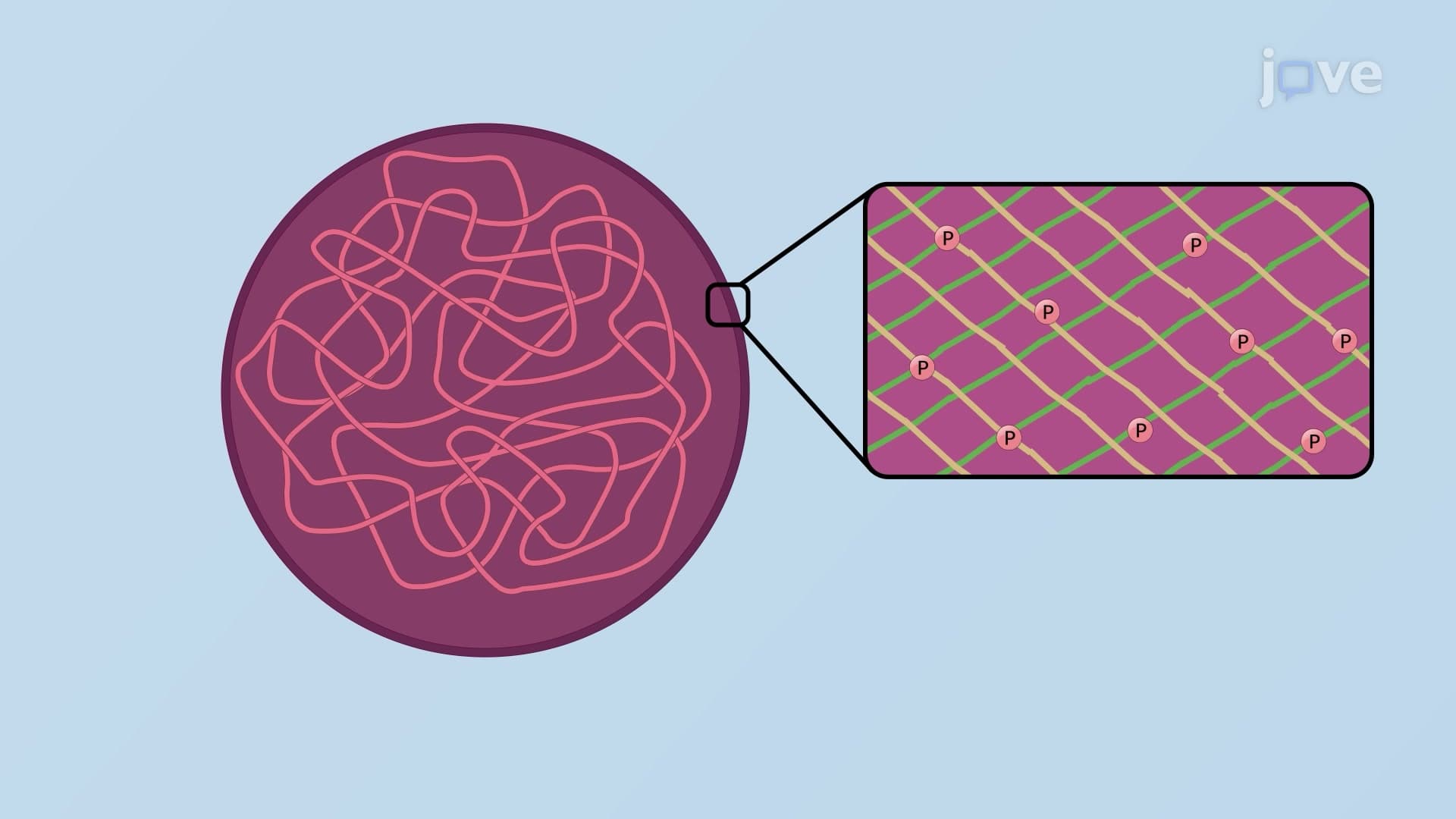
This stabilization mechanism becomes clinically relevant in tauopathies, where abnormal Tau phosphorylation disrupts microtubule binding. The resulting neurodegeneration illustrates why understanding MAP function is essential for pre-med students preparing for the MCAT's biochemistry section.
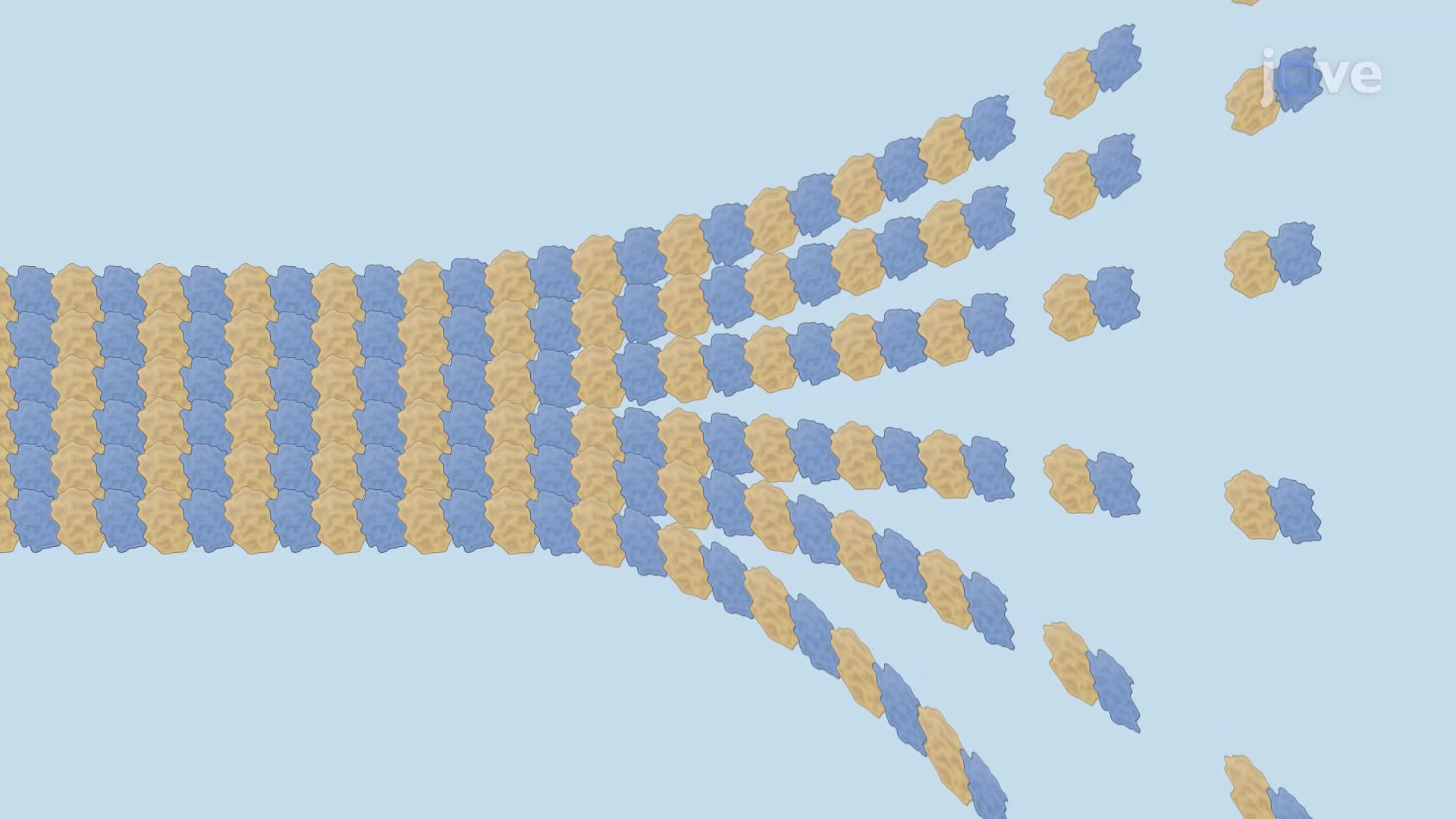
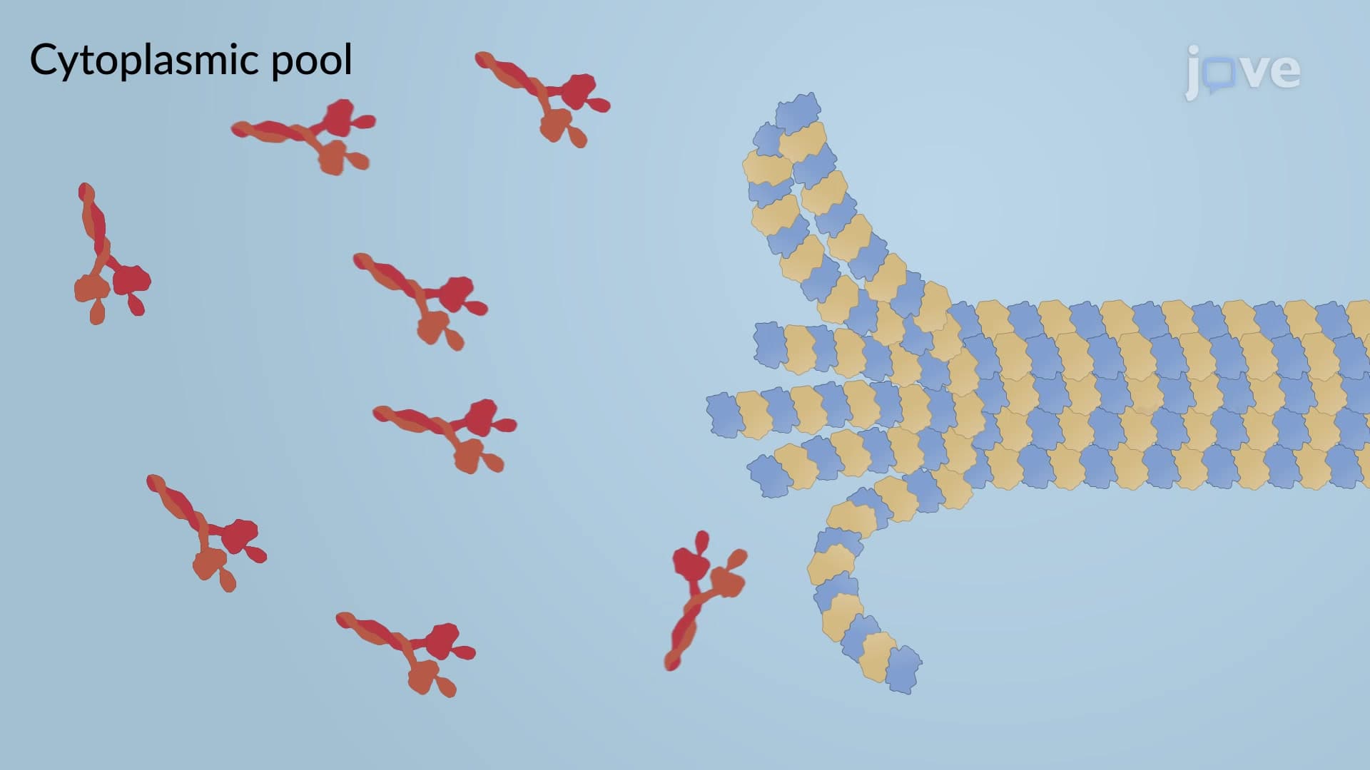
Destabilizing MAPs like stathmin provide the counterbalance to stabilization, ensuring microtubules remain responsive to cellular needs. Stathmin binds directly to tubulin dimers, preventing their incorporation into growing microtubules. This mechanism becomes particularly important during cell division, when rapid microtubule turnover is essential for proper chromosome segregation.
Cancer researchers at Memorial Sloan Kettering have identified stathmin overexpression in various tumors, where excessive microtubule destabilization contributes to chromosomal instability. This connection between MAP dysfunction and disease makes these proteins important targets for therapeutic intervention.
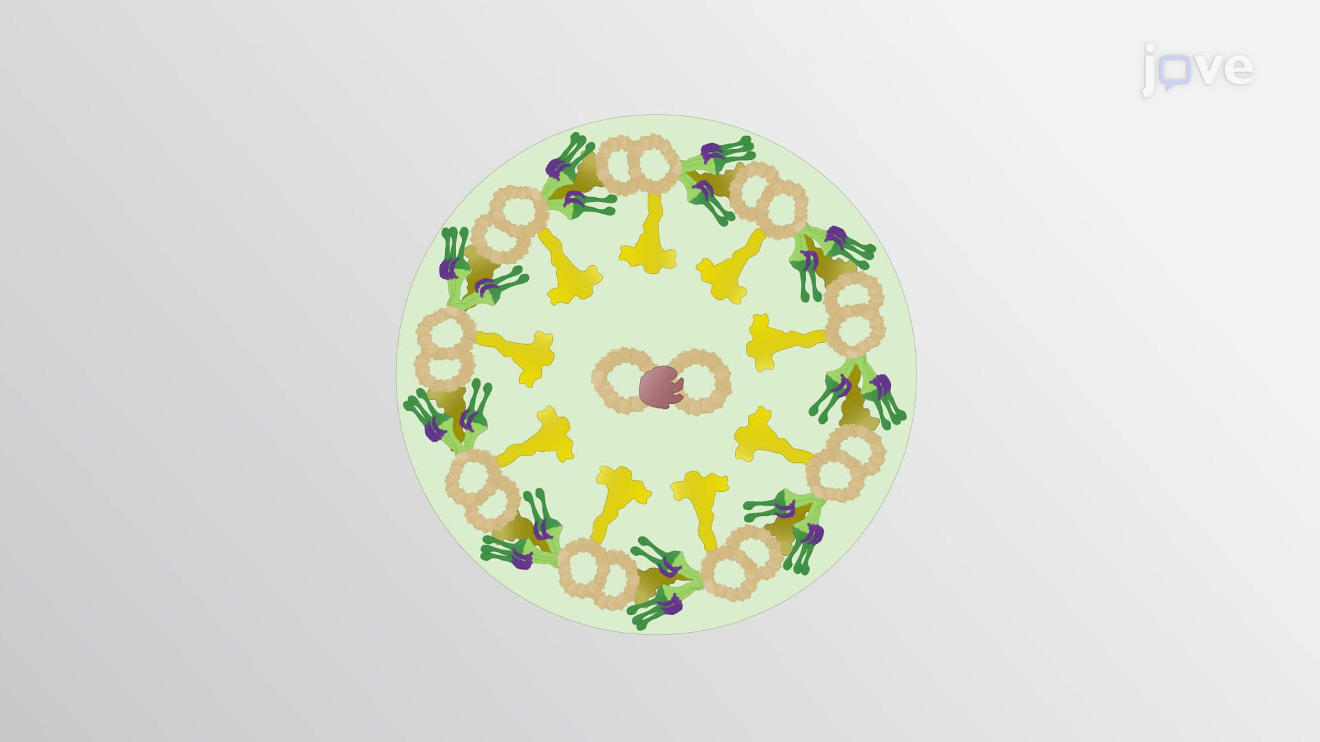
Capping proteins like the gamma-tubulin ring complex (γ-TURC) control microtubule nucleation sites, typically at centrosomes. By binding to microtubule minus-ends, γ-TURC ensures unidirectional growth toward the cell periphery. This asymmetric organization is crucial for establishing cellular polarity, a concept frequently tested in AP Biology exams.
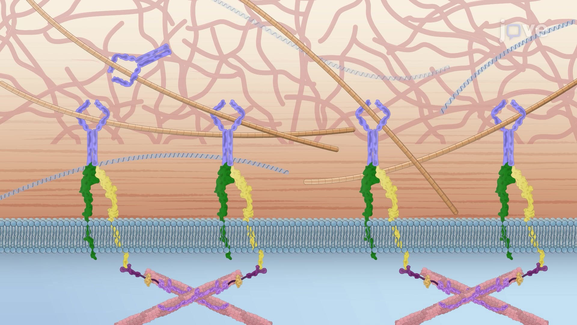
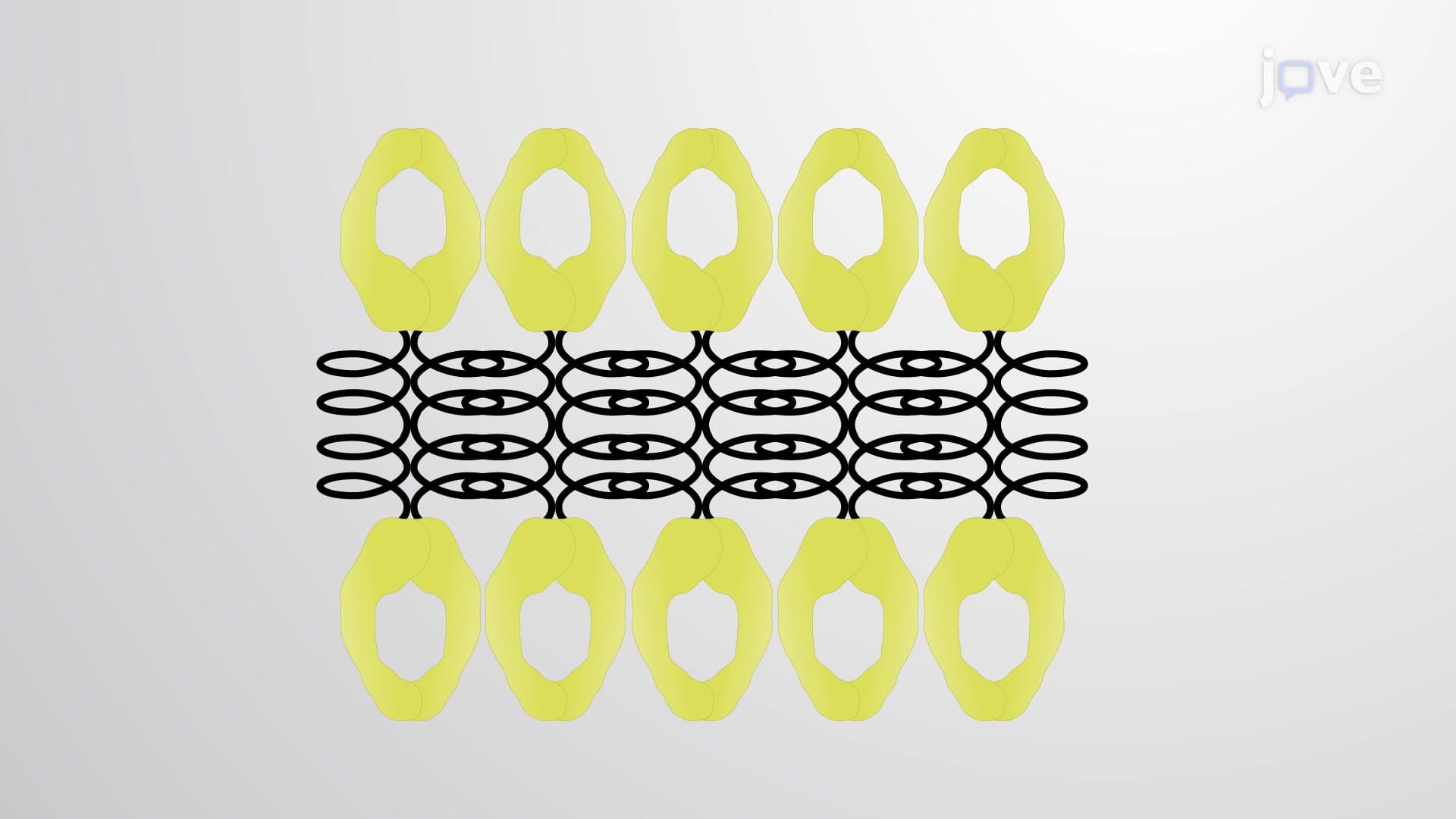
Crosslinker MAPs and cytoskeletal integrators create higher-order organization. MAP65 bundles antiparallel microtubules during mitosis, while plakin proteins connect microtubules to intermediate filaments. These interactions create the integrated cytoskeletal network that maintains cell shape and enables coordinated cellular movements.
Related Micro-courses