Loading video…


 19 Concepts
 78.1K views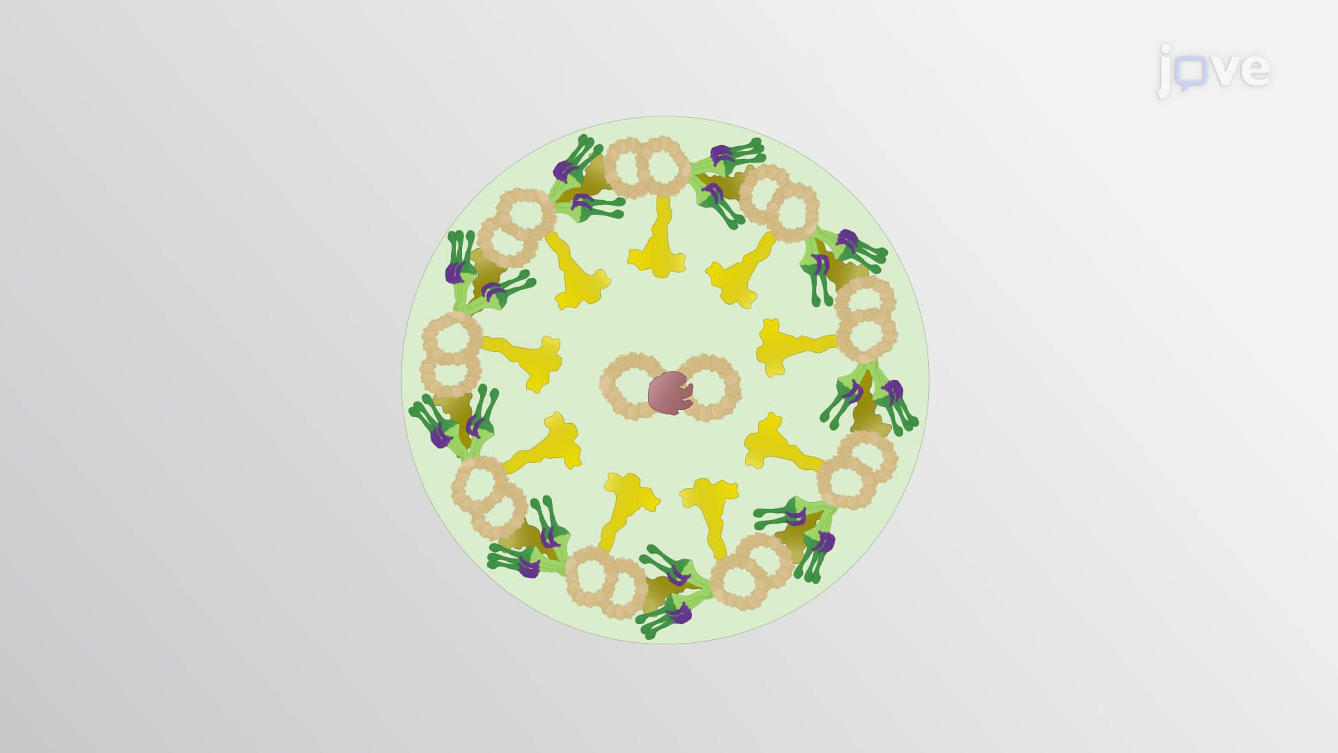

Video Summary: What Is Microtubules in Cell Motility
Ever wonder how sperm cells swim toward an egg or how bacteria propel themselves through liquid? Microtubules cell motility mechanisms power these remarkable movements through specialized structures called cilia and flagella. These hair-like projections contain a precise 9+2 arrangement of microtubules working with motor proteins like dynein to generate the coordinated beating patterns that drive cellular movement. In human reproduction, sperm flagella use this exact system to navigate the female reproductive tract. Watch the full video on JoVE Coach to master this concept with expert-led visuals and step-by-step explanations.
Microtubules cell motility represents one of biology's most elegant engineering solutions. These cylindrical protein structures serve as both the skeleton and motor system for cellular movement, particularly in specialized appendages like cilia and flagella. Unlike simple passive structures, microtubules actively participate in generating the forces needed for coordinated cellular locomotion.
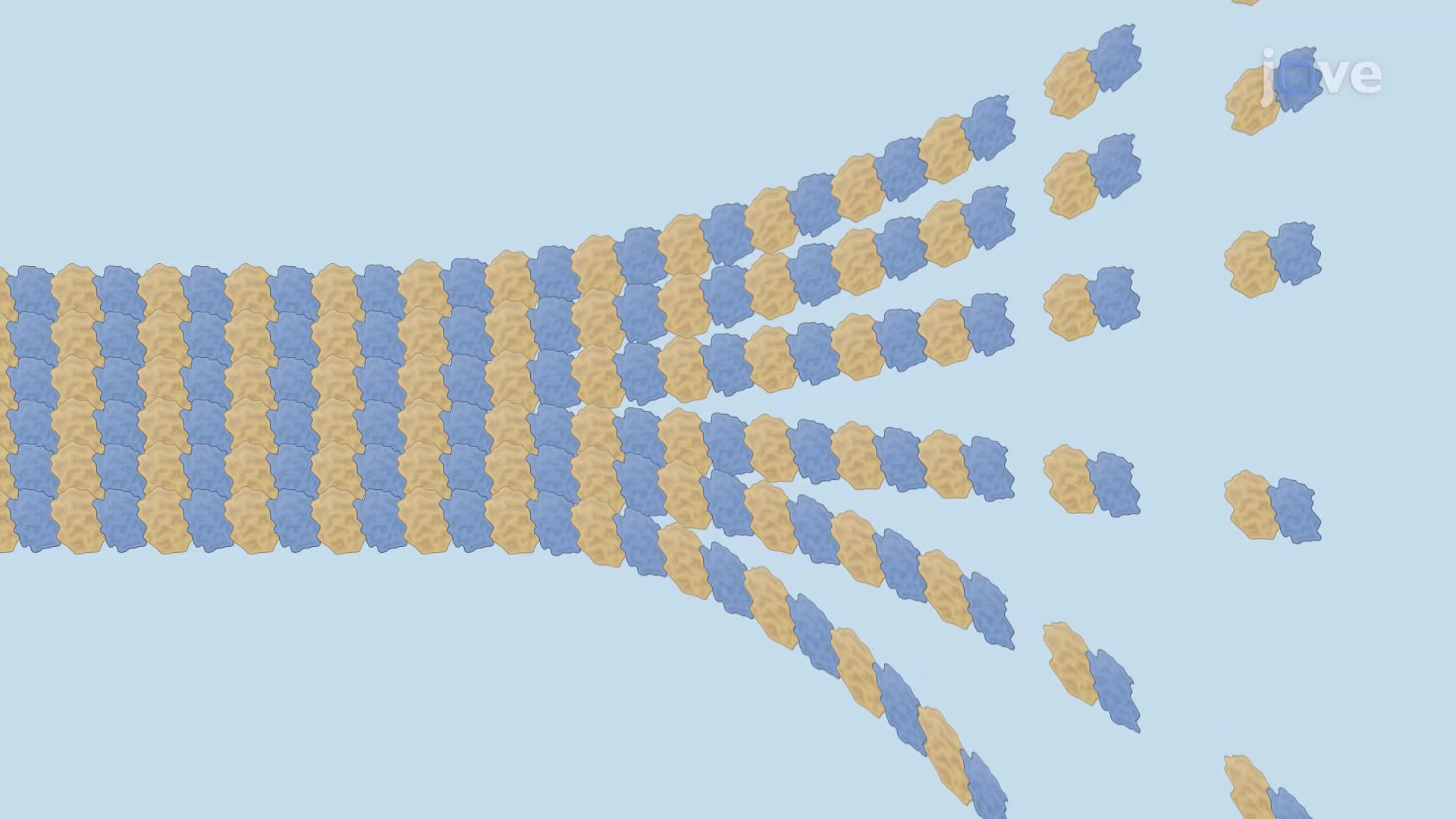
The microtubule role in cell movement becomes clear when examining the axoneme's precise architecture. Nine doublet microtubules form an outer ring surrounding two central singlet microtubules-a pattern so consistent it appears in organisms from algae to humans. Each doublet consists of a complete A-tubule (13 protofilaments) fused to an incomplete B-tubule (10 protofilaments). This asymmetric design creates the structural foundation for directional force generation.
AP Biology students frequently encounter this topic when studying cellular structure and function. The 9+2 pattern exemplifies how molecular organization directly relates to biological function-a key concept tested on college entrance exams and introductory biology courses at institutions like UCLA or Harvard.
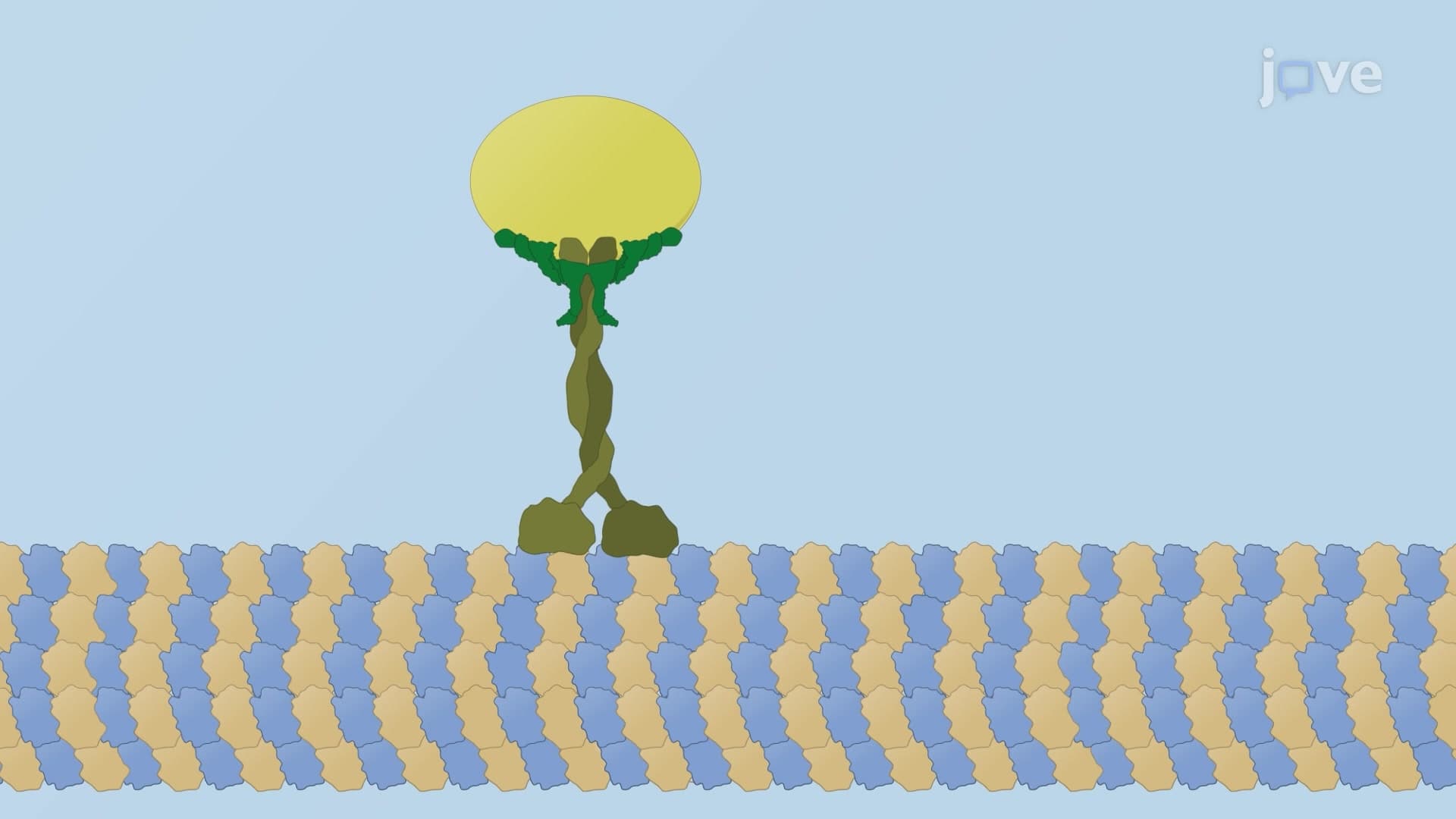
How microtubules contribute to cell motility depends critically on dynein motor proteins. These molecular machines have stems anchored in A-tubules while their globular heads reach toward neighboring B-tubules. When dynein "walks" along microtubules using ATP energy, it creates sliding forces between adjacent doublets. However, nexin links prevent complete sliding, converting linear motion into the bending waves characteristic of ciliary and flagellar beating.
This mechanism explains sperm motility disorders studied in US fertility clinics. Patients with defective dynein arms experience reduced sperm movement, directly linking molecular structure to reproductive health outcomes.
Understanding cytoskeletal motility microtubule function helps explain primary ciliary dyskinesia (PCD), affecting approximately 1 in 15,000 Americans. Patients with this genetic disorder have defective cilia in their respiratory tracts, leading to chronic infections and breathing difficulties. Medical schools like Johns Hopkins emphasize these connections when teaching future physicians about the molecular basis of disease.
Related Micro-courses