Loading video…


 19 Concepts
 78.1K views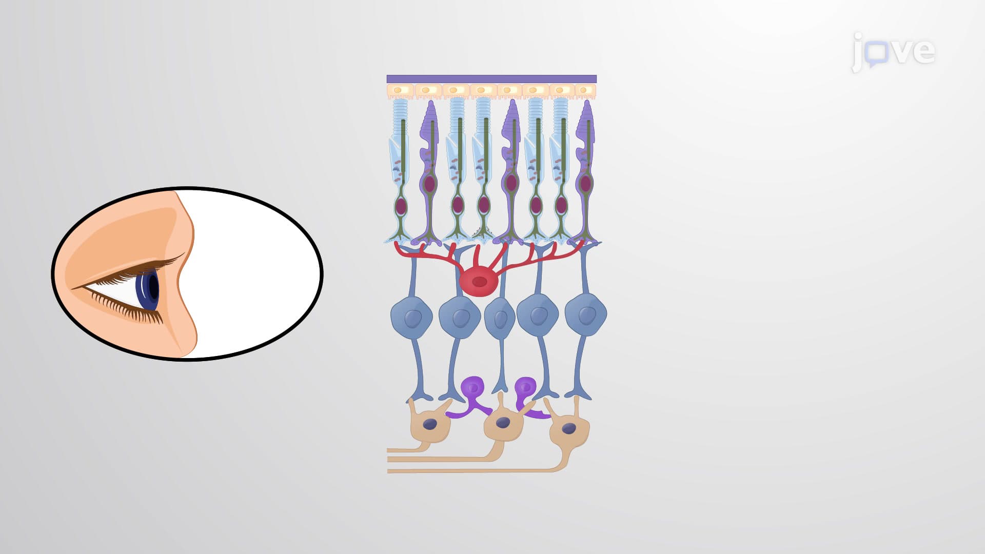

Video Summary: What Is Microtubules in Signaling
Did you know that every cell in your body has tiny "antenna" structures that help detect changes in your environment? Microtubules in signaling serve as cellular communication highways, with specialized structures called primary cilia acting like microscopic sensors. In your eye's retina, these microtubule-based structures in rod cells capture light and convert it into the electrical signals that let you see. Watch the full video on JoVE Coach to master this concept with expert-led visuals and step-by-step explanations.
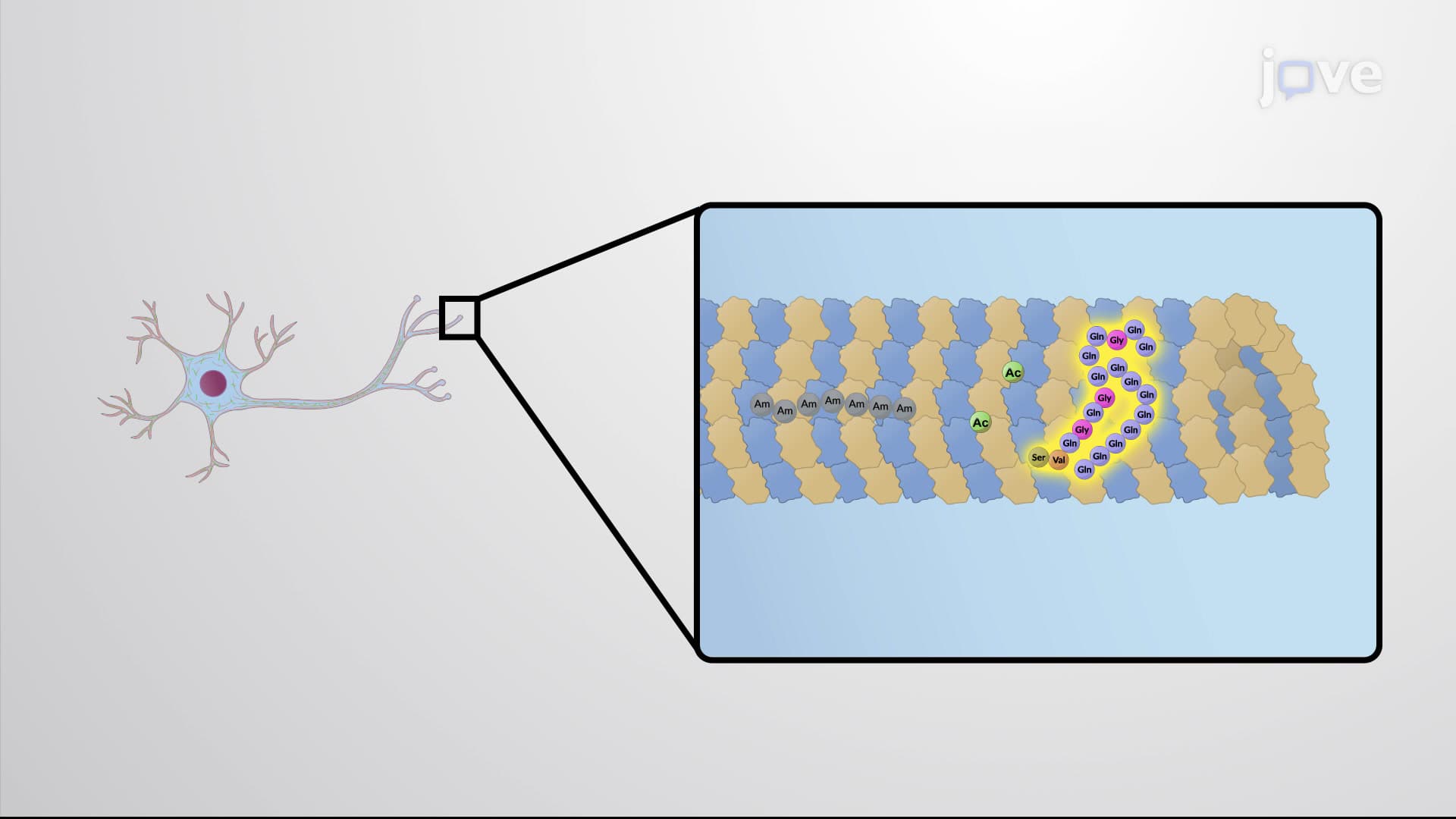
Microtubules in signaling represents a sophisticated cellular communication system where these dynamic protein filaments serve dual roles as structural supports and information highways. Unlike their well-known function in cell division, signaling microtubules create specialized sensory structures that detect environmental changes and coordinate cellular responses. This concept frequently appears on AP Biology exams and college cell biology courses, particularly when discussing cellular communication mechanisms.
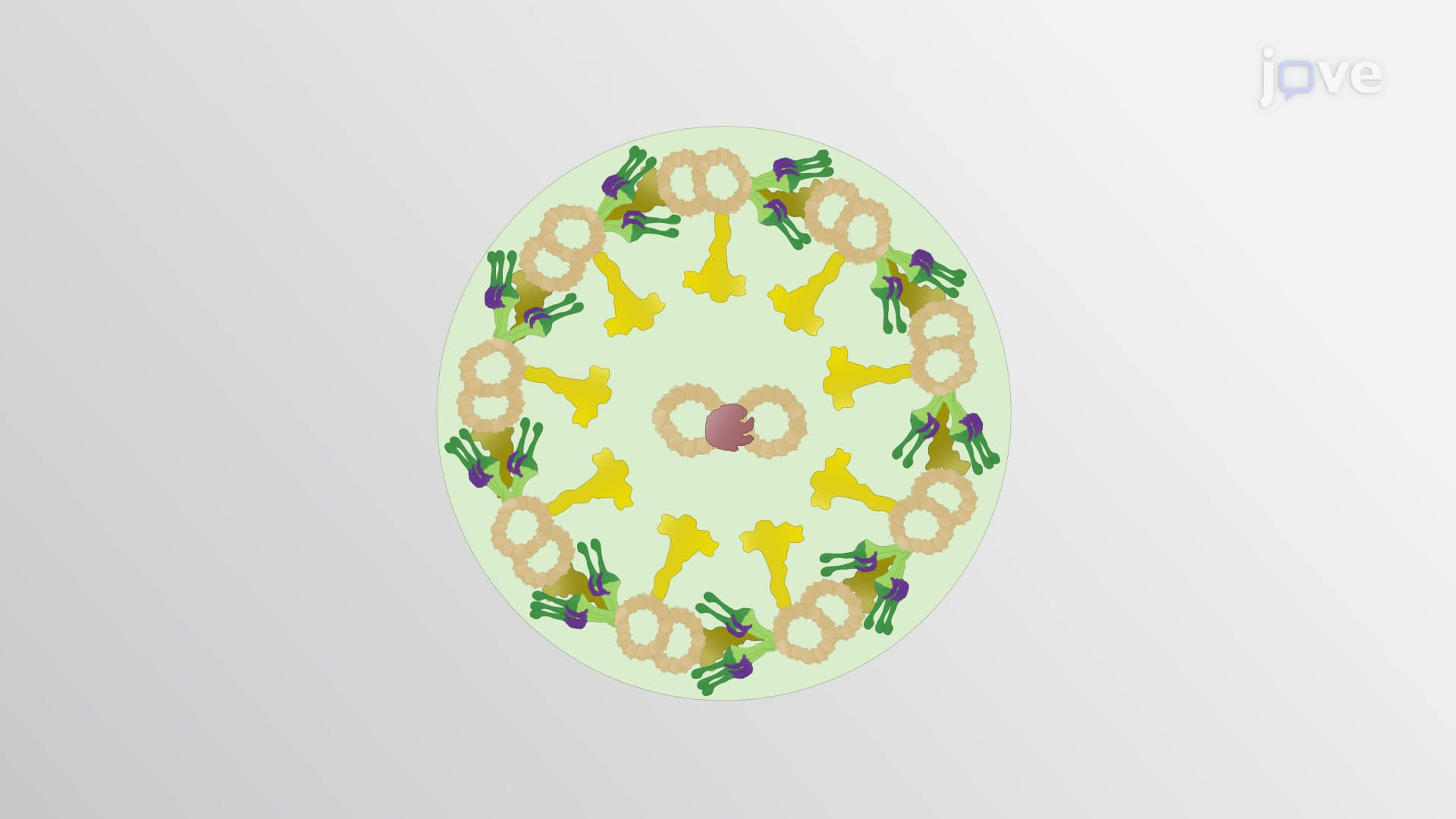
The most remarkable signaling structure involving microtubules is the primary cilium-a hair-like projection that acts as the cell's environmental sensor. These structures feature a unique 9+0 microtubule arrangement, meaning nine doublet microtubules form a circle with no central pair. This differs from motile cilia found in your respiratory tract, which have a 9+2 arrangement and move to clear mucus. Primary cilia lack the motor proteins (dyneins) that create movement, instead specializing in signal detection and transmission.
In the human visual system, microtubules in signaling demonstrate their critical importance through retinal photoreception. Rod cells in your retina contain primary cilia that connect the outer segment (containing light-detecting rhodopsin) to the inner segment (containing cellular machinery). When light hits rhodopsin molecules, the signal travels through microtubule-organized pathways to eventually release glutamate neurotransmitters. This process, essential for vision, can be disrupted in conditions like Leber congenital amaurosis, where ciliary defects cause blindness.
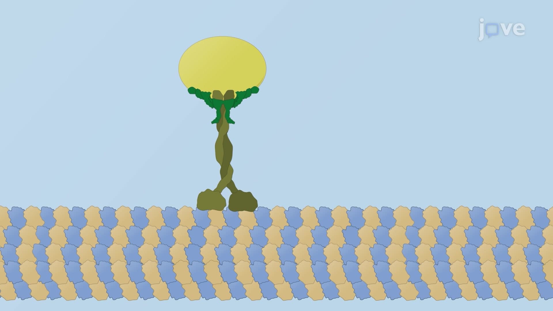
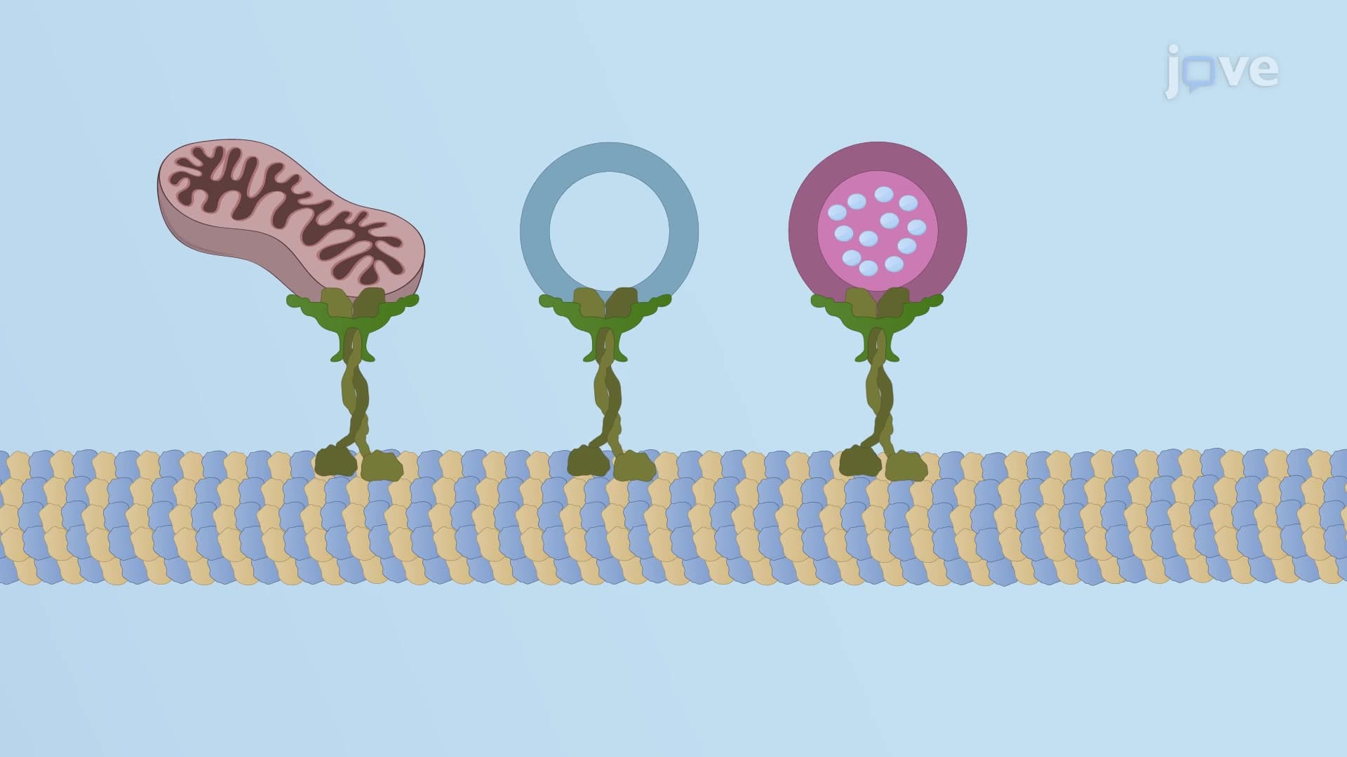
At nerve terminals, microtubules coordinate multiple signaling functions simultaneously. They anchor mitochondria in optimal positions to provide energy for neurotransmitter synthesis while also guiding synaptic vesicles to release sites. This organization is crucial for proper neural communication and appears frequently in MCAT neurophysiology questions. Understanding how microtubules facilitate both structural organization and signal transmission helps explain complex neurological processes tested in medical school curricula.
Related Micro-courses