Loading subject page…


145,116 views

Video Summary: Electron Transport Chain Complex I and Ii Explained
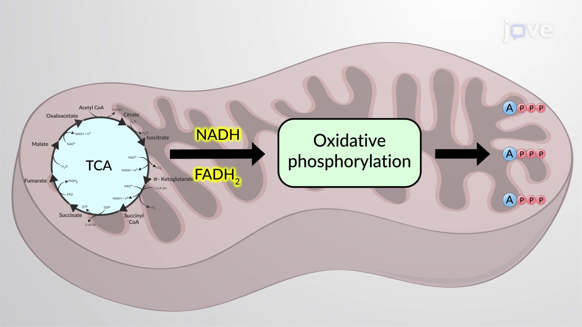
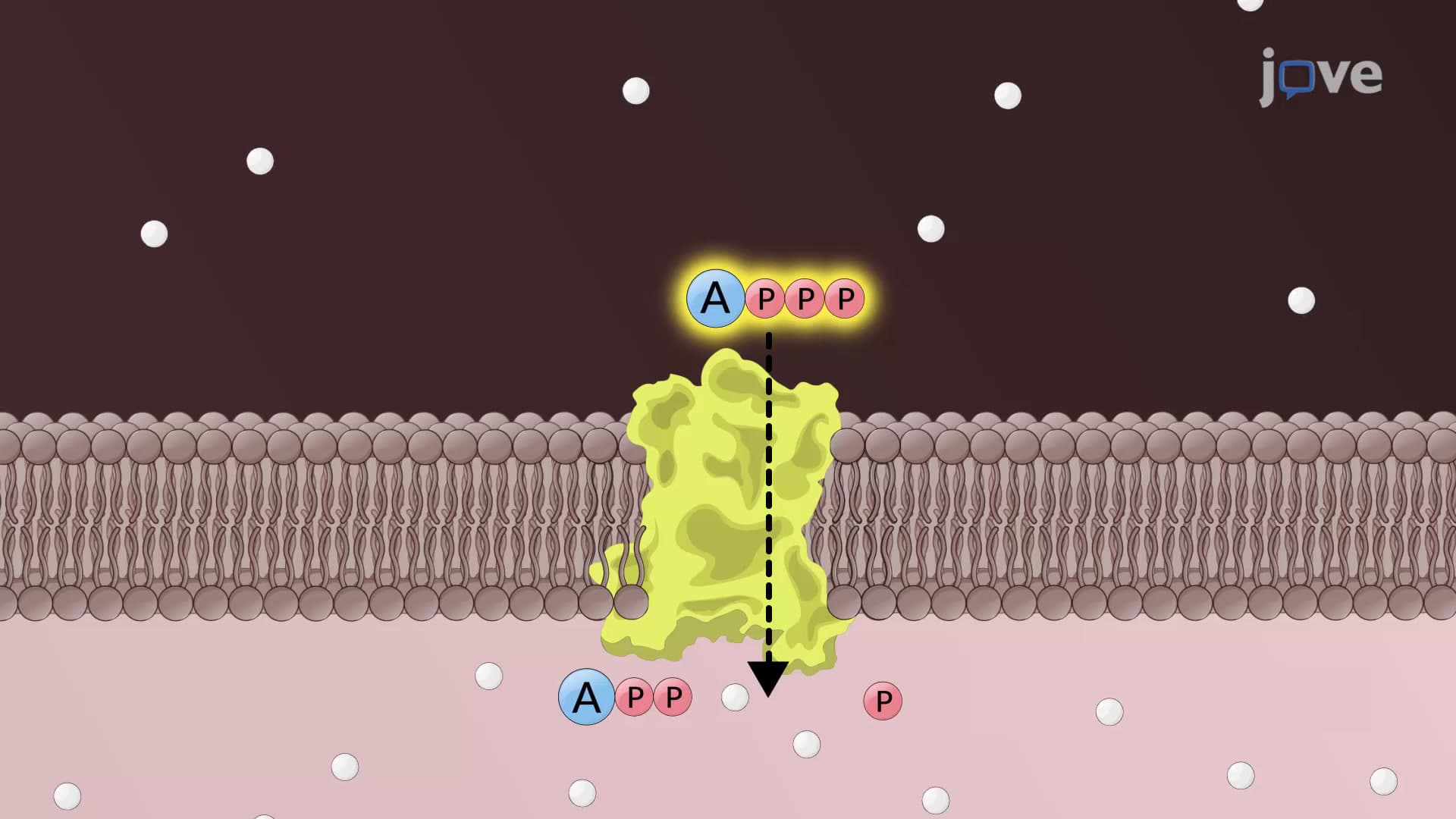
Did you know that every cell in your body contains molecular machines that generate ATP with the precision of a power plant turbine? The electron transport chain complex I and II are the first two critical components in this cellular energy factory, working together to capture energy from food molecules and convert it into usable cellular fuel. Consider how medications for Parkinson's disease specifically target Complex I dysfunction in brain cells, highlighting the clinical importance of these mitochondrial complexes. These protein complexes orchestrate the initial steps of electron transfer that ultimately power everything from muscle contractions to brain function. Watch the full video on JoVE Coach to master this concept with expert-led visuals and step-by-step explanations.
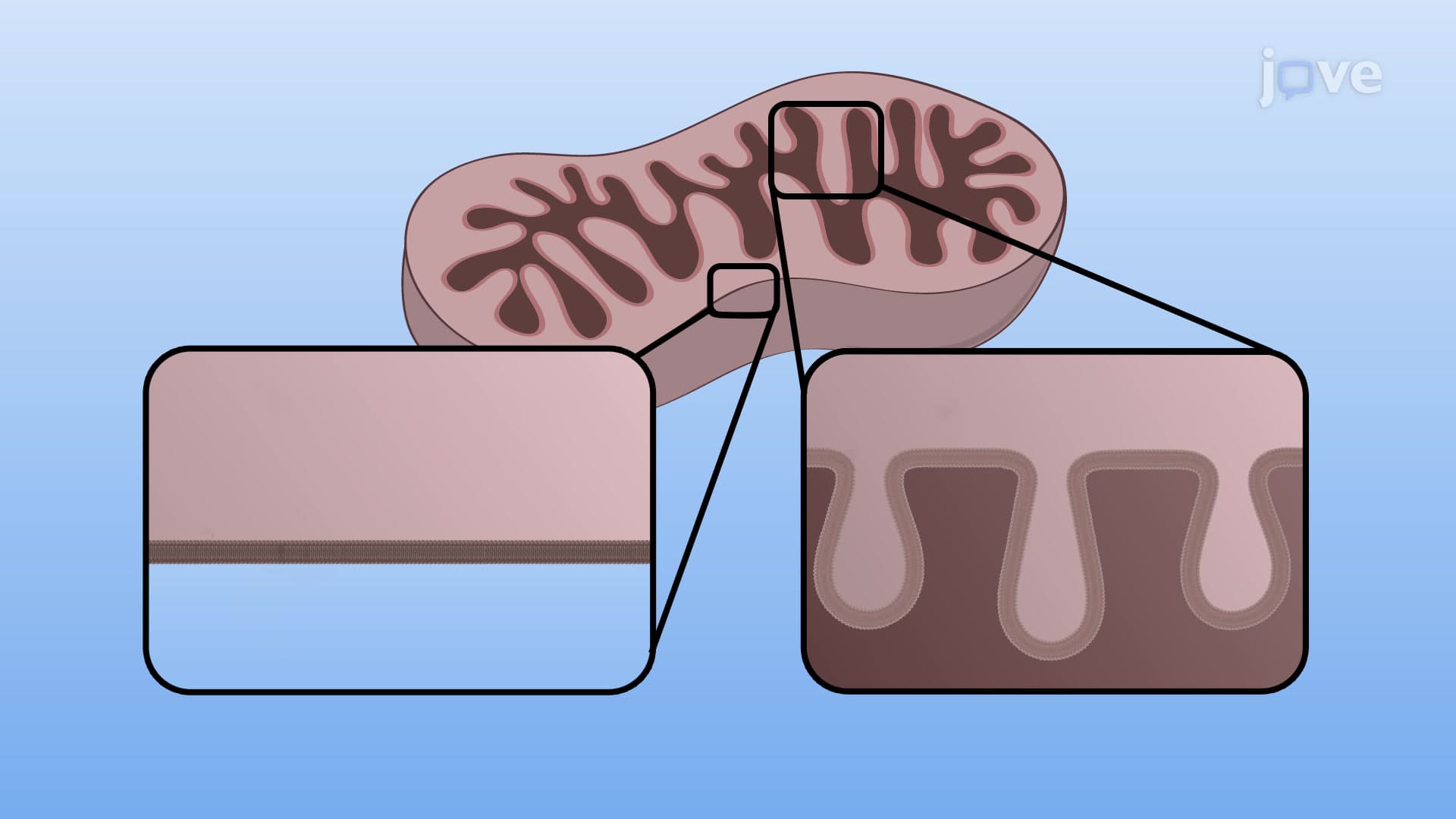
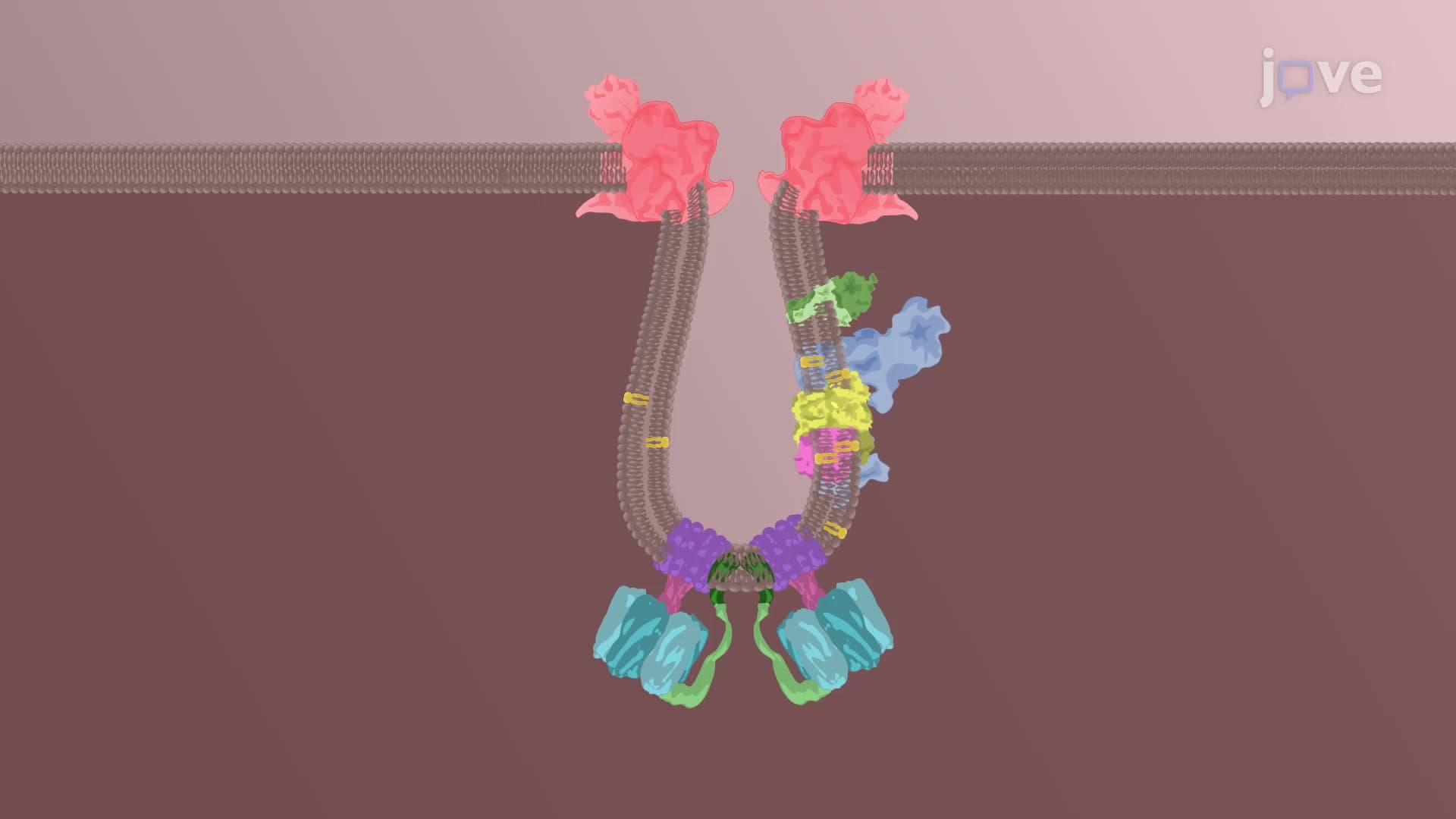
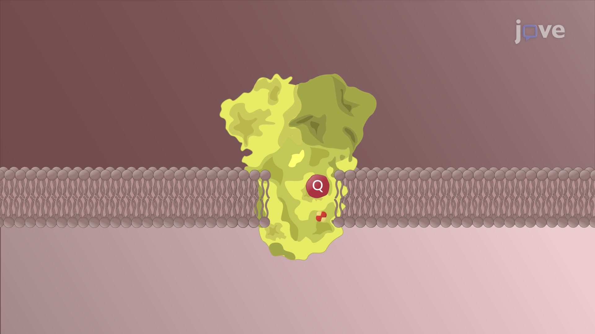
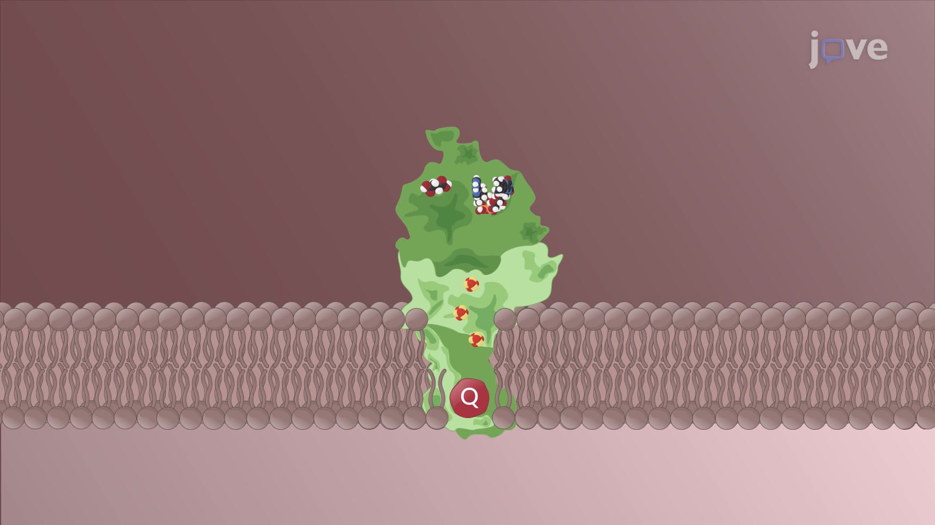
The electron transport chain complex I and II represent sophisticated molecular machines embedded in the inner mitochondrial membrane, each with distinct structural features that enable precise electron transfer. Complex I, formally known as NADH-Q oxidoreductase, stands as the largest protein complex in the respiratory chain, containing 45 subunits arranged in an characteristic L-shaped configuration. This massive assembly spans both the mitochondrial matrix and inner membrane, with seven subunits encoded by mitochondrial DNA-a critical point tested on the MCAT and AP Biology exams.
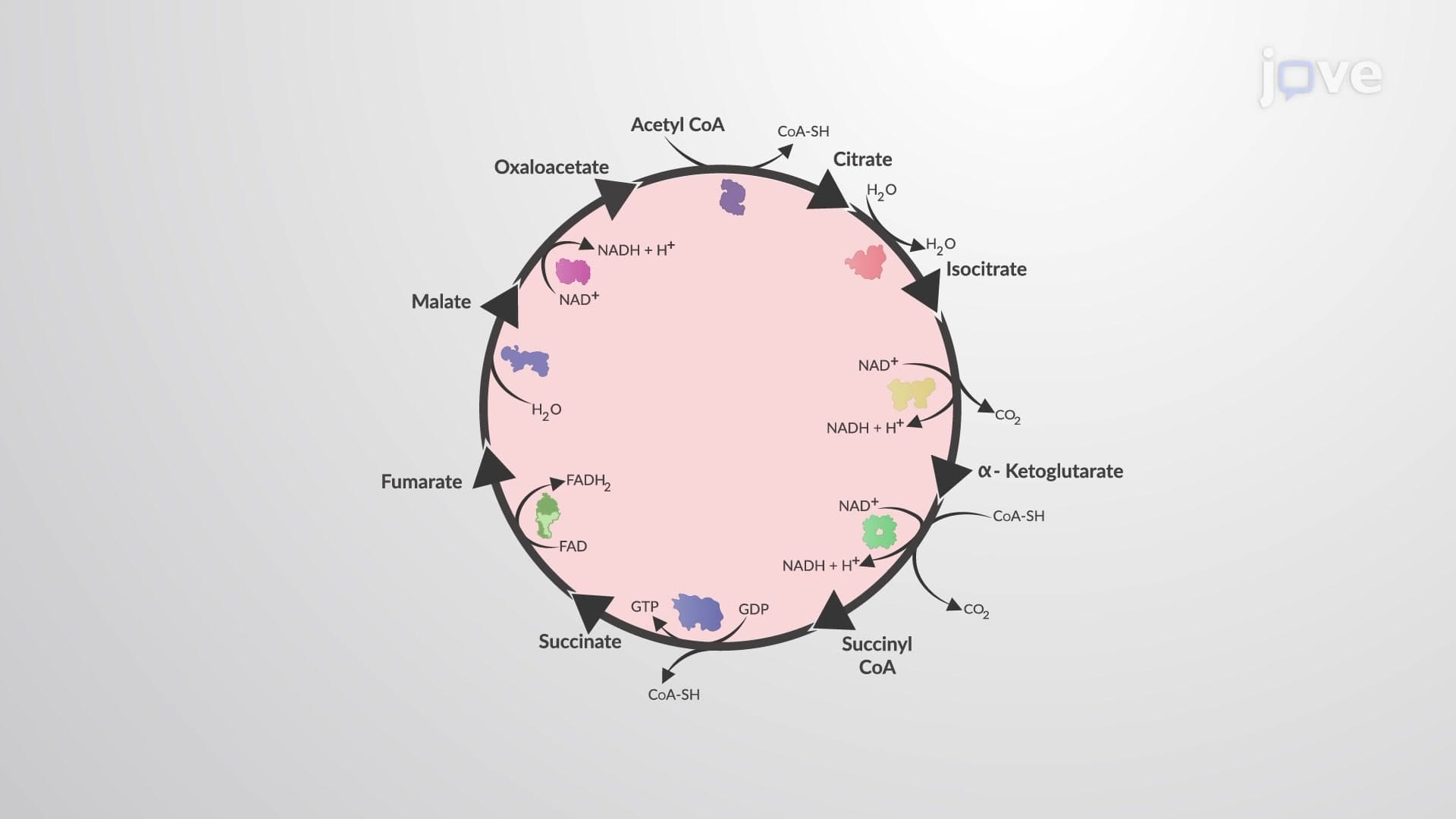
Complex I functions as the primary entry point for electrons derived from glycolysis and the citric acid cycle. The complex contains three essential functional domains: the NADH-binding site that accepts electrons from this high-energy carrier, flavin mononucleotide (FMN) serving as the immediate electron acceptor, and a series of iron-sulfur clusters that facilitate electron transfer to coenzyme Q (ubiquinone). This electron flow follows thermodynamically favorable energy gradients, with each transfer step releasing energy that Complex I harnesses to pump protons across the inner mitochondrial membrane.
Students preparing for college biochemistry courses should recognize that Complex I dysfunction underlies several human diseases, including Leber hereditary optic neuropathy and certain forms of Parkinson's disease. These clinical examples frequently appear on USMLE Step 1 examinations, emphasizing the medical relevance of understanding electron transport mechanisms.
Complex II, designated succinate-Q reductase, uniquely participates in both the citric acid cycle and electron transport chain, making it a favorite topic for AP Biology and college-level biochemistry exams. Unlike Complex I, Complex II is entirely encoded by nuclear DNA and consists of four subunits organized into two functional pairs. Subunits A and B form the catalytic core, with subunit A containing the FAD cofactor and succinate binding site, while subunit B houses three iron-sulfur clusters essential for electron transfer.
The hydrophobic subunits C and D anchor the complex within the inner mitochondrial membrane and contain the coenzyme Q binding site. This structural organization allows Complex II to oxidize succinate to fumarate while simultaneously reducing FAD to FADH2 and transferring electrons to coenzyme Q. Understanding this dual functionality helps students tackle complex metabolic pathway questions commonly found on standardized exams.
Both complexes serve as critical control points for cellular energy production, and their malfunction can lead to severe metabolic consequences. Complex II mutations cause certain forms of paraganglioma and pheochromocytoma, conditions frequently discussed in medical school pathology courses. For high school students, focusing on the basic electron transfer mechanisms and cofactor requirements provides excellent preparation for advanced coursework in biochemistry and cell biology.
Related Micro-courses