Loading subject page…


145,119 views

Video Summary: What Is the Inner Mitochondrial Membrane
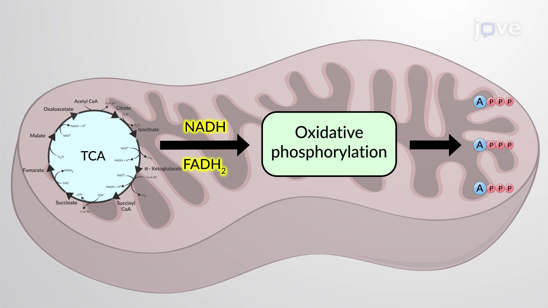
Did you know that mitochondria in a single human heart muscle cell can contain over 1,000 specialized internal folds? The inner mitochondrial membrane creates these intricate structures through distinct functional domains that power cellular energy production. From the electron transport chains studied in AP Biology to the ATP synthesis mechanisms crucial for medical school entrance exams like the MCAT, this membrane system drives the biochemical processes keeping every cell in your body alive. Watch the full video on JoVE Coach to master this concept with expert-led visuals and step-by-step explanations.
The inner mitochondrial membrane represents one of biology's most sophisticated membrane systems, featuring specialized domains that work together to power cellular metabolism. Unlike simple cellular membranes, this structure contains functionally distinct regions that create compartments with unique biochemical environments essential for energy production.
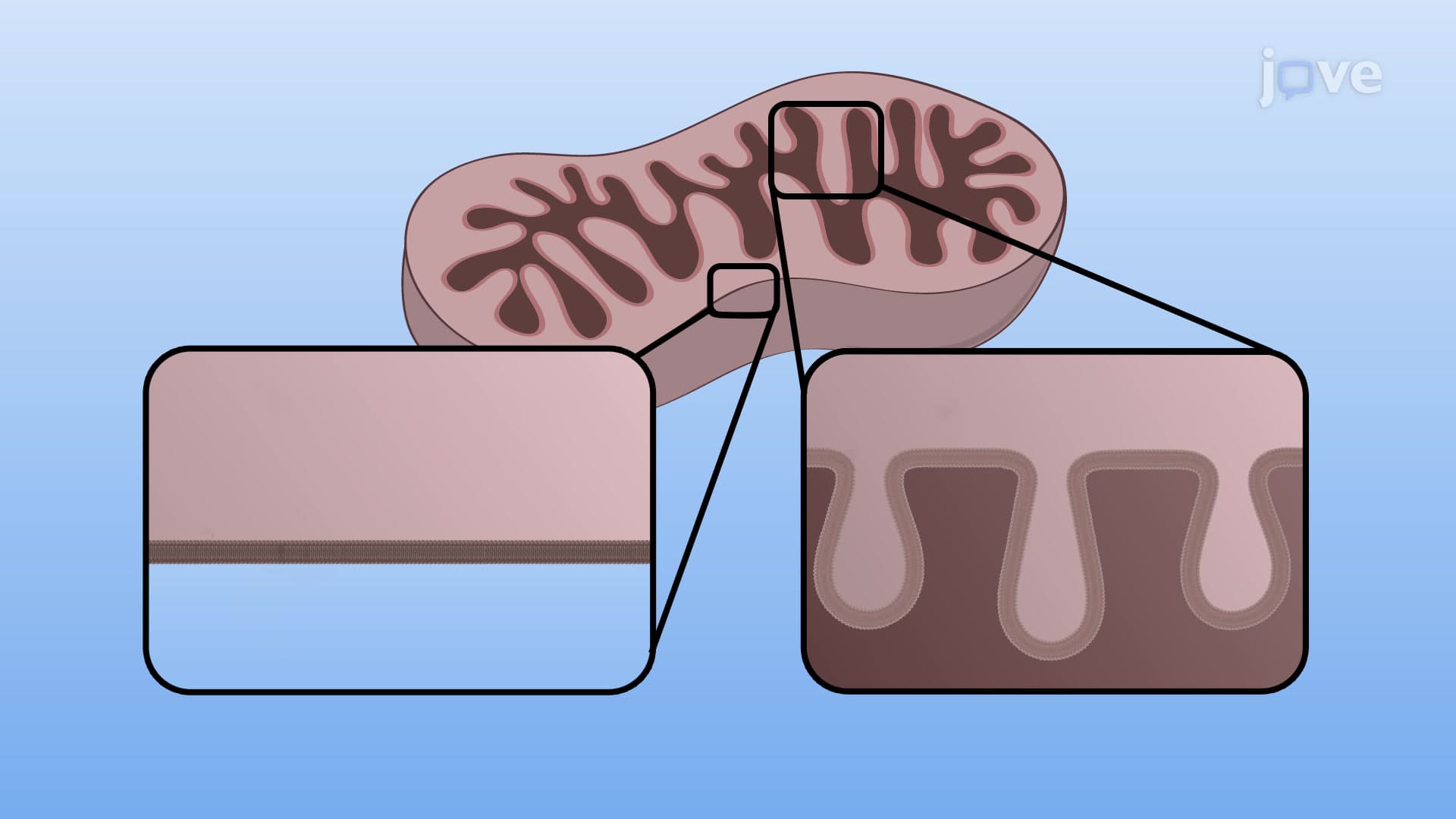
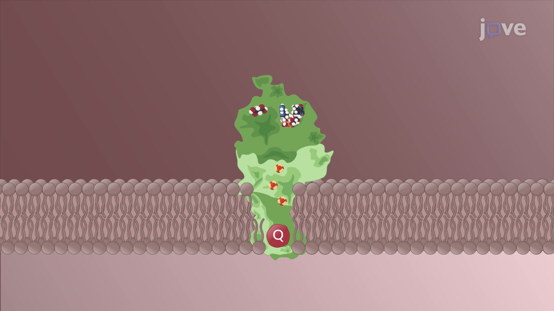
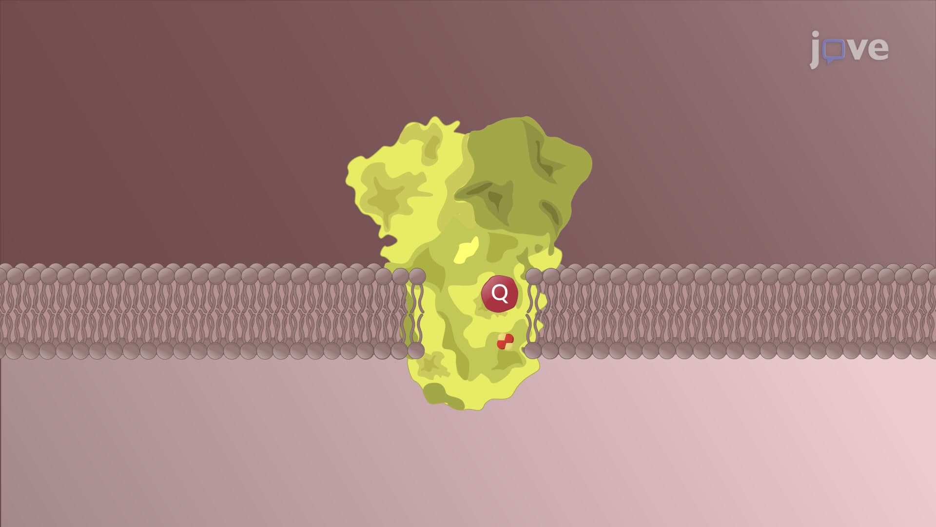
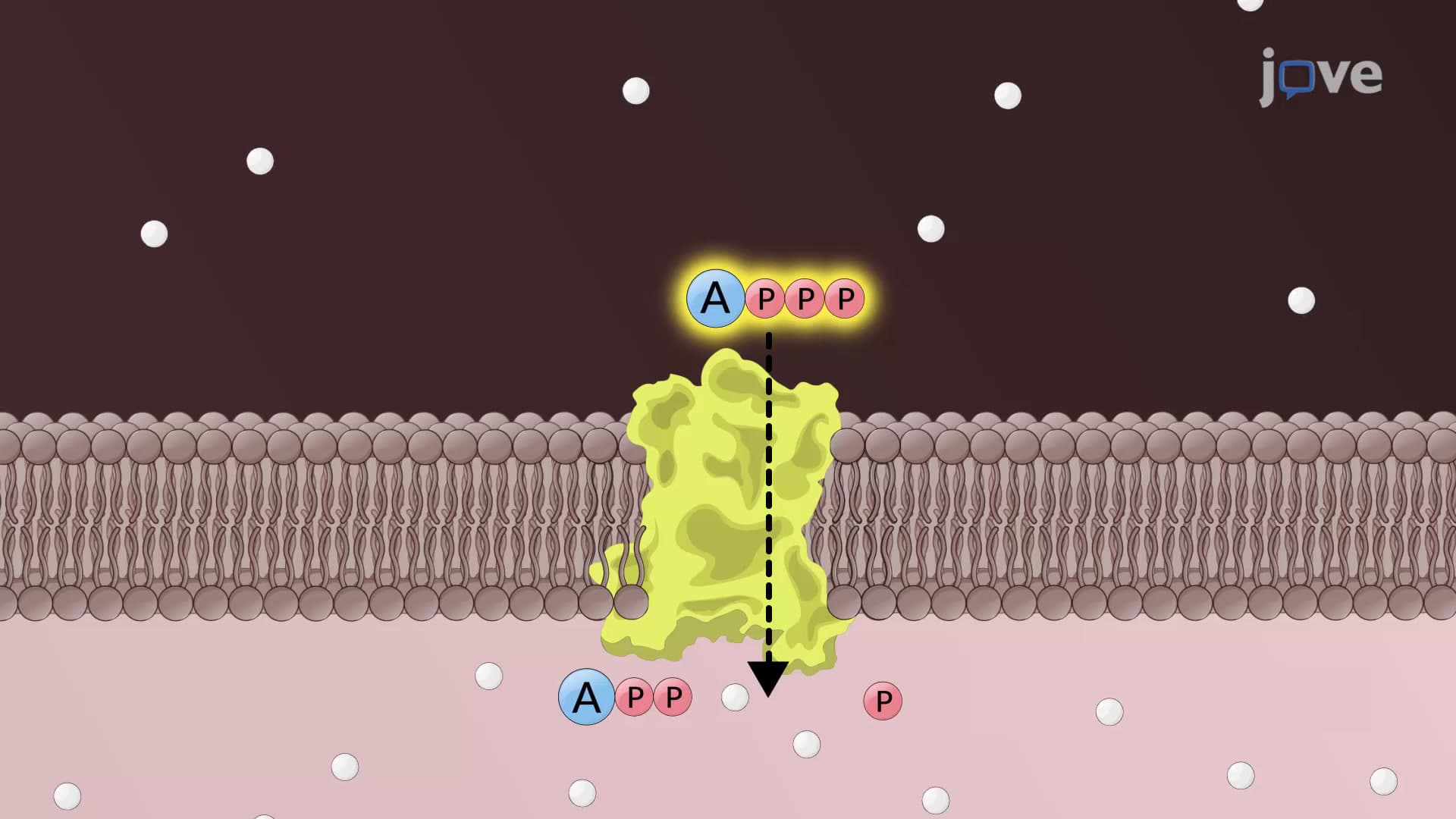
The inner boundary membrane forms the foundational layer directly adjacent to the outer mitochondrial membrane. This region serves as the organizational hub for electron transport chain complexes, containing assembly factors that arrange individual protein complexes into large, efficient supercomplexes. Students preparing for the MCAT or AP Biology exams should understand that this membrane contains heterogeneous compositions, with localized concentrations of specialized proteins like the mitochondrial contact site and cristae organizing system (MICOS) and ATP synthase.
The presence of cardiolipin, a unique four-chain phospholipid found almost exclusively in mitochondrial membranes, plays a crucial role in maintaining membrane curvature and supporting protein complex function. This lipid composition becomes particularly important when studying cardiac muscle cells, where mitochondria must maintain high-energy output to support continuous heart contractions.
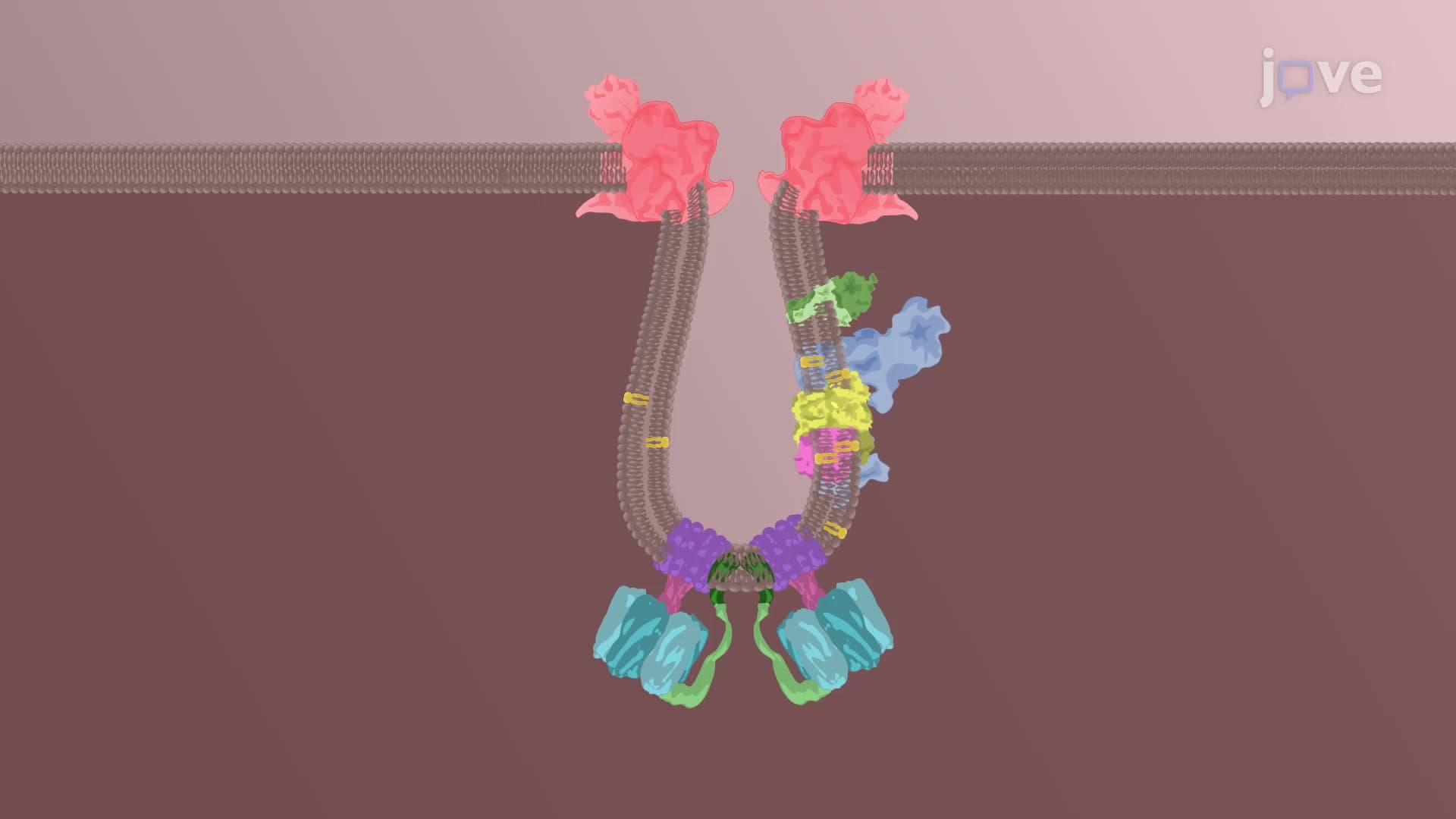
The inner membrane's ability to fold inward creates cristae, dramatically increasing the surface area available for biochemical reactions. This invagination process, promoted by MICOS complexes and specialized lipids, forms what researchers call the cristae membrane network. Each crista functions as a semi-independent compartment optimized for specific aspects of cellular respiration.
For college biochemistry students, understanding cristae structure proves essential when calculating ATP yield from glucose metabolism. The increased surface area allows for more electron transport chains and ATP synthase complexes, directly correlating to enhanced energy production capacity.
Crista junctions represent narrow, tubular connections between the cristae membrane and inner boundary membrane. These structures, supported by MICOS complexes, act as selective barriers that prevent random mixing of proteins and lipids between compartments. This compartmentalization ensures that each membrane domain maintains its specialized composition necessary for optimal mitochondrial function.
Students studying for medical school entrance exams should recognize that crista junction dysfunction contributes to various metabolic disorders, making this concept clinically relevant for future healthcare professionals.
Related Micro-courses