Loading subject page…


33,104 views

Video Summary: What Is Liver Regeneration
Did you know the liver is the only human organ that can completely rebuild itself after losing up to 75% of its tissue? Liver regeneration is this remarkable biological process where hepatocytes exit their dormant state and rapidly multiply to restore damaged tissue. When patients at Johns Hopkins receive partial liver transplants, both the donor's remaining liver and the recipient's transplanted portion regenerate to full size within months. This extraordinary healing mechanism involves complex cellular signaling between Kupffer cells, stellate cells, and growth factors that orchestrate precise tissue repair. Watch the full video on JoVE Coach to master this concept with expert-led visuals and step-by-step explanations.
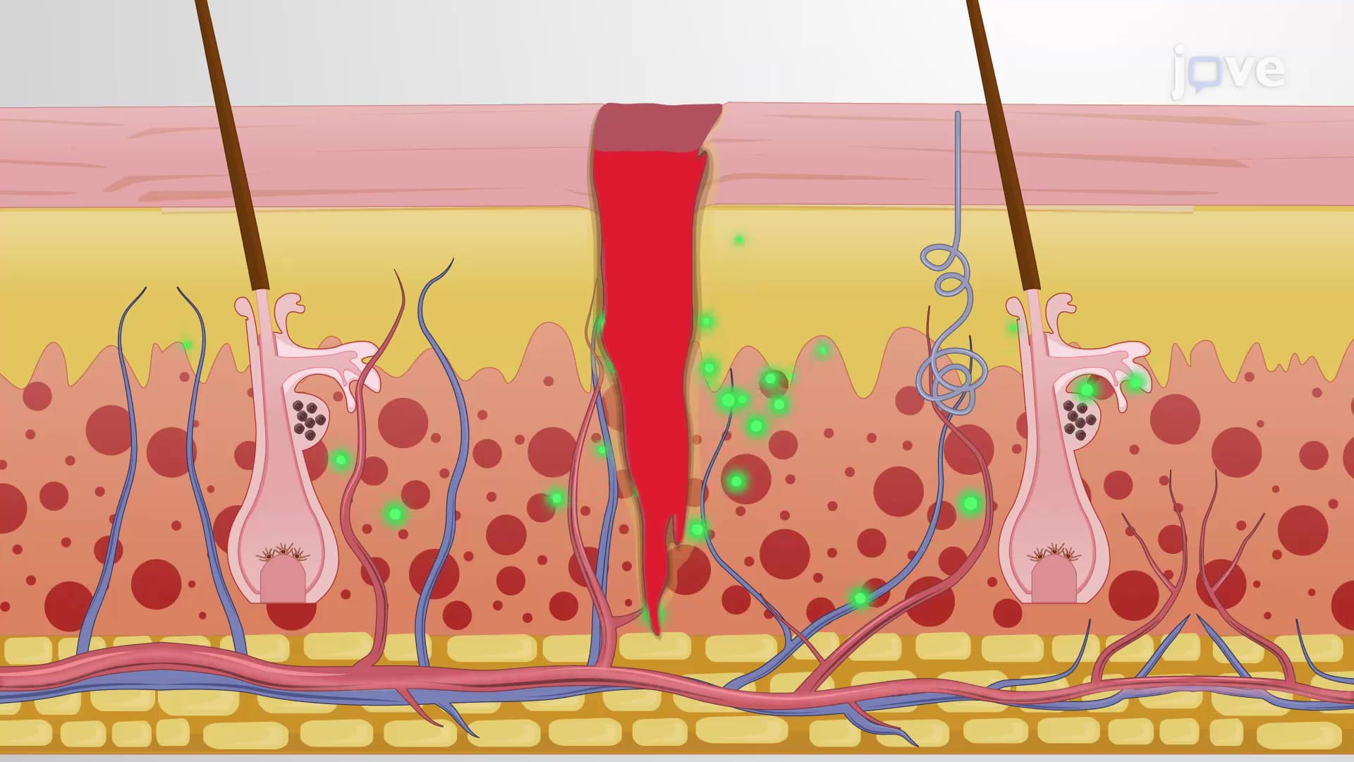
Liver regeneration represents one of biology's most impressive healing mechanisms, enabling complete functional and structural restoration after significant tissue loss. Unlike other organs that form scar tissue when damaged, the liver rebuilds itself with fully functional hepatocytes, maintaining its critical metabolic, detoxification, and synthetic functions. This process is clinically vital for patients undergoing liver resection at major medical centers like Mayo Clinic or receiving living donor transplants.
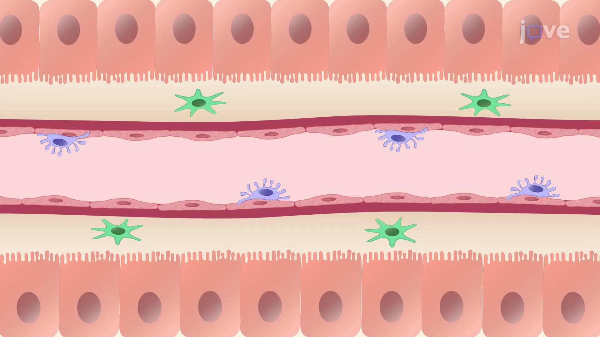
The liver's regenerative power stems from its unique cellular composition and organization. Hepatocytes comprise approximately 80% of liver mass and normally remain in the quiescent G0 phase, performing routine metabolic functions without dividing. This dormant state changes dramatically following injury, infection, or surgical removal. The liver's specialized architecture, including sinusoidal capillaries lined with Kupffer cells and scattered stellate cells, creates an ideal environment for coordinated regenerative responses.
The regenerative process begins when Kupffer cells-the liver's resident macrophages-detect tissue damage and release inflammatory cytokines IL-6 and TNF-α. These signaling molecules prime hepatocytes by inducing expression of growth factor receptors c-MET and EGFR on their surface. Simultaneously, stellate cells and duodenal tissues release hepatocyte growth factor (HGF) and epidermal growth factor (EGF), which travel through the bloodstream to bind their respective receptors on primed hepatocytes.
This receptor binding triggers intracellular signaling pathways that override the G0/G1 cell cycle checkpoint, compelling normally dormant hepatocytes to enter active proliferation. The precision of this system ensures that cell division occurs specifically at injury sites rather than throughout the entire organ.
The regenerative process includes sophisticated termination mechanisms preventing excessive growth. Once appropriate organ size and tissue architecture are restored, stellate cells release transforming growth factor-β (TGF-β), which blocks further hepatocyte proliferation and returns cells to their quiescent state. This regulatory control distinguishes healthy regeneration from pathological overgrowth.
Understanding liver regeneration is crucial for pre-med students preparing for the MCAT's biological systems section and appears frequently in AP Biology discussions of cell cycle regulation. Medical students encounter this concept extensively in pathology and surgery courses, while nursing students studying for the NCLEX must understand regeneration's role in patient recovery following liver procedures.
Related Micro-courses