Loading video…


 20 Concepts
 158.1K views

Video Summary: Insertion of Single Pass Transmembrane Explained
Did you know that insulin receptors on muscle cells rely on precise protein insertion to function properly? The insertion of single pass transmembrane proteins is a critical cellular process where proteins are strategically positioned across cell membranes. For example, the insulin receptor in pancreatic beta cells must be correctly oriented to detect blood glucose levels and trigger insulin release. This complex mechanism involves signal sequences, specialized channels, and precise molecular machinery working together to create functional membrane proteins. Watch the full video on JoVE Coach to master this concept with expert-led visuals and step-by-step explanations.
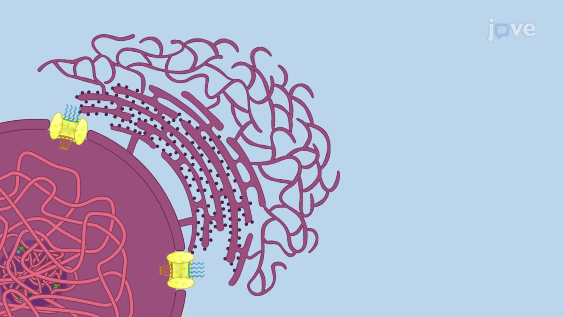
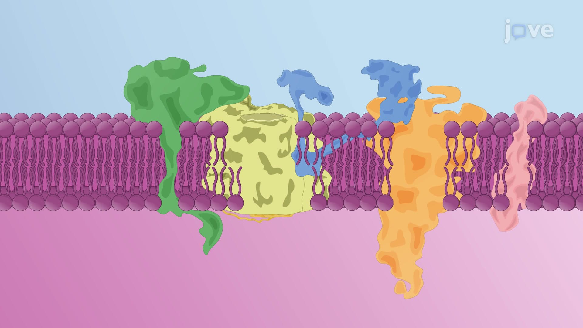
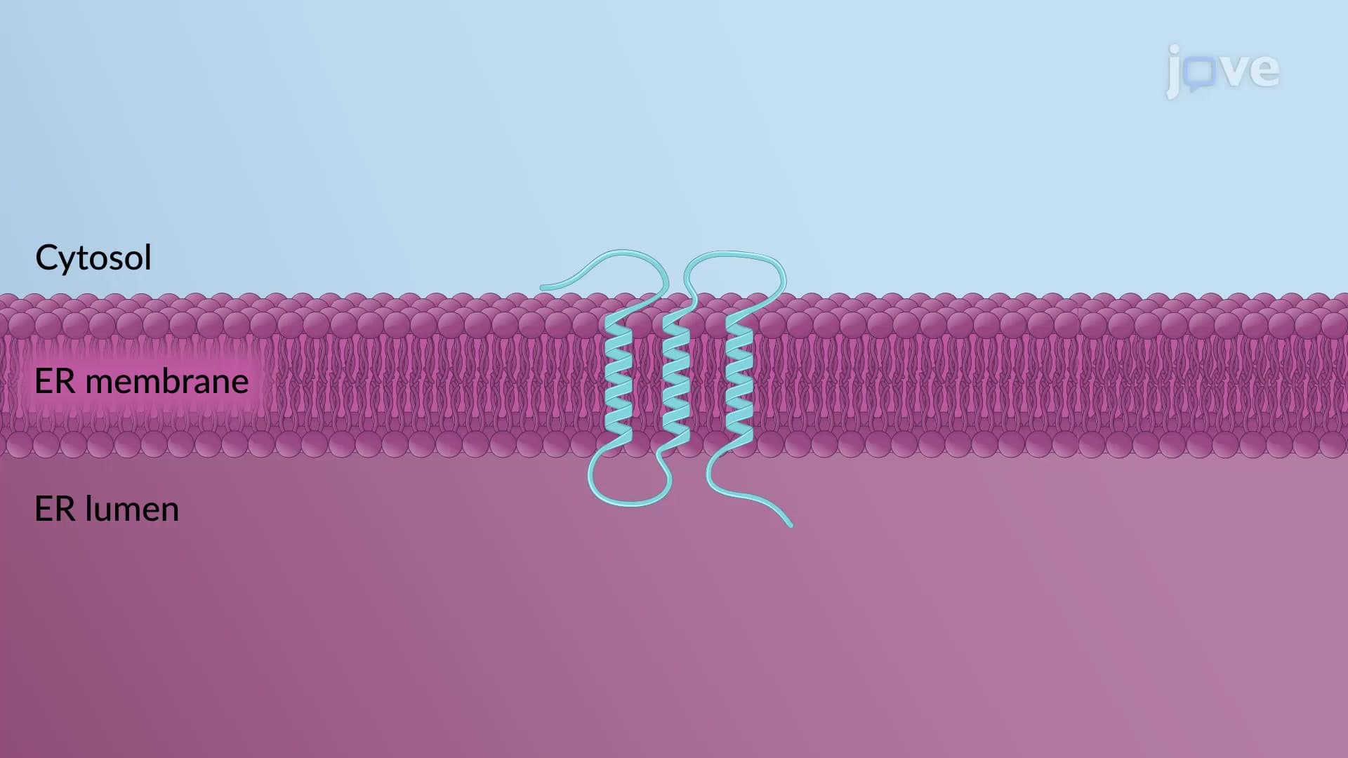
The insertion of single pass transmembrane represents one of the most sophisticated quality control mechanisms in cell biology. This process ensures that membrane proteins achieve their correct orientation and topology within cellular membranes, particularly the endoplasmic reticulum (ER). Unlike soluble proteins that are completely translocated across membranes, transmembrane proteins must be precisely positioned to span the membrane while maintaining specific domains on either side.
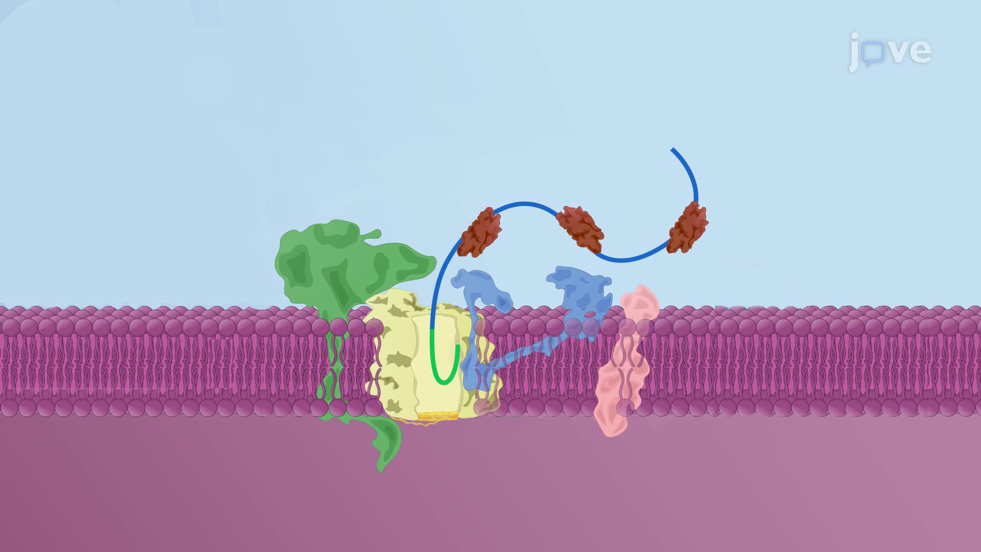
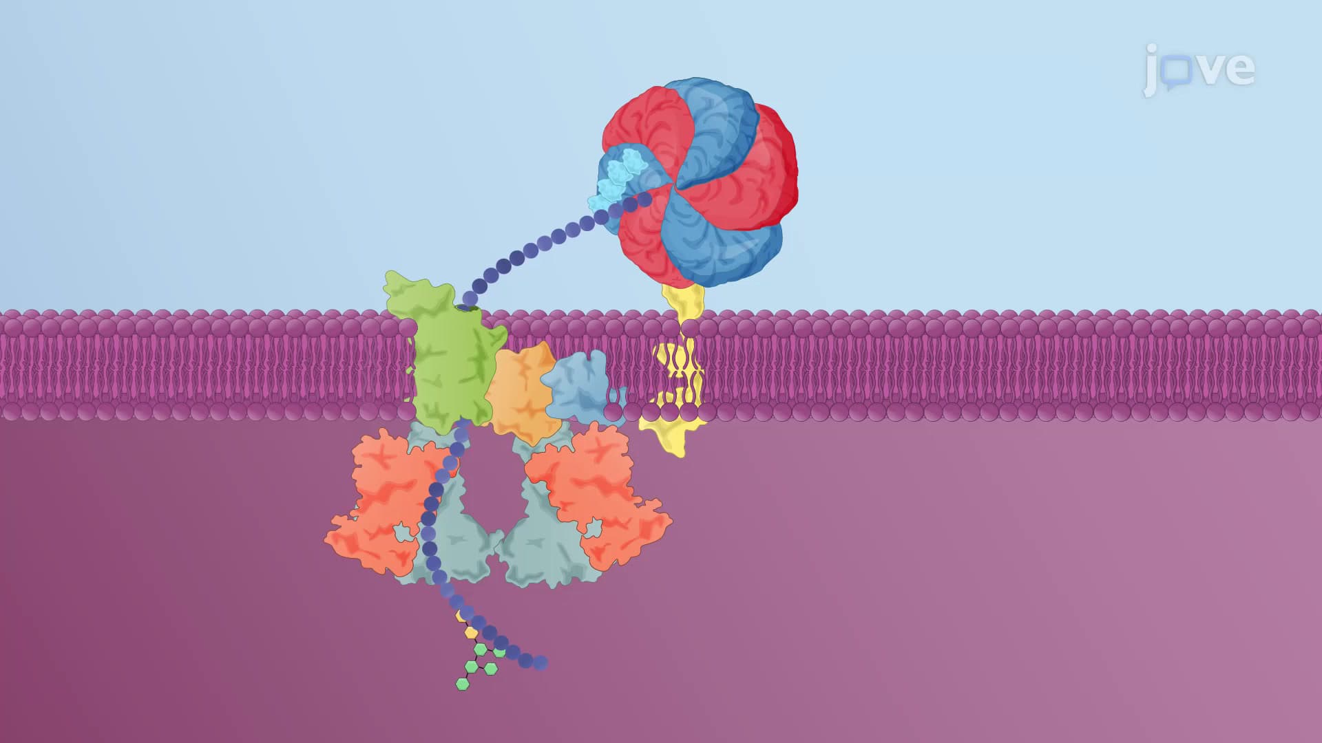
The process begins when ribosomes synthesizing transmembrane proteins recognize specific signal sequences. The ER signal sequence acts as a molecular address label, directing the ribosome-nascent protein complex to the ER membrane. The Sec61 translocon channel serves as the gateway, forming a protein-conducting channel that can open both vertically (for complete translocation) and laterally (for membrane integration). This dual functionality is crucial for accommodating different protein destinies.
Signal peptidase complexes work alongside the translocon, cleaving signal sequences at precise locations to release protein domains into the ER lumen. Meanwhile, the lateral gate mechanism allows hydrophobic transmembrane domains to escape into the lipid bilayer rather than continuing through the aqueous channel. This sophisticated sorting system ensures that each protein domain reaches its intended cellular compartment.
The final orientation of transmembrane proteins depends on the distribution of charged residues, particularly the "positive-inside rule." Type I proteins have their N-terminus in the ER lumen (extracellular space) and C-terminus in the cytoplasm. Classic examples include growth factor receptors like EGFR, which are crucial for cancer research at institutions like MD Anderson Cancer Center in Houston.
Type II proteins display the opposite orientation, with N-terminus in the cytoplasm and C-terminus in the lumen. This configuration often results from positively charged residues preceding the transmembrane domain, which favors cytoplasmic retention according to the positive-inside rule. Understanding these orientations is essential for MCAT preparation and advanced cell biology courses at universities like Stanford and MIT.
Proper transmembrane protein insertion is vital for cellular function. Defects in this process contribute to diseases like cystic fibrosis, where the CFTR protein fails to reach the cell surface properly. Pharmaceutical companies such as Vertex Pharmaceuticals have developed drugs targeting these insertion defects, highlighting the clinical relevance of understanding transmembrane protein biogenesis. Students preparing for the USMLE should recognize how insertion defects can lead to protein misfolding diseases and therapeutic opportunities.
Related Micro-courses