Loading video…


 20 Concepts
 158.1K views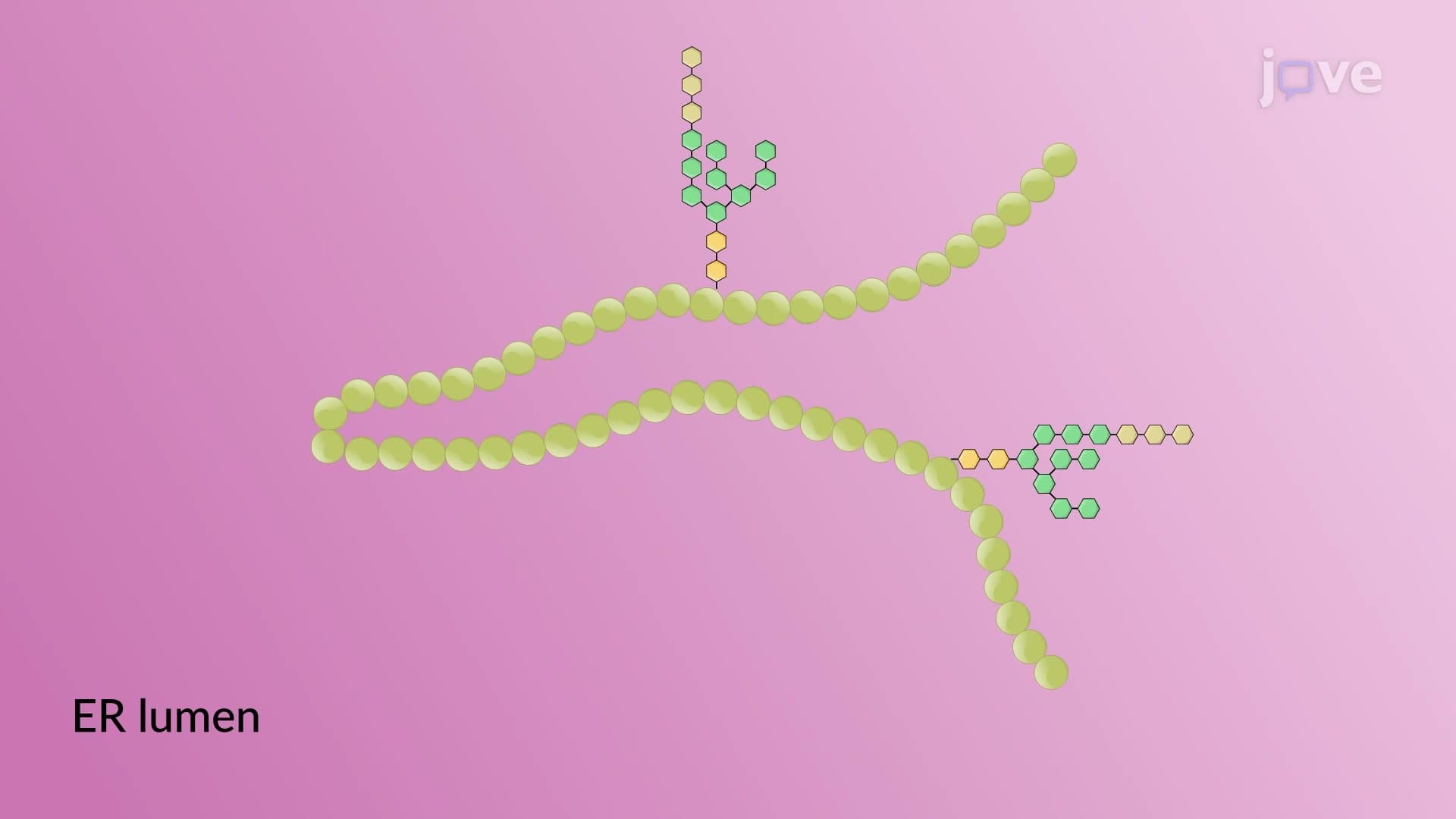

Video Summary: What Is Protein Folding Quality Check
Ever wonder how your body ensures that critical proteins like insulin are properly shaped before being released into your bloodstream? The protein folding quality check is a sophisticated cellular mechanism that acts like a molecular assembly line inspector, ensuring proteins achieve their correct three-dimensional structure in the endoplasmic reticulum. This quality control system prevents misfolded proteins-which can cause diseases like cystic fibrosis-from leaving the cellular factory. Watch the full video on JoVE Coach to master this concept with expert-led visuals and step-by-step explanations.
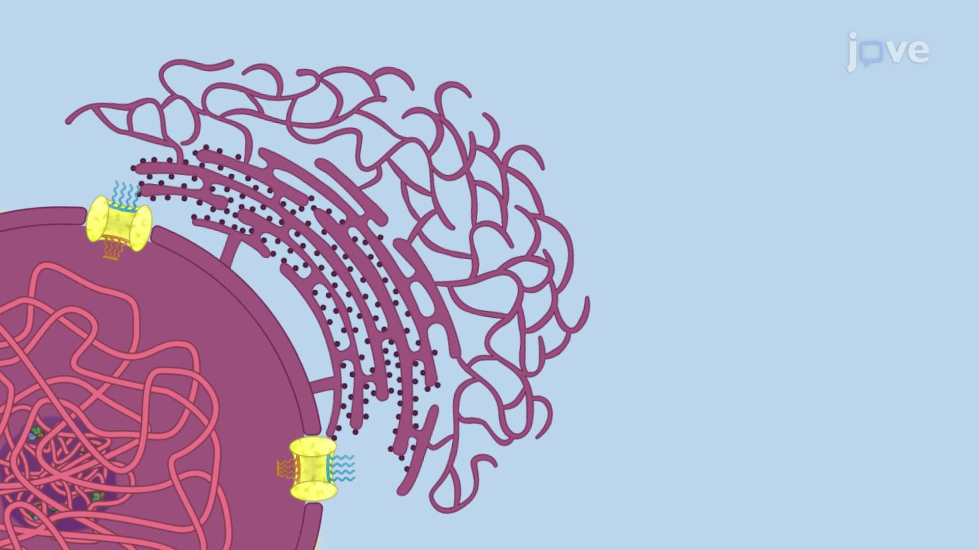
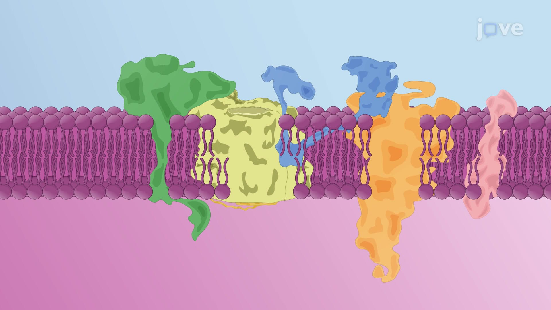
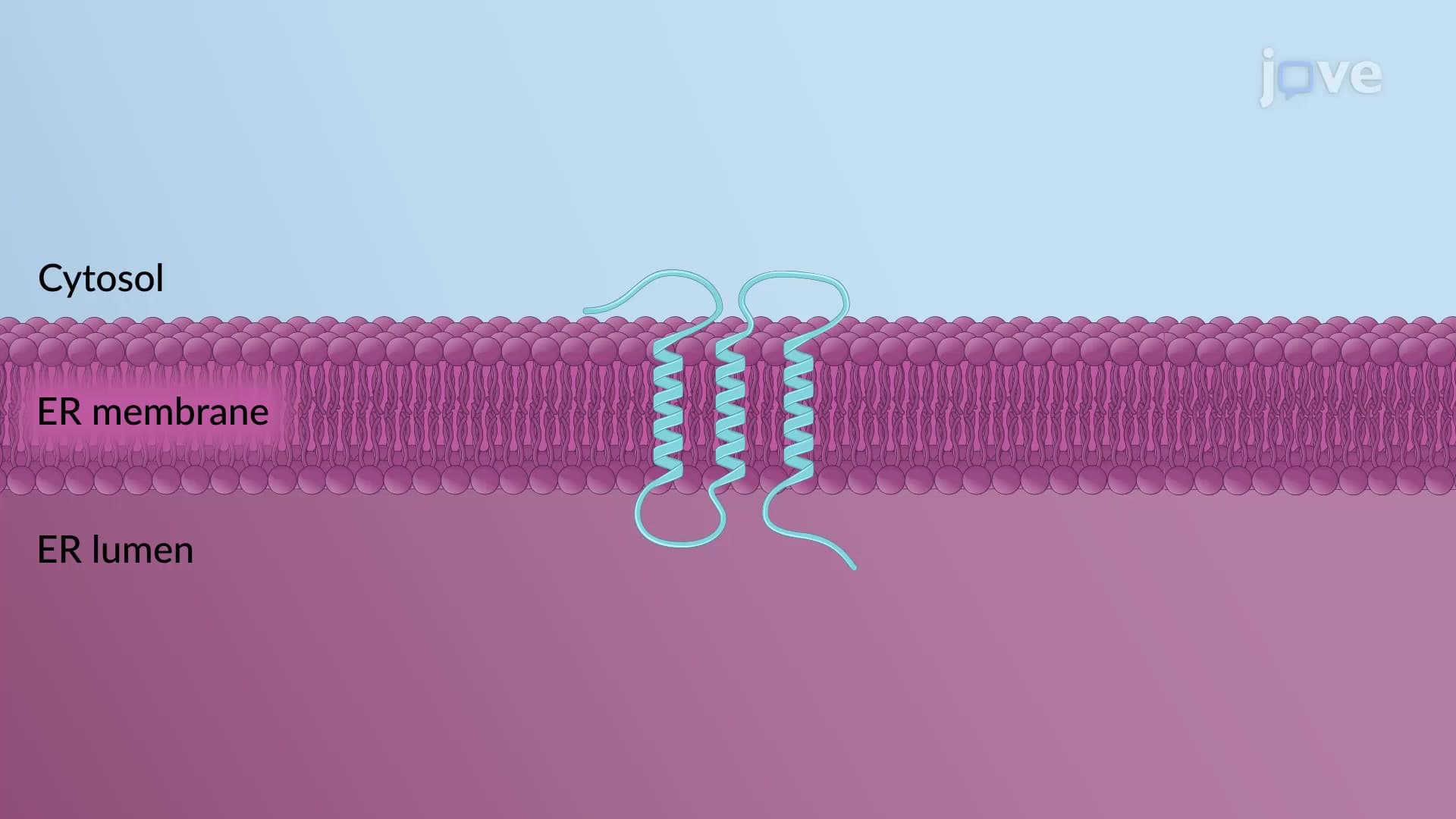
The protein folding quality check represents one of biology's most sophisticated quality control mechanisms, operating within the endoplasmic reticulum (ER) to ensure proteins achieve their functional three-dimensional structure. This process is particularly crucial for secreted proteins and membrane proteins that must maintain precise shapes to function properly in the human body.
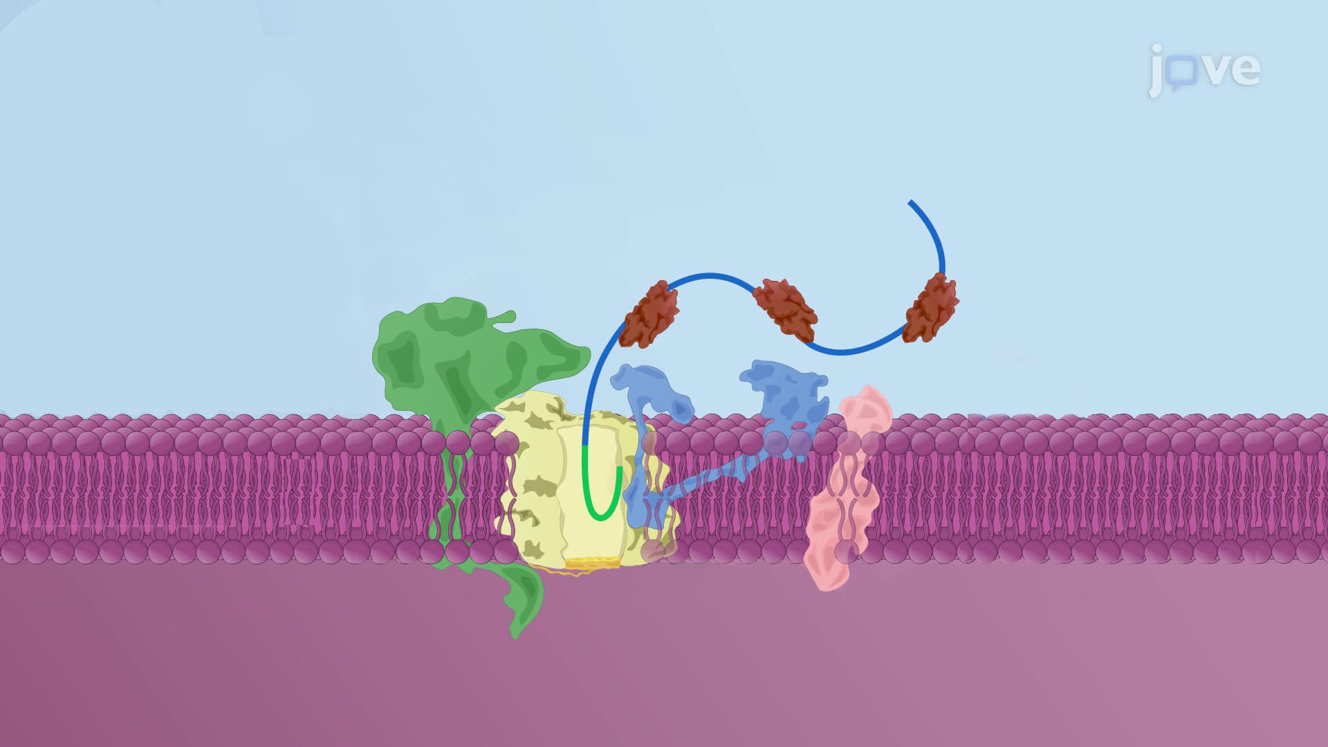
The quality check system relies on a series of carefully orchestrated modifications to N-linked glycans-complex carbohydrate structures attached to asparagine residues on newly synthesized proteins. Initially, these glycans contain three terminal glucose residues that serve as molecular barcodes indicating the protein's folding status.
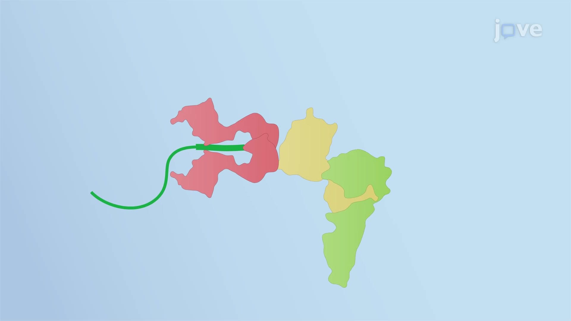
The process begins when glucosidase I removes the outermost glucose, followed by glucosidase II removing a second glucose residue. This creates a monoglucosylated glycan that serves as a recognition signal for the lectin chaperones calnexin (membrane-bound) and calreticulin (soluble). These chaperones act like molecular assistants, recruiting additional folding machinery to help the protein achieve its proper conformation.
The most fascinating aspect of this system involves the UDP-glucose:glycoprotein glucosyltransferase 1 (UGGT1) enzyme, which functions as the primary quality inspector. After glucosidase II removes the final glucose residue, UGGT1 performs a sophisticated structural assessment of the protein. If the protein has achieved proper folding, it receives clearance to exit the ER and continue to its cellular destination.
However, if UGGT1 detects structural problems-such as exposed hydrophobic regions or incorrect disulfide bonds-it adds a glucose residue back to the glycan. This reglucosylation effectively sends the protein back to the "folding department" for another round of chaperone-assisted folding attempts.
Understanding protein folding quality check mechanisms is essential for students pursuing careers in medicine, biotechnology, or pharmaceutical research. Defects in this system contribute to numerous diseases, including cystic fibrosis (caused by misfolded CFTR protein), certain forms of diabetes (involving misfolded insulin), and neurodegenerative diseases like Alzheimer's.
For students preparing for the MCAT or AP Biology exams, this topic frequently appears in questions about cellular biology, protein structure, and disease mechanisms. The concept also relates directly to current therapeutic approaches, such as pharmacological chaperones being developed by companies like Amicus Therapeutics to treat lysosomal storage disorders.
Related Micro-courses